. 2014 Apr 25;114(9):1435-45.
doi: 10.1161/CIRCRESAHA.114.303634. Epub 2014 Mar 17.

Targeted overexpression of tissue inhibitor of matrix metalloproteinase-4 modifies post-myocardial infarction remodeling in mice

Affiliations

Targeted overexpression of tissue inhibitor of matrix metalloproteinase-4 modifies post-myocardial infarction remodeling in mice

Juozas A Zavadzkas et al. Circ Res. .

Abstract

Rationale: Myocardial infarction (MI) causes an imbalance between matrix metalloproteinases and tissue inhibitors of matrix metalloproteinases (TIMPs) and is associated with adverse left ventricular (LV) remodeling. A uniform reduction in TIMP-4 post-MI has been observed.

Objective: To examine post-MI remodeling with cardiac-restricted overexpression of TIMP-4, either through a transgenic or viral delivery approach.

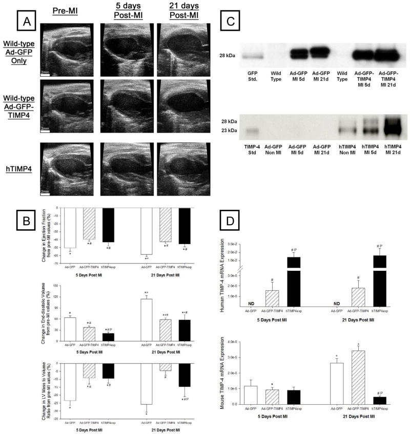
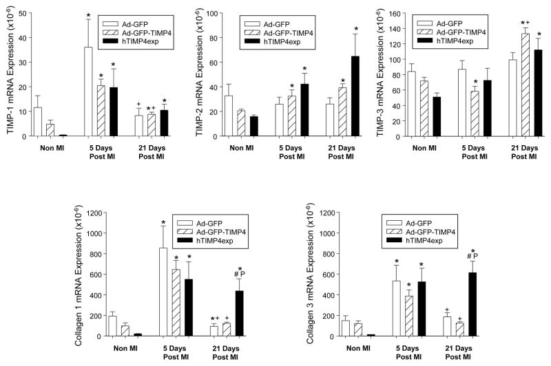
Methods and results: MI was induced in mice and then randomized to targeted injection of an adenoviral construct (10 μL; 8×10(9) plaque forming units/mL) encoding green fluorescent protein (GFP) and the full-length human TIMP-4 (Ad-GFP-TIMP4) or GFP. A transgenic construct with cardiac-restricted overexpression TIMP-4 (hTIMP-4exp) was used in a parallel set of studies. LV end-diastolic volume, an index of LV remodeling, increased by >60% from baseline at 5 days post-MI and by >100% at 21 days post-MI in the Ad-GFP only group. However, LV dilation was reduced by ≈50% in both the Ad-GFP-TIMP4 and hTIMP-4exp groups at these post-MI time points. LV ejection fraction was improved with either Ad-GFP-TIMP-4 or hTIMP-4exp. Fibrillar collagen expression and content were increased within the MI region with both TIMP-4 interventions, suggestive of matrix stabilization.

Conclusions: This study is the first to demonstrate that selective myocardial targeting for TIMP-4 induction through either a viral or transgenic approach favorably altered the course of adverse LV remodeling post-MI. Thus, localized induction of endogenous matrix metalloproteinase inhibitors, such as TIMP-4, holds promise as a means to interrupt the progression of post-MI remodeling.

Keywords: extracellular matrix; myocardial infarction; tissue inhibitor of metalloproteinases; ventricular function.


Figures

Figure 1
Figure 1
(A) Representative echocardiograms of the LV long axis taken at end diastole at the baseline study (pre-MI time point) and in terminal studies performed at 5 or 21 days post-MI in mice injected with either Ad-GFP only (Top Panel) or the Ad-GFP-TIMP-4 construct (Middle Panel). In a second set of studies, mice with transgenic overexpression of human TIMP-4 (hTIMP-4exp) were treated in identical fashion (Bottom Panel). Significant LV dilation was observed in all groups at 5 and 21 days post-MI, but the relative degree of LV dilation appeared attenuated in both the Ad-GFP-TIMP4 and hTIMP-4exp groups. Summary data are presented in Table 1. (B) Key LV response variables as a function from baseline. LV ejection fraction significantly fell in all groups at 5 and 21 days post-MI, but this relative reduction was attenuated in the Ad-GFP-TIMP4 or hTIMP-4exp groups. LV end-diastolic volume and LV mass/volume ratio, both indices of post-MI remodeling, were significantly reduced in the Ad-GFP-TIMP-4 or hTIMP-4exp groups. (C) LV myocardial levels of GFP were expressed at equivalent levels in both adenoviral injection groups at both 5 and 21 days post-MI. In the hTIMP-4exp line, consistent human TIMP-4 levels were obtained in the non-MI referent control mice and these levels were higher at 5 and 21 days post MI. (D) LV myocardial mRNA levels for human TIMP-4 were detectable in the Ad-GFP-TIMP4 and hTIMP-4exp groups at 5 and 21 days post-MI. While endogenous mouse TIMP-4 was reduced at 5 days post-MI, expression levels were higher at 21 days post-MI with the notable exception in the hTIMP-4 group. [*p<0.05 compared to baseline (or referent non-MI values), # compared to Ad-GFP only group, Pp<0.05 vs Ad-GFP-TIMP-4 group].
Figure 2
Figure 2
(A) Distribution plots for myocyte cross sectional area (CSA) for referent control and within the LV of the remote, viable myocardium at 5 and 21 days post-MI in the Ad-GFP, Ad-GFP-TIMP-4, and the hTIMP-4exp groups. A significant increase in myocyte CSA occurred post-MI in all groups, which appeared higher in the Ad-GFP-TIMP-4 group at 21 days post-MI. (B) LV relative collagen content was computed in both the MI and remote regions in all groups at 5 and 21 days post-MI using morphometric measurements on PSR stained sections. While collagen content was increased within the MI region as well as in the remote region in all groups, consistent with a post-MI fibrotic response, these values were higher in both TIMP-4 induction groups. [*p<0.05 compared to referent non-MI values, +p<0.05 vs respective 5 day values, # compared to Ad-GFP only group, $p<0.05 vs Ad-GFP-TIMP-4 group; sample sizes are n>10/group/time point].
Figure 3
Figure 3
Relative myocardial mRNA levels for TIMP-1, TIMP-2, and TIMP-3, as well as for the fibrillar collagens, collagen type I and III, were computed in referent non-MI mice immediately following myocardial injection of Ad-GFP, Ad-GFP-TIMP4, or in hTIMP-4exp mice and at 5 and 21 days post-MI. TIMP-1 levels increased at 5 days post-MI and then fell at 21 days post-MI with no significant differences between groups. Relative TIMP-2 mRNA levels increased in the Ad-GFP-TIMP4 and hTIMP-4exp groups at 5 and 21 days post-MI. At 21 days post-MI, TIMP-3 mRNA levels were higher in the Ad-GFP-TIMP4 and hTIMP-4exp groups. At 5 days post-MI, increased mRNA levels for collagen I and III were equivalently increased in all groups. However, a persistent induction of collagen I and III occurred at 21 days post-MI in the hTIMP-4exp group. [*p<0.05 compared to referent non-MI values, +p<0.05 vs respective 5 day values, # compared to Ad-GFP only group, Pp<0.05 vs Ad-GFP-TIMP-4 group; sample sizes are n>10/group/time point].
Figure 4
Figure 4
(Left Panels) LV myocardial mRNA levels for MT1-MMP increased in all groups at 5 days post-MI and remained elevated at 21 days post-MI in the hTIMP-4exp group. Relative DDR2 mRNA levels appeared lower in the Ad-GFP-TIMP-4 group with no MI and at 5 days post-MI, whereas DDR2 mRNA levels were increased in the hTIMP-4exp group at 21 days post-MI. TGFbeta-R1 mRNA levels increased in all post-MI time points in all groups, but was highest in the hTIMP-4exp groups. (Middle Panels) LV myocardial IL-6 content increased in all groups at 5 days post-MI and reduced to referent control values by 21 days post-MI. However, IL-6 levels were lower in the hTIMP-4exp group at 5 days post-MI. LV myocardial IL-10 levels were detectable in referent control and Ad-GFP post-MI time points but were significantly reduced in the Ad-GFP-TIMP-4 group and non-detectable (ND) in the hTIMP-4exp group. MCP-1 was increased at 5 days post-MI in all groups but reduced in the hTIMP-4exp group. By 21 days post-MI, MCP-1 levels had fallen below referent control values. (Right Panels) LV myocardial expression levels for Bcl2 were higher at the non-MI and 21 day post-MI time point in the hTIMP-4exp group. A significant induction of Apaf1 and Ripk1 occurred at 5 days post-MI in the Ad-GFP group but not in either the Ad-GFP-TIMP-4 or hTIMP-4exp groups. [*p<0.05 compared to baseline (or referent non-MI values), +p<0.05 vs respective 5 day values, # compared to Ad-GFP only group, Pp<0.05 vs Ad-GFP-TIMP-4 group; sample sizes are n>6/group/time point].
Figure 5
Figure 5
(Top) Representative myocardial zymograms performed in referent non-MI mice immediately following myocardial injection of Ad-GFP, Ad-GFP-TIMP4, or in hTIMP-4exp mice and at 5 and 21 days post-MI. The latent form of MMP-9 (92 kDa) and the latent and active forms of MMP-2 (72/68 kDa, respectively) were identified and quantified by densitometry. (Bottom) Summary results from zymography were computed as a percent of referent non-MI values, whereby referent values were set to 100% (dashed line). In the Ad-GFP group, MMP-9 values increased at 5 days post-MI and returned to within normal ranges by 21 days post-MI. In the Ad-GFP-TIMP4 and hTIMP-4exp group, relative MMP-9 levels did not increase at 5 days post-MI and were actually reduced at specific post-MI time points. For MMP-2, a robust increase in active MMP-2 occurred in the Ad-GFP group at 5 and 21 days post-MI, which was significantly blunted in both the Ad-GFP-TIMP4 and hTIMP-4exp groups. [*p<0.05 compared to referent non-MI values, +p<0.05 vs respective 5 day values, # compared to Ad-GFP only group; sample sizes are n>10/group/time point]
Figure 6
Figure 6
(Left Panels) LV murine myocardial fibroblasts were transduced with either Ad-GFP or Ad-GFP-TIMP4, which yielded a uniform transduction efficiency of 70% with both vectors. LV fibroblasts were imaged for GFP fluorescence using confocal microscopy (Ziess LSM 510 Meta Confocal, Plan-Neofluar 43X, GFP excitation/emission 488/505–530 nm). (Right Panels) LV fibroblast cell density, indicative of cell proliferation, was higher at 24 hours post-transduction with TIMP-4. In addition, transduction of TIMP-4 in these LV fibroblast cultures resulted in significantly higher mRNA levels for Bcl2, collagen I, DDR2, and TGFbetaR1. [*p<0.05 compared to Ad-GFP only values, #p<0.05 fold change compared to Ad-GFP; n=3 with all studies performed in triplicate]

References

    1. Matsumura S, Iwanaga S, Mochizuki S, Okamoto H, Ogawa S, Okada Y. Targeted deletion or pharmacological inhibition of MMP-2 prevents cardiac rupture after myocardial infarction in mice. J Clin Invest. 2005;115:599–609. - PMC - PubMed
    1. Ikonomidis JS, Hendrick JW, Parkhurst AM, Herron AR, Escobar PG, Dowdy KB, Stroud RE, Hapke E, Zile MR, Spinale FG. Accelerated LV remodeling after myocardial infarction in TIMP-1-deficient mice: effects of exogenous MMP inhibition. Am J Physiol Heart Circ Physiol. 2005;288:H149–58. - PubMed
    1. Spinale FG, Escobar GP, Hendrick JW, Clark LL, Camens SS, Mingoia JP, Squires CG, Stroud RE, Ikonomidis JS. Chronic matrix metalloproteinase inhibition following myocardial infarction in mice: differential effects on short and long-term survival. J Pharmacol Exp Ther. 2006;318:966–73. - PubMed
    1. Vanhoutte D, Schellings M, Pinto Y, Heymans S. Relevance of matrix metalloproteinases and their inhibitors after myocardial infarction: a temporal and spatial window. Cardiovasc Res. 2006;69:604–13. - PubMed
    1. Yarbrough WM, Mukherjee R, Escobar GP, Mingoia JT, Sample JA, Hendrick JW, Dowdy KB, McLean JE, Lowry AS, O’Neill TP, Spinale FG. Selective targeting and timing of matrix metalloproteinase inhibition in post-myocardial infarction remodeling. Circulation. 2003;108:1753–9. - PubMed

Publication types

MeSH terms