. 2014 May 15;123(20):3195-9.
doi: 10.1182/blood-2013-10-534032. Epub 2014 Mar 17.

Computationally designed liver-specific transcriptional modules and hyperactive factor IX improve hepatic gene therapy

Affiliations

Computationally designed liver-specific transcriptional modules and hyperactive factor IX improve hepatic gene therapy

Nisha Nair et al. Blood. .

Erratum in

  • Blood. 2015 Mar 19;125(12):2007. Dosage error in article text

Abstract

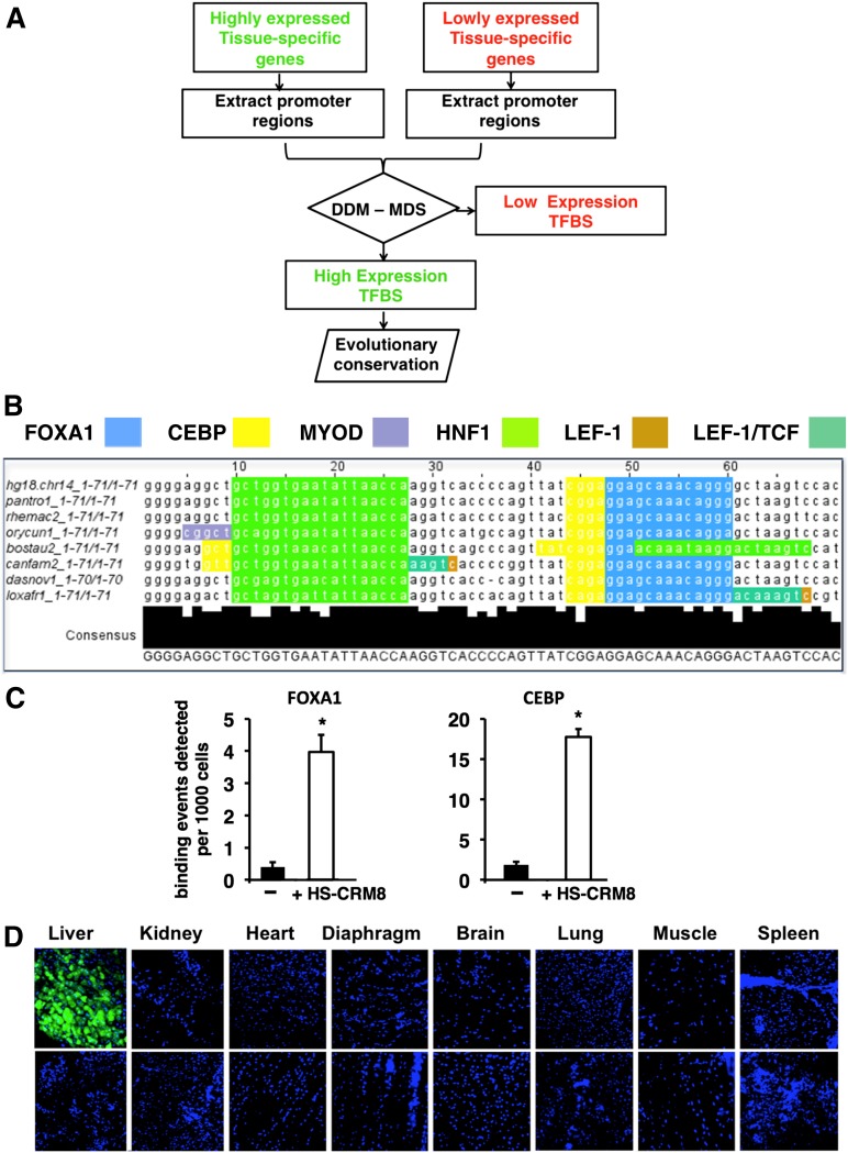
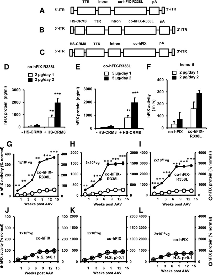
The development of the next-generation gene therapy vectors for hemophilia requires using lower and thus potentially safer vector doses and augmenting their therapeutic efficacy. We have identified hepatocyte-specific transcriptional cis-regulatory modules (CRMs) by using a computational strategy that increased factor IX (FIX) levels 11- to 15-fold. Vector efficacy could be enhanced by combining these hepatocyte-specific CRMs with a synthetic codon-optimized hyperfunctional FIX-R338L Padua transgene. This Padua mutation boosted FIX activity up to sevenfold, with no apparent increase in thrombotic risk. We then validated this combination approach using self-complementary adenoassociated virus serotype 9 (scAAV9) vectors in hemophilia B mice. This resulted in sustained supraphysiologic FIX activity (400%), correction of the bleeding diathesis at clinically relevant, low vector doses (5 × 10(10) vector genomes [vg]/kg) that are considered safe in patients undergoing gene therapy. Moreover, immune tolerance could be induced that precluded induction of inhibitory antibodies to FIX upon immunization with recombinant FIX protein.


Figures

Figure 1
Figure 1
Computational approach to identifying tissue-specific CRM. (A) The algorithm is based on the following steps: (1) identification of tissue-specific genes that are highly or lowly expressed based on statistical analysis of microarray expression data of normal human tissues; (2) extraction of the corresponding promoter sequences from publicly available databases; (3) mapping transcription factor binding sites (TFBSs) to these promoters by using the TRANSFAC database and identification of the tissue-specific CRM using a differential distance matrix (DDM)/multidimensional scaling (MDS) approach; and (4) searching the genomic context of the highly expressed genes for evolutionary conserved CRM. (B) Evolutionary conservation, nucleotide sequence, and TFBSs located within the 71-bp HS-CRM8 element from human SERPINA1 identified by the aforementioned algorithm. The TFBSs include binding sites for FOXA1 (blue), CEBP (yellow), HNF1 (light green), MyoD (purple), LEF-1 (dark green), and LEF-1/TCF (brown). Some of these TFBSs are partially overlapping. (C) Chromatin immunoprecipitation assay confirming the binding of FOXA1 and CEBP on HS-CRM8. Antibodies specific to FOXA1 and CEBP and polymerase chain reaction (PCR) primers specific for the corresponding TFBS were used. In particular, PCR primers were designed to amplify a region within the vector corresponding to HS-CRM8 (that binds FOXA1 and CEBP), an untranscribed region on chromosome 6 was used as negative control (–). Binding events per 103 cells (mean + standard deviation) were determined for each of the corresponding primer pairs. Significant differences compared with the negative control were indicated (Student t test, *P ≤ .05). (D) Confocal microscopy of different organs of mice injected with AAV9-HS-CRM8-TTR-GFP (5 × 10¹¹ vg/mouse; n = 4) with 4′,6 diamidino-2-phenylindole nuclear staining (top panels). A representative confocal scan is shown. Noninjected mice were used as controls (bottom panels). Pictures were taken at ×20 magnification.
Figure 2
Figure 2
Vector design and functional validation.Schematic representation of pAAV-TTR-co-hFIX-R338L (A), pAAV-HS-CRM8-TTR-co-hFIX-R338L (B) and pAAV-HS-CRM8-TTR-co-hFIX (C) plasmids used in this study. The liver-specific minimal transthyretin (TTR) promoter drives the codon-optimized human FIX (co-hFIX) complementary DNA (cDNA) with or without the Padua R338L mutation (co-hFIX-R338L). The HS-CRM8 is located upstream of the TTR promoter. The minute virus of mouse (MVM) mini-intron and bovine growth hormone polyadenylation site (pA) are also indicated. The expression cassettes were cloned in an scAAV backbone, flanked by the 5′ and 3′ AAV inverted terminal repeats (ITRs), as indicated, and were used to generate the cognate scAAV9-HS-CRM8-TTR-co-hFIX-R338L and scAAV9-HS-CRM8-TTR-co-hFIX vectors. The effect of the HSCRM8 element was assessed by hydrodynamic transfection of the pAAV-HS-CRM8-TTR-co-hFIX-R338L (indicated as +HS-CRM8) (B) and control pAAV-TTR-cohFIX-R338L (indicated as –HS-CRM8) (A) plasmids in C57BL/6 mice at doses of 2 µg/mouse and 5 µg/mouse (D-E). FIX expression was measured by using a validated hFIX-specific enzyme-linked immunosorbent assay (ELISA) (n = 4) on plasma samples collected at day 1 or 2 posttransfection. Similarly, to assess the impact of the Padua R338L mutation, hemophilic mice were hydrodynamically transfected with pAAV-HS-CRM8-TTR-cohFIX-R338L (indicated as co-hFIX-R338L) compared with the pAAV-HS-CRM8-TTR-co-hFIX control (indicated as co-hFIX) (F). The clotting factor activity was measured by using a functional chromogenic FIX assay. Subsequently, we injected the cognate scAAV9-HS-CRM8-TTR-co-hFIX-R338L (designated as co-hFIX-R338L) (G-I) and scAAV9-HS-CRM8-TTR-co-hFIX (designated as co-hFIX) (J-L) in FIX-deficient hemophilic mice at a dose of 1 × 10⁹ vg/mouse (5 × 10¹⁰ vg/kg), 5 × 10⁹ vg/mouse (2.5 × 10¹¹ vg/kg), and 2 × 10¹⁰ vg per mouse (1012 vg/kg) (n = 3 per group) (G-L). FIX activity and antigen levels were determined at the indicated times after AAV administration by using a chromogenic FIX activity assay and hFIX-specific ELISA, respectively. Results are presented as mean ± standard error of the mean. *P < .05; **P < .01; ***P < .001 (Student t test); N.S., not significant (P > .1).

Comment in

References

    1. Manno CS, Pierce GF, Arruda VR, et al. Successful transduction of liver in hemophilia by AAV-Factor IX and limitations imposed by the host immune response. Nat Med. 2006;12(3):342–347. - PubMed
    1. Nathwani AC, Tuddenham EG, Rangarajan S, et al. Adenovirus-associated virus vector-mediated gene transfer in hemophilia B. N Engl J Med. 2011;365(25):2357–2365. - PMC - PubMed
    1. Mingozzi F, Maus MV, Hui DJ, et al. CD8(+) T-cell responses to adeno-associated virus capsid in humans. Nat Med. 2007;13(4):419–422. - PubMed
    1. Mingozzi F, High KA. Therapeutic in vivo gene transfer for genetic disease using AAV: progress and challenges. Nat Rev Genet. 2011;12(5):341–355. - PubMed
    1. De Bleser P, Hooghe B, Vlieghe D, van Roy F. A distance difference matrix approach to identifying transcription factors that regulate differential gene expression. Genome Biol. 2007;8(5):R83. - PMC - PubMed

Publication types