. 2014 Mar 17;9(3):e91850.
doi: 10.1371/journal.pone.0091850. eCollection 2014.

Identification of novel genes associated with renal tertiary lymphoid organ formation in aging mice

Affiliations

Identification of novel genes associated with renal tertiary lymphoid organ formation in aging mice

Yuan Huang et al. PLoS One. .

Abstract

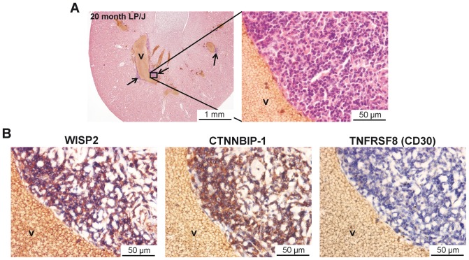
A hallmark of aging-related organ deterioration is a dysregulated immune response characterized by pathologic leukocyte infiltration of affected tissues. Mechanisms and genes involved are as yet unknown. To identify genes associated with aging-related renal infiltration, we analyzed kidneys from aged mice (≥20 strains) for infiltrating leukocytes followed by Haplotype Association Mapping (HAM) analysis. Immunohistochemistry revealed CD45+ cell clusters (predominantly T and B cells) in perivascular areas coinciding with PNAd+ high endothelial venules and podoplanin+ lymph vessels indicative of tertiary lymphoid organs. Cumulative cluster size increased with age (analyzed at 6, 12 and 20 months). Based on the presence or absence of clusters in male and female mice at 20 months, HAM analysis revealed significant associations with loci on Chr1, Chr2, Chr8 and Chr14 in male mice, and with loci on Chr4, Chr7, Chr13 and Chr14 in female mice. Wisp2 (Chr2) showed the strongest association (P = 5.00×10(-137)) in male mice; Ctnnbip1 (P = 6.42×10(-267)) and Tnfrsf8 (P = 5.42×10(-245)) (both on Chr4) showed the strongest association in female mice. Both Wisp2 and Ctnnbip1 are part of the Wnt-signaling pathway and the encoded proteins were expressed within the tertiary lymphoid organs. In conclusion, this study revealed differential lymphocytic infiltration and tertiary lymphoid organ formation in aged mouse kidneys across different inbred mouse strains. HAM analysis identified candidate genes involved in the Wnt-signaling pathway that may be causally linked to tertiary lymphoid organ formation.


Conflict of interest statement

Competing Interests: The authors have declared that no competing interests exist.

Figures

Figure 1
Figure 1. Phenotypic characterization of perivascular immune cell clusters in the aged mouse kidney.
(A) PAS staining was used for computerized morphometric analysis. Left panel: low-power magnification (40x) showing representative perivascular infiltrates (arrows) in a male C57L/J mouse. Middle panel: higher-power magnification (100x) of the left panel showing three individually measured perivascular cell clusters. The cell clusters were encircled and the surface area was calculated and expressed in μm2 as indicated. Right panel: high-power magnification (400×) of the middle panel. (B) The immune cell clusters consisted of CD45+ cells of which the majority was CD3+ T cells (C) and B220+ B cells (D). Panels B, C & D display serial sections (magnification: 200×). Insets show high-power magnifications (500x) of the indicated areas. Arrows indicate immune cell clusters. a: arteriole, v: vein.
Figure 2
Figure 2. Distribution of relative immune cell cluster size in 20-months-old male and female mice across the different mouse strains.
Perivascular immune cell clusters were measured in 20-months-old male and female mice and expressed as relative cluster size (as described in Materials & Methods ). Only those strains containing both female mice and male mice analyzed were listed (see also Table 1). Data are expressed as mean ± SEM. Numbers indicated above each bar represent the number of mice analyzed.
Figure 3
Figure 3. Perivascular immune cell clusters in aged mice have characteristics of tertiary lymphoid organs (TLOs).
Representative photomicrographs of a perivascular cell cluster from C57L/J mouse serial sections containing (A) Ki67+ proliferating cells (magnification 200x) and (B) peripheral node addressin (PNAd) expressing high endothelial venules (HEVs) (magnification 400×). Insets show higher-power magnifications of the indicated areas. Arrowheads indicate proliferating lymphocytes (A) and PNAd+ HEVs (B). (C) Podoplanin expression on lymphatic endothelial cells in a lymph vessel in the close proximity of a perivascular cell cluster (left panel: magnification 200x, right panel: 400x). (D) Immunofluorescent double labeling for LYVE-1/podoplanin (upper row) and VEGFR3/podoplanin (bottom row) on C57Bl/6 mouse kidney sections. Abbreviations: i: infiltrate; lv: lymph vessel; v: vein.
Figure 4
Figure 4. The size of renal perivascular immune cell clusters increase with age.
(A) Representative photomicrographs (PAS staining) of perivascular cell clusters in kidneys obtained from C57L/J mice at the age of 6, 12 and 20 months. Cell cluster surface area was calculated and expressed in μm2 as indicated (magnification 100x). Arrows indicate individual perivascular infiltrates. Abbreviation: v: vein. (B) Perivascular immune cell clusters were measured in 6-, 12- and 20-months-old male (C57L/J, NON/LtJ, and P/J strains) and female (C57L/J, C57BR/cdJ and P/J strains) mice and expressed as relative cluster size (as described in Materials & Methods ). Data expressed as mean ± SEM. Numbers indicated above each bar represent the number of mice analyzed. Strains from which no tissues were available are marked as nd (not determined). No differences between 6-months-old and 12-months-old mice in both sexes were observed. However, a significant increase in relative cluster size was observed in 20-months-old mice compared with both 6-months-old and 12-months-old mice (**P<0.01).
Figure 5
Figure 5. Strains with TLO formation are characterized by reduced numbers of lymph vessels.
(A) Representative photomicrograph of peri-arteriolar lymph vessels in a C57BLKS/J mouse kidney (left panel). The right panel shows a high-power magnification of the indicated framed area. Arrowheads indicate podoplanin+ lymph vessels. Abbreviations: a: arteriole; g: glomerulus; lv: lymph vessel; v: vein. (B) The number of podoplanin+ lymph vessels was quantified in selected strains based on the relative absence (BALB/cByJ, C3H/HeJ, RIIIS/J, C57BLKS/J [black bars]) and abundance (BTBR T+ tf/J, C57L/J, P/J, LP/J [white bars]) of TLOs at the age of 20 months. Numbers indicated within each bar represent the number of mice analyzed. (C) The mean number of lymph vessels in the 4 strains without TLOs (BALB/cByJ, C3H/HeJ, RIIIS/J, C57BLKS/J [black bars]) and with TLOs (BTBR T+ tf/J, C57L/J, P/J, LP/J [white bars]) was calculated. Strains with TLO formation had overall significantly lower numbers of lymph vessels compared with strains without TLOs (*P<0.05).
Figure 6
Figure 6. Renal TLO formation is associated with perivascular infiltrates in the liver.
Four strains with relative absence (BALB/cByJ [n = 9], C3H/HeJ [n = 10], RIIIS/J [n = 10], C57BLKS/J [n = 10]) and four strains with relative abundance (BTBR T+ tf/J [n = 7], C57L/J [n = 9], P/J [n = 8], LP/J [n = 4]) of renal TLOs were analyzed for the presence of perivascular infiltrates in the liver. (A) PAS staining on C3H/HeJ kidney and liver without perivascular infiltrates (magnifications: 40x and 200x). (B) PAS staining on P/J kidney and liver with perivascular infiltrates (magnifications: 40x and 200x). (C) 21/31 (67.7%) of mice with renal TLOs contained liver TLOs, whereas 8/27 (29.6%) of mice without renal TLOs contained liver TLOs (Pearson's χ2 test, P<0.0001). (D) The perivascular infiltrates in liver consisted of CD3+ T cells and B220+ B cells (magnification: 40x). Insets show high-power magnifications of the indicated areas. Arrowheads indicate positively stained cell clusters. Abbreviations: a: arteriole, bd: bile duct; i: infiltrate; pv: portal vein, v: vein.
Figure 7
Figure 7. Genome-wide haplotype association mapping in aged mice.
In both female (A) and male (B) mice, binary data were used based on the threshold at 0.15 of relative cluster size. Strains with relative cluster size less than 0.15 were marked as “0”, and those higher than 0.15 were marked as “1”. Associations with a P-value of less than 10−6 were considered significant. Results are displayed in Manhattan plots (left graphs) and Q-Q plots (right graphs).
Figure 8
Figure 8. Lymphocytes in renal TLOs express WISP2 and CTNNBIP1, but not TNFRSF8.
(A) H&E staining on a kidney section from a 20 month old male LP/J mouse with perivascular infiltrates (TLOs). Magnification: 20x (left panel) and 400x (right panel). (B) Immunohistochemistry revealed expression of WISP2 (left panel) and CTNNBIP1 (middle panel), but not TNFRSF8 (right panel). Magnification: 400x. v: vein.

References

    1. Hoang K, Tan JC, Derby G, Blouch KL, Masek M, et al. (2003) Determinants of glomerular hypofiltration in aging humans. Kidney Int 64: 1417–1424. - PubMed
    1. Choudhury D, Levi M (2011) Kidney aging—inevitable or preventable? Nat Rev Nephrol 7: 706–717. - PubMed
    1. Lindeman RD, Tobin J, Shock NW (1985) Longitudinal studies on the rate of decline in renal function with age. J Am Geriatr Soc 33: 278–285. - PubMed
    1. Melk A, Mansfield ES, Hsieh SC, Hernandez-Boussard T, Grimm P, et al. (2005) Transcriptional analysis of the molecular basis of human kidney aging using cDNA microarray profiling. Kidney Int 68: 2667–2679. - PubMed
    1. Venkatachalam MA, Griffin KA, Lan R, Geng H, Saikumar P, et al. (2010) Acute kidney injury: A springboard for progression in chronic kidney disease. Am J Physiol Renal Physiol 298: F1078–94. - PMC - PubMed

Publication types

MeSH terms

Substances