. 2014 Jan-Feb;5(1):28-39.
doi: 10.4161/gmic.26489. Epub 2013 Sep 25.

Proteobacteria-specific IgA regulates maturation of the intestinal microbiota

Affiliations

Proteobacteria-specific IgA regulates maturation of the intestinal microbiota

Julie Mirpuri et al. Gut Microbes. 2014 Jan-Feb.

Abstract

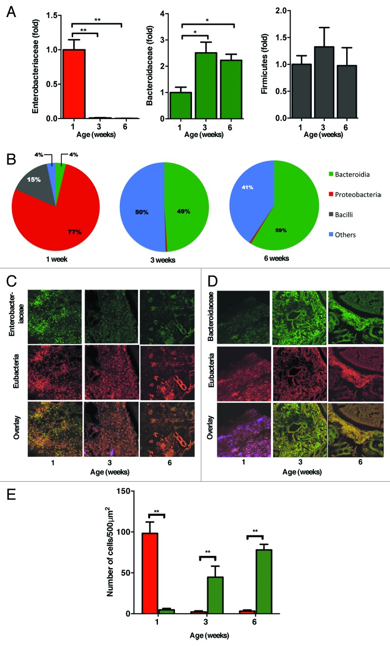
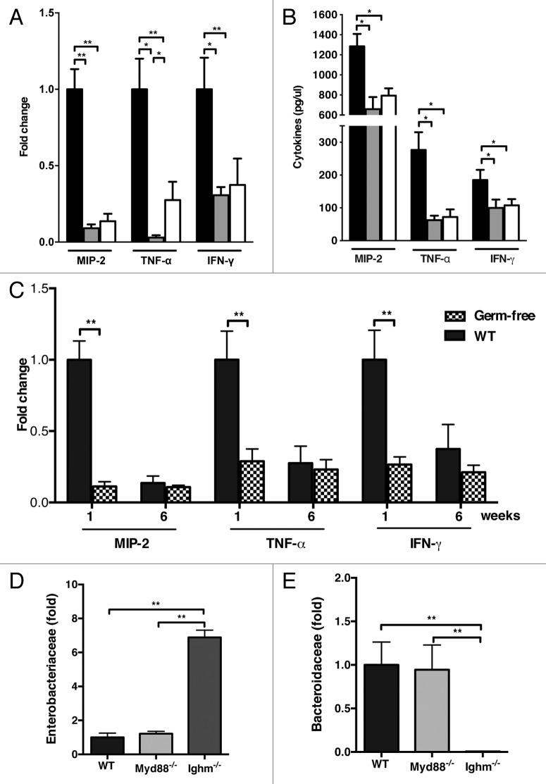
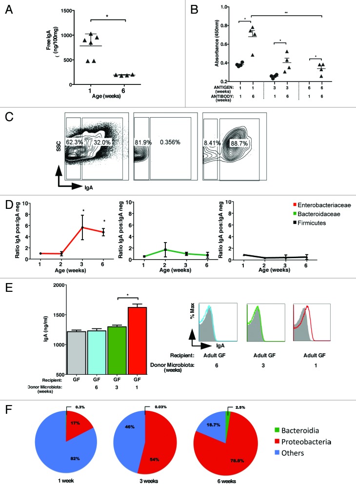
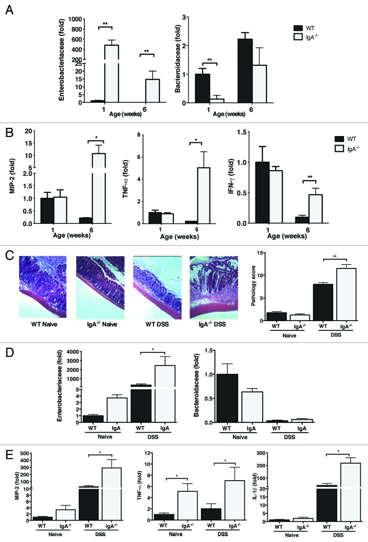
The intestinal microbiota changes dynamically from birth to adulthood. In this study we identified γ-Proteobacteria as a dominant phylum present in newborn mice that is suppressed in normal adult microbiota. The transition from a neonatal to a mature microbiota was in part regulated by induction of a γ-Proteobacteria-specific IgA response. Neocolonization experiments in germ-free mice further revealed a dominant Proteobacteria-specific IgA response triggered by the immature microbiota. Finally, a role for B cells in the regulation of microbiota maturation was confirmed in IgA-deficient mice. Mice lacking IgA had persistent intestinal colonization with γ-Proteobacteria that resulted in sustained intestinal inflammation and increased susceptibility to neonatal and adult models of intestinal injury. Collectively, these results identify an IgA-dependent mechanism responsible for the maturation of the intestinal microbiota.

Keywords: IgA; colitis; microbiota; necrotizing enterocolitis; proteobacteria.


Figures

None
Figure 1. Immature microbiota in newborn mice is characterized by a predominance of Proteobacteria. (A) The relative abundance of Proteobacteria (red), Bacteroidetes (green) and Firmicutes (black) present in colonic contents of 1, 3, and 6-week-old mice was analyzed by qRT-PCR. Data are represented as fold change relative to 1-week old mice. (B) The proportion of major bacterial phyla seen in 1, 3, and 6 weeks old mice was analyzed by 454-pyrosequencing. (C) Fluorescence in situ hybridization detection of Enterobactericeae (green) and Eubacteria (red) or (D) Bacteroidaceae (green) and Eubacteria (red) in colons of 1, 3, and 6 week old mice. (E) Quantification of the FISH positive cells per 500 μm2 section from a minimum of 10 sections from 3‒4 mice at each age group shown in (C) and (D). The data shown are representative of 3 experiments with 5‒7 mice in each group. Data are depicted as mean ± SEM (* p < 0.05, ** p < 0.01).
None
Figure 2. B cells restrict the immature microbiota responsible for early intestinal inflammation. Quantification of MIP-2, TNF-α and IFN-γ by (A) qRT-PCR and (B) ELISA. Data represented as fold change relative to 1-week old mice. (C) Comparative expression of colonic inflammatory cytokines in conventional and GF mice was analyzed by qRT-PCR in 1 and 6 week old mice. (D) qRT-PCR-based quantification of Enterobacteriaceae and (E) Bacteroidaceae in adult WT, Myd88−/−, and Ighm−/− mice. Data represented as fold change relative to WT adult mice. The data shown are representative of 3 experiments for conventional and 2 experiments for GF mice, with 5‒7 mice in each group. Data are depicted as mean ± SEM (* p < 0.05, ** p < 0.01).
None
Figure 3. Preferential induction of Proteobacteria specific IgA. (A) Quantification of total colonic IgA in 1 and 6 week old mice by ELISA. (B) Detection of bacterial antigen specific IgA in 1 and 6 week old mice by ELISA. Bacterial antigens were prepared from colonic contents of 1, 3, and 6- week old mice by sonication as described in the Materials and Methods section. (C) Cell-sort based purification of IgA positive and IgA negative bacteria and their post sort analysis. The percentages shown indicate purity of IgA positive and negative populations achieved. (D) qRT-PCR analysis of IgA positive and negative Enterobacteriaceae (red), Bacteroidaceae (green) and Firmicutes (black) shown as ratio of IgA positive to IgA negative bacteria. (E) Total IgA measured in GF mice gavage fed with 1-, 3-, or 6-week microbiota measured on day 5 post-recolonization (left). Enterobacteriaceae-specific IgA in the recolonized GF mice was measured by flow-cytometry in the same sample (right). (F) Proportion of major bacterial phyla seen in pooled APCs from 4 mice per group isolated from lamina propria in 1, 3, and 6 week old mice were analyzed by 454-pyrosequencing. The data shown are representative of three experiments with 4–6 mice in each group. Data are depicted as mean ± SEM (* p < 0.05 and ** p < 0.01).
None
Figure 4. IgA deficiency leads to persistence of Proteobacteria and enhanced susceptibility to DSS colitis. (A) Quantification of Enterobacteriaceae and Bacteroidaceae in the colonic contents of 1- and 6- week old IgA deficient mice by qRT-PCR in comparison to their WT controls. The data are represented as fold change from 1-week-old WT mice. (B) Expression of MIP-2, TNF-α, and IFN-γ in colon of 1- and 6-week old WT and IgA deficient mice. (C) WT and IgA deficient mice were treated with 2% DSS in their drinking water for 10 d. Representative images of colons from control and DSS treated WT and IgA- deficient mice (left) and their respective colitis scores (right) are shown. (D) Relative expression of Enterobacteriaceae and Bacteroidaceae in Naïve and DSS exposed mice. (E) Expression of pro-inflammatory cytokines from colon in DSS treated mice. The data shown are representative of three experiments with 4–6 mice in each group. Data are depicted as mean ± SEM (* p < 0.05 and ** p < 0.01).

References

    1. Palmer C, Bik EM, DiGiulio DB, Relman DA, Brown PO. Development of the human infant intestinal microbiota. PLoS Biol. 2007;5:e177. doi: 10.1371/journal.pbio.0050177. - DOI - PMC - PubMed
    1. Ley RE, Peterson DA, Gordon JI. Ecological and evolutionary forces shaping microbial diversity in the human intestine. Cell. 2006;124:837–48. doi: 10.1016/j.cell.2006.02.017. - DOI - PubMed
    1. Hooper LV, Gordon JI. Commensal host-bacterial relationships in the gut. Science. 2001;292:1115–8. doi: 10.1126/science.1058709. - DOI - PubMed
    1. Carvalho FA, Koren O, Goodrich JK, Johansson ME, Nalbantoglu I, Aitken JD, Su Y, Chassaing B, Walters WA, González A, et al. Transient inability to manage proteobacteria promotes chronic gut inflammation in TLR5-deficient mice. Cell Host Microbe. 2012;12:139–52. doi: 10.1016/j.chom.2012.07.004. - DOI - PMC - PubMed
    1. Kirkland D, Benson A, Mirpuri J, Pifer R, Hou B, DeFranco AL, Yarovinsky F. B cell-intrinsic MyD88 signaling prevents the lethal dissemination of commensal bacteria during colonic damage. Immunity. 2012;36:228–38. doi: 10.1016/j.immuni.2011.11.019. - DOI - PMC - PubMed

Publication types

MeSH terms

LinkOut - more resources