. 2014 Mar 17;9(3):e92349.
doi: 10.1371/journal.pone.0092349. eCollection 2014.

Actin and myosin-dependent localization of mRNA to dendrites

Affiliations

Actin and myosin-dependent localization of mRNA to dendrites

Varuzhan Balasanyan et al. PLoS One. .

Abstract

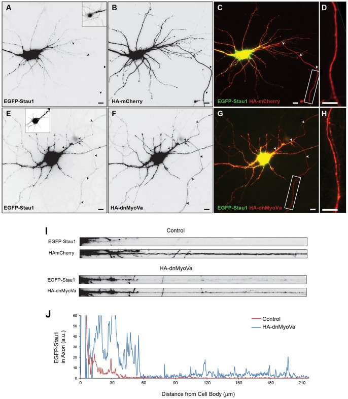
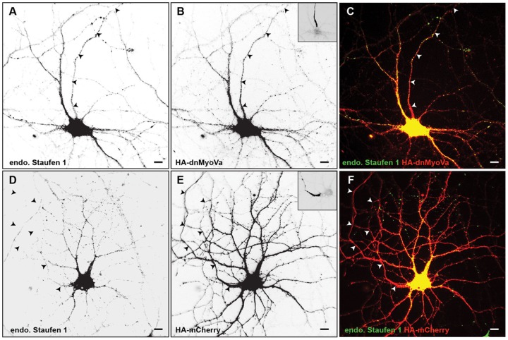
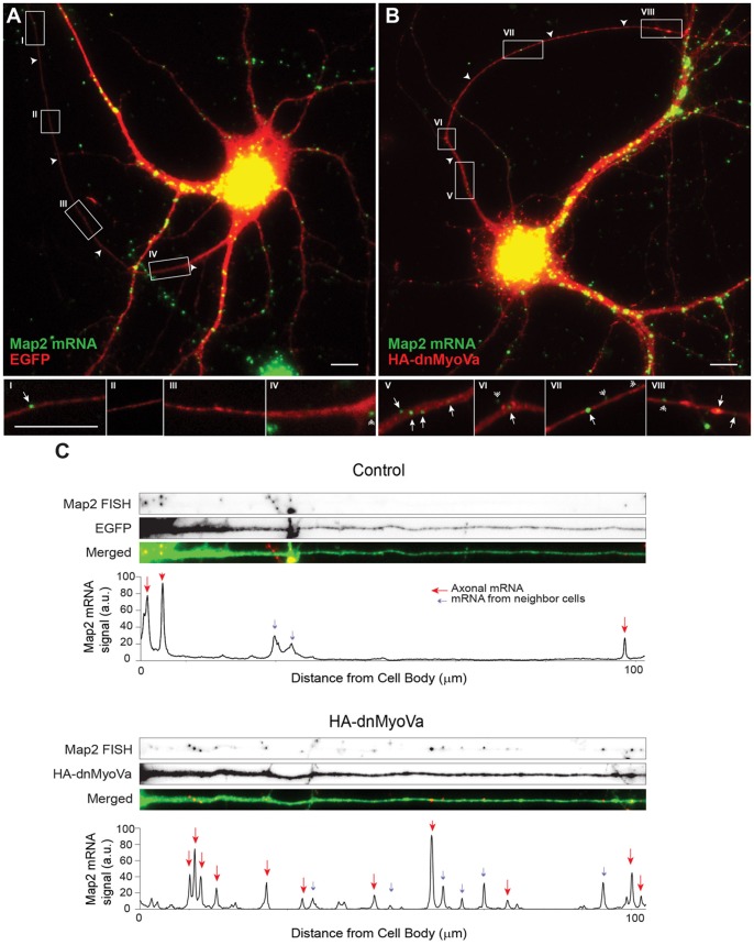
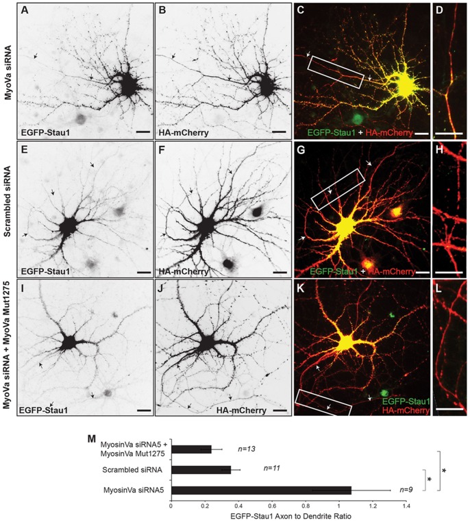
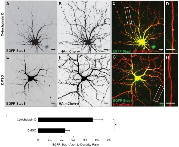
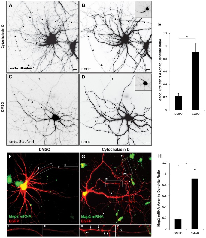
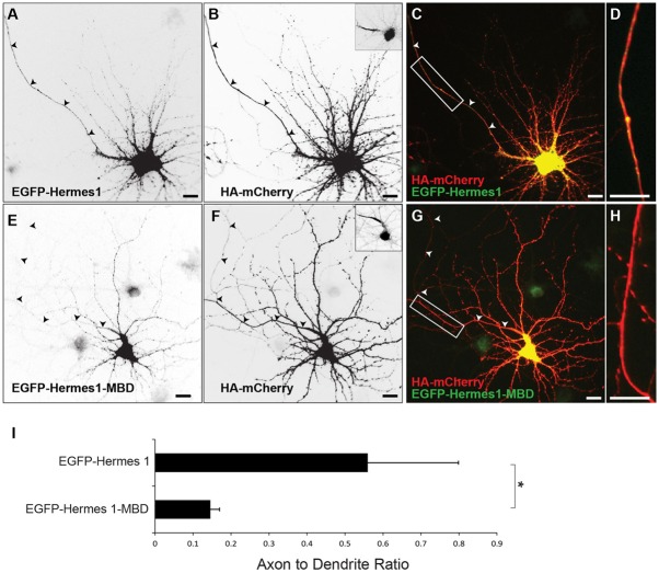
The localization of mRNAs within axons and dendrites allows neurons to manipulate protein levels in a time and location dependent manner and is essential for processes such as synaptic plasticity and axon guidance. However, an essential step in the process of mRNA localization, the decision to traffic to dendrites and/or axons, remains poorly understood. Here we show that Myosin Va and actin filaments are necessary for the dendritic localization of the mRNA binding protein Staufen 1 and of mRNA encoding the microtubule binding protein Map2. Blocking the function or expression of Myosin Va or depolymerizing actin filaments leads to localization of Staufen 1 and of Map2 mRNA in both axons and dendrites. Furthermore, interaction with Myosin Va plays an instructive role in the dendritic localization of Hermes 1, an RNA binding protein. Wild-type Hermes 1 localizes to both axons and dendrites, whereas Hermes 1 fused with a Myosin Va binding peptide, localizes specifically to dendrites. Thus, our results suggest that targeting of mRNAs to the dendrites is mediated by a mechanism that is dependent on actin and Myosin Va.


Conflict of interest statement

Competing Interests: The authors have declared that no competing interests exist.

Figures

Figure 1
Figure 1. Myosin Va is necessary for somatodentritic targeting of EGFP-Stau1.
A. Following expression in a cortical neuron in dissociated culture EGFP-Stau1 is present in the soma and dendrites, but not in the axon. B. Co-expressed HA-mCherry localizes to the axon as well as to the soma and dendrites in the same cell as in (A). C. Merge of HA-mCherry (red) and EGFP-Stau1 (green). D. High power image of boxed region from (C). E. EGFP-Stau1 is present in the axon as well as in the soma and dendrites when co-expressed with HA-dnMyoVa (F), a dominant-negative variant of Myosin Va. Arrowheads point to the axon. Scale bars represent 10 μm. G. Merge of HA-dnMyoVa (red) and EGFP-Stau1 (green). H. High power image of boxed region from (G). I. A straightened axon from the neuron in (A-D) co-expressing HA-mCherry (upper panel) shows a relative absence of EGFP-Stau1 staining. In contrast, a straightened axon from the neuron in (E-H) expressing HA-dnMyoVa (lower panel) shows EGFP-Stau1 staining. J. EGFP-Stau1 staining vs. distance in the distal axon for the neuron in (A-D; red) and the neuron in (E-H; blue).
Figure 2
Figure 2. Myosin Va is necessary for somatodentritic targeting of endogenous Staufen1.
A. Endogenous Staufen 1 is present in the axon as well as in the soma and dendrites when co-expressed with HA-dnMyoVa (B), a dominant-negative variant of Myosin Va. C. Merge of endogenous Staufen 1 (green) and HA-dnMyoVa (red). D. Endogenous Staufen 1 is completely absent from the axonal compartment, but is expressed in the somatodendritic compartment of cortical neurons, expressing HA-mCherry (E). F. Merge of endogenous Staufen 1 (green) and HA-mCherry (red). Arrowheads point to the axon. Scale bars represent 10 μm. Insets show staining for Ankyrin G, a marker for the axon initial segment.
Figure 3
Figure 3. Myosin Va is necessary for somatodendritic targeting of endogenous Map2 mRNA.
A. Map2 mRNA labeled by fluorescent in situ hybridization (green) in a cortical neuron in dissociated culture expressing EGFP (red). Map2 mRNA is present in the somatodendritic compartment, but largely absent from the axon (arrowheads). Insets I-IV are high power images corresponding to boxes drawn in (A). B. In a cortical neuron expressing HA-dnMyoVa, staining for endogenous Map2 mRNA is present in the axon as well as in the somatodendritic compartment. Insets V-VIII are high power images corresponding to boxes in (B). Arrowheads point to axons, arrows to Map2 mRNAs that are present in the axon, double arrowheads to Map2 mRNAs from neighboring untransfected neurons. Scale bar, 10 μm. C. Signal intensity plots of Map2 mRNA staining in straightened axons of the control neuron in (A) and the neuron expressing HA-dnMyoVa in (B). The presence of HA-dnMyoVa is correlated with an increase in the amount of Map2 mRNA in the axon. Red arrows point to Map2 mRNA puncta in the axon. Blue arrows point to Map2 mRNA puncta from neighboring neurons.
Figure 4
Figure 4. Knockdown of Myosin Va disrupts somatodendritic targeting of EGFP-Stau1.
A. EGFP-Stau1 localizes to both the axon and the somatodendritic compartment in a cortical neuron transfected with MyoVa siRNA and HA-mCherry (B). C. Merge of EGFP-Stau1 (green) and HA-mCherry (red). D. High power image of boxed area in (C). E. EGFP-Stau1 localizes in the soma and dendrites, but not in the axon, of a neuron expressing scrambled siRNA and HA-mCherry (F). G. Merge of EGFP-Stau1 (green) and HA-mCherry (red). H. High power image of boxed area in (G). I. EGFP-Stau1 localizes in the soma and dendrites, but not in the axon, of a neuron expressing MyoVa siRNA as well as MyoVa Mut1275 and HA-mCherry (J). K. Merge of EGFP-Stau1 (green) and HA-mCherry (red). L. High power image of boxed area in (K). Scale bar, 10 μm. Note that this result indicates that blocking of somatodendritic localization of EGFP-Stau1 by MyoVa siRNA is not due to off-target effects. M. Axon to dendrite ratio of EGFP-Stau1 in neuron expressing MyoVa siRNA is significantly different from those in neurons expressing either scrambled siRNA or MyoVa siRNA and MyoVa Mut1275. * indicates p<0.001 (Kruskal-Wallis).
Figure 5
Figure 5. Intact actin filaments are necessary for somatodendritic localization of EGFP-Stau1.
A. Following exposure to Cytochalasin D (4 μM) for 3 hours EGFP-Stau1 is present in the axon as well as in the dendritic compartment. B HA-mCherry staining in the same cell as in A. C. Merge of EGFP-Stau1 (green) and HA-mCherry (red). D. High power image of boxed area in (C). E. EGFP-Stau1 is absent from the axonal compartment of cortical neurons expressing HA-mCherry (F) and exposed to DMSO. G. Merge of EGFP-Stau1 (green) and HA-mCherry (red). H. High power image of boxed area in (G). I. EGFP-Stau1 axon to dendrite ratio is significantly higher in cells exposed to Cytochalasin D than in cells exposed only to DMSO. * indicates p<0.03 (Wilcoxon). Scale bar 10 μm.
Figure 6
Figure 6. Intact actin filaments are necessary for somatodendritic localization of both endogenous Staufen 1 and Map2 mRNA.
A. Following exposure to Cytochalasin D (4 μM) for 6 hours endogenous Staufen 1 is present in the axon as well as in the dendritic compartment. B. EGFP staining in the same cell as in (A). C. Endogenous Staufen 1 is absent from the axonal compartment of cortical neurons expressing EGFP (D) and exposed to DMSO. Insets show Ankyrin G staining, which labels the axon initial segment of the corresponding cells. E. The axon to dendrite ratio of endogenous Staufen 1 is significantly higher in cells exposed to Cytochalasin D than in cells exposed to DMSO alone. F. In a cortical neuron exposed to DMSO FISH labeling reveals Map2 expression in dendrites, but not in the axon. Insets, I and II, which are high power images corresponding to labeled boxes in (F), show a relative absence of Map2 mRNA staining in the axon. G. In a cortical neuron exposed to 4 μM Cytochalasin D for 6 hours Map 2 mRNA staining is present in both the axon and the somatodendritic compartment. Insets III and IV show the presence of Map2 mRNA staining in axons. H. Map2 mRNA axon to dendrite ratio is significantly higher in cells exposed to Cytochalasin D than in cells exposed to DMSO alone. * indicates p<0.001 (Wilcoxon). Scale bar is 10 μm.
Figure 7
Figure 7. Association with Myosin Va is sufficient for somatodendritic targeting of a non-specifically localized mRNA binding protein, Hermes 1.
A. EGFP-Hermes1 localizes both to the axon and dendrites of neurons co-expressing HA-mCherry (B). Insets show Ankyrin G staining, which labels the axon initial segment of the corresponding cell. C. Merge of HA-mCherry (red) and EGFP-Hermes1 (green). D. High power image of boxed area in (C). E. EGFP-Hermes1 fused to a Myosin Va binding domain (EGFP-Hermes1-MBD) localizes specifically to the somatodendritic compartment of a cortical neuron co-expressing HA-mCherry (F). G. Merge of HA-mCherry (red) and EGFP-Hermes1-MBD (green). H. High power image of boxed area in (G). I. The axon to dendrite ratio of Hermes 1 is significantly higher than that of Hermes 1-MBD. * indicates p<0.02 (Wilcoxon). Scale bar is 10 μm.

References

    1. Sidorov MS, Auerbach BD, Bear MF (2013) Fragile X mental retardation protein and synaptic plasticity. Mol Brain 6: 15. - PMC - PubMed
    1. Richter JD (2010) Translational control of synaptic plasticity. Biochem Soc Trans 38: 1527–1530. - PubMed
    1. Martin KC (2004) Local protein synthesis during axon guidance and synaptic plasticity. Curr Opin Neurobiol 14: 305–310. - PubMed
    1. Jung H, Yoon BC, Holt CE (2012) Axonal mRNA localization and local protein synthesis in nervous system assembly, maintenance and repair. Nat Rev Neurosci 13: 308–324. - PMC - PubMed
    1. Santini E, Huynh TN, MacAskill AF, Carter AG, Pierre P, et al. (2013) Exaggerated translation causes synaptic and behavioural aberrations associated with autism. Nature 493: 411–415. - PMC - PubMed