. 2014 Mar 17;9(3):e90924.
doi: 10.1371/journal.pone.0090924. eCollection 2014.

On the antiquity of cancer: evidence for metastatic carcinoma in a young man from ancient Nubia (c. 1200 BC)

Affiliations

On the antiquity of cancer: evidence for metastatic carcinoma in a young man from ancient Nubia (c. 1200 BC)

Michaela Binder et al. PLoS One. .

Abstract

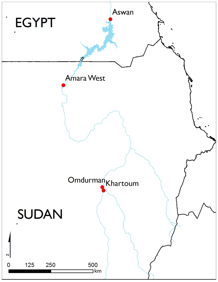
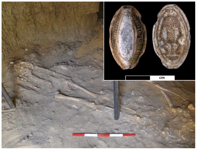
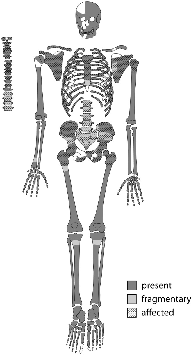
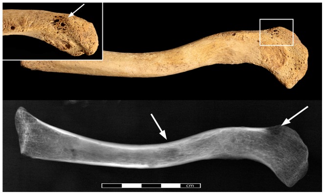
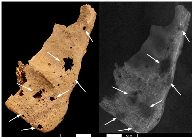
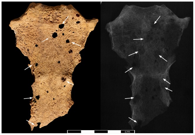
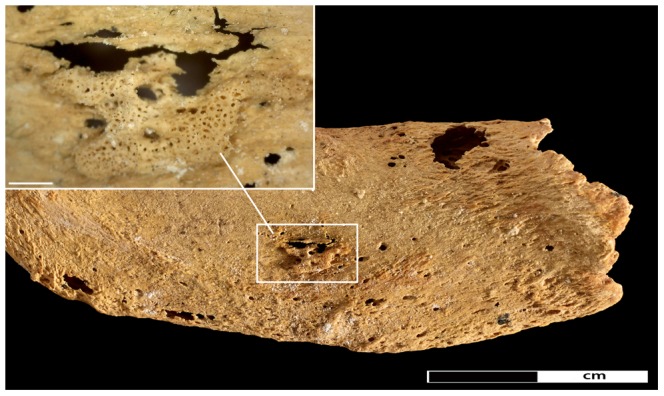
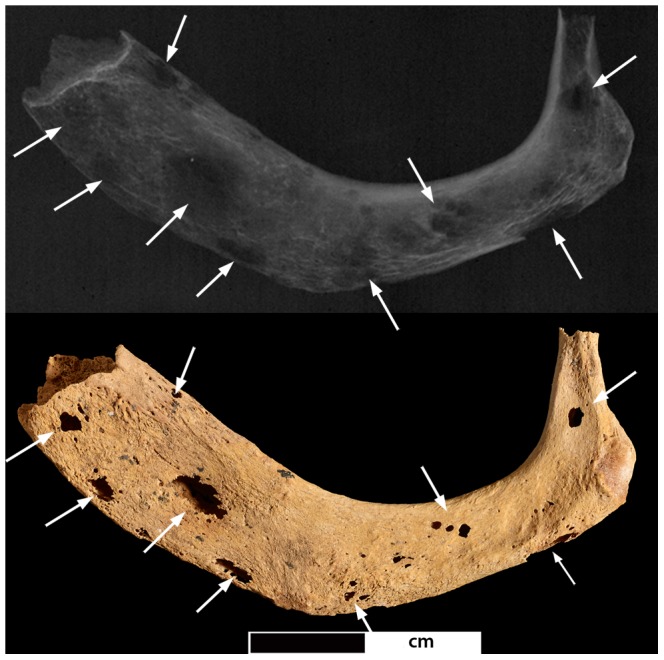
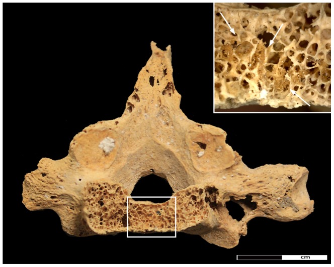
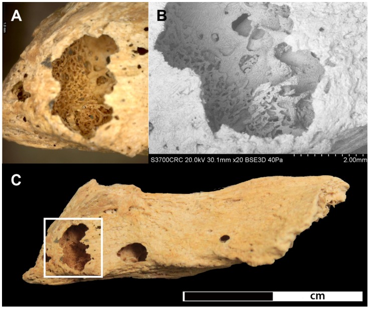
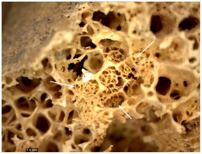
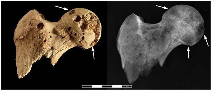
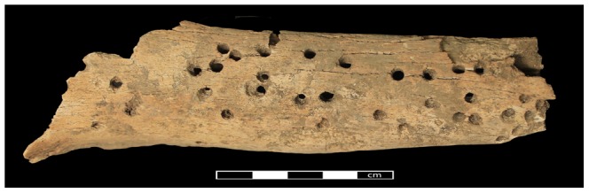
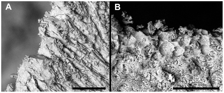
Cancer, one of the world's leading causes of death today, remains almost absent relative to other pathological conditions, in the archaeological record, giving rise to the conclusion that the disease is mainly a product of modern living and increased longevity. This paper presents a male, young-adult individual from the archaeological site of Amara West in northern Sudan (c. 1200 BC) displaying multiple, mainly osteolytic, lesions on the vertebrae, ribs, sternum, clavicles, scapulae, pelvis, and humeral and femoral heads. Following radiographic, microscopic and scanning electron microscopic (SEM) imaging of the lesions, and a consideration of differential diagnoses, a diagnosis of metastatic carcinoma secondary to an unknown soft tissue cancer is suggested. This represents the earliest complete example in the world of a human who suffered metastatic cancer to date. The study further draws its strength from modern analytical techniques applied to differential diagnoses and the fact that it is firmly rooted within a well-documented archaeological and historical context, thus providing new insights into the history and antiquity of the disease as well as its underlying causes and progression.


Conflict of interest statement

Competing Interests: The authors have declared that no competing interests exist.

Figures

Figure 1
Figure 1. Location of Amara West.
Map of modern Sudan showing the location of the archaeological site (Map drawn by M. Binder, source: ESRI).
Figure 2
Figure 2. The skeleton Sk244-8.
Skeleton Sk244-8 in its original burial position in the western chamber of G244. The insert shows faience amulet F9273 found associated with the individual from both sides. The Egyptian god Bes (right side) is depicted on the reverse side.
Figure 3
Figure 3. Preservation of Sk244-8.
Preserved elements of the skeleton and elements affected by pathological changes. Dark areas indicate full preservation, light areas indicate fragmented areas. Hatched areas are the bones affected by lytic lesions.
Figure 4
Figure 4. The left clavicle of Sk244-8.
General view of the left clavicle with the pathological lesions indicated by arrows. Radiograph of the same bone on the bottom. Insert shows close-up of the lesion on the superior margin indicated by rectangle.
Figure 5
Figure 5. Pathological lesions in the right scapula.
Photo- and radiograph of the right scapular blade (view from anterior, arrows indicate location of lytic lesions).
Figure 6
Figure 6. The sternum of Sk244-8.
Anterior aspect of the sternum. Arrows indicate location of lytic lesions.
Figure 7
Figure 7. Pathological changes in the first rib.
New bone formation on the inferior aspect of the left first rib. Insert shows close-up of area of new bone formation (45× magnification).
Figure 8
Figure 8. Lytic lesions in the first rib.
Photo- and radiograph of the left first rib (superior surface. Arrows indicate location of the lesions.
Figure 9
Figure 9. Destructive lesion in vertebral body of the 7th thoracic vertebrae.
Detail of the pathological changes in the 7th thoracic vertebra. Rectangle indicates area of new bone infill of the spongiosa. Close-up of new bone formation indicated by arrows is shown in the insert; arrows indicate new bone formation.
Figure 10
Figure 10. Lytic lesion in the spinous process of the 5th thoracic vertebra.
A) shows a close-up of bone formation at 35x magnification located within the lytic focus, B) SEM image of the lytic focus C) shows the complete spinous process with the location of the lytic focus highlighted in the rectangle.
Figure 11
Figure 11. Detail of new bone formation in the iliac crest.
The close-up shows a focus of new bone formation indicated by arrows in a lytic lesion in the iliac crest (40x magnification).
Figure 12
Figure 12. Pathological changes in the right femoral head.
Photo- and radiograph of lytic lesions in the right femoral head (arrows indicate areas of pathological lesions).
Figure 13
Figure 13. Post-mortem damage caused by insects.
Tibia of a skeleton from Amara West showing extensive damage caused by dermestid beetles.
Figure 14
Figure 14. Comparison of SEM-images of taphonomic and pathological lesions.
A) shows the margin of a defect caused by post-mortem damage. B) shows pathological changes on the margin of a lytic lesion in the cortical surface (bar on the bottom of each image indicates 100 µm).

References

    1. Boyle P, Levin P, editors (2008) World Cancer Report 2008. Lyon: International Agency for Research on Cancer.
    1. WHO (2013) Fact sheet N°297: Cancer.
    1. David AR, Zimmerman MR (2010) Cancer: an old disease, a new disease or something in between? Nature Reviews: Cancer 10: 728–733. - PubMed
    1. McKeown RE (2009) The Epidemiologic Transition: Changing Patterns of Mortality and Population Dynamics. American Journal of Lifestyle Medicine 3: 19S–26S. - PMC - PubMed
    1. Karpozilos A, Pavlidis N (2004) The treatment of cancer in Greek antiquity. European Journal of Cancer 40: 2033–2040. - PubMed

Publication types

LinkOut - more resources