

In multiple myeloma, 14q32 translocations are nonrandom chromosomal fusions driving high expression levels of the respective partner genes
- PMID: 24638926
- PMCID: PMC4016160
- DOI: 10.1002/gcc.22165
In multiple myeloma, 14q32 translocations are nonrandom chromosomal fusions driving high expression levels of the respective partner genes
Abstract
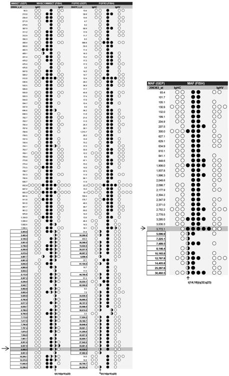
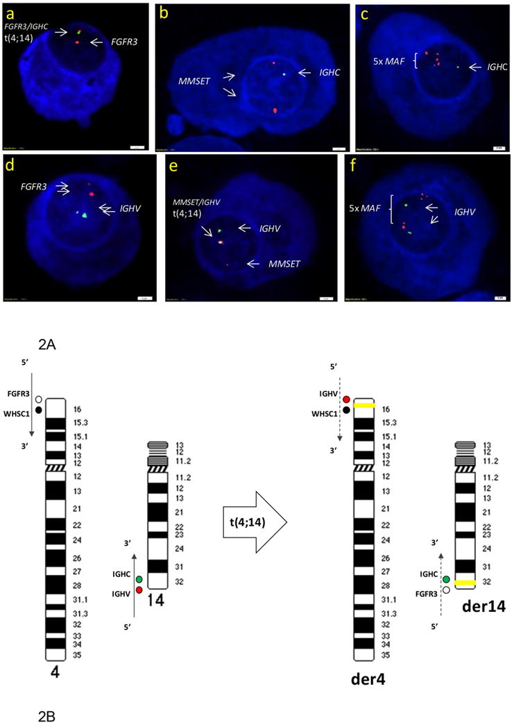
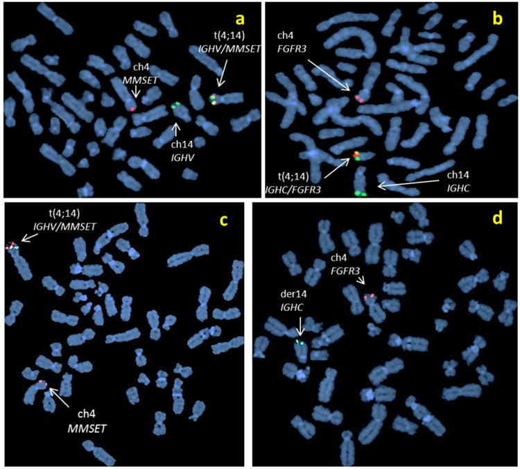
In studies of patients with multiple myeloma (MM), gene expression profiling (GEP) of myeloma cells demonstrates substantially higher expression of MMSET, FGFR3, CCND3, CCND1, MAF, and MAFB--the partner genes of 14q32 translocations--than GEP of plasma cells from healthy individuals. Interphase fluorescent in situ hybridization (FISH) was used to discriminate between chromosomal translocations involving different regions of the immunoglobulin heavy chain (IGH) genes at 14q32. With special probes designed for the constant region (IGHC) and the variable region (IGHV), IGH translocations were shown to be definite, nonrandom chromosomal fusions of IGHC with the loci of FGFR3, CCND1, CCND3, MAF, and MAFB genes; and IGHV with the locus of MMSET gene. When correlated with GEP results, the IGH translocations were found to drive expression levels of the partner genes to significantly higher levels (spikes) than copy-number variations. Hence, 42% of IGH translocations were identified among newly diagnosed MM patients (448/1,060). As GEP has become essential for assessing cancer risk, this novel approach is highly consistent with the cytogenetic features of the chromosomal translocations to effectively stratify molecular subgroups of MM on the basis of gene expression profiles of the IGH translocation partner genes in myeloma cells. © 2014 Wiley Periodicals, Inc.
Copyright © 2014 Wiley Periodicals, Inc.
Figures
References
-
- Avet-Loiseau H, Brigaudeau C, Morineau N, Talmant P, Laï JL, Daviet A, Li JY, Praloran V, Rapp MJ, Harousseau JL, Facon T, Bataille R. High incidence of cryptic translocations involving the Ig heavy chain gene in multiple myeloma, as shown by fluorescence in situ hybridization. Genes Chromosomes Cancer. 1999;24:9–15. - PubMed
-
- Bergsagel PL, Kuehl WM. Chromosome translocations in multiple myeloma. Oncogene. 2001;20:5611–5622. - PubMed
-
- Boersma-Vreugdenhil GR, Kuipers J, Van Stralen E, Peeters T, Michaux L, Hagemeijer A, Pearson PL, Clevers HC, Bast BJ. The recurrent translocation t(14;20)(q32;q12) in multiple myeloma results in aberrant expression of MAFB: A molecular and genetic analysis of the chromosomal breakpoint. Br J Haematol. 2004;126:355–363. - PubMed
-
- Chesi M, Bergsagel PL, Shonukan OO, Martelli ML, Brents LA, Chen T, Schröck E, Ried T, Kuehl WM. Frequent dysregulation of the c-maf proto-oncogene at 16q23 by translocation to an Ig locus in multiple myeloma. Blood. 1998;91:4457–4463. - PubMed
Publication types
MeSH terms
Grants and funding
LinkOut - more resources
Full Text Sources
Other Literature Sources
Medical
Research Materials
