

Tracking changes over time in retinal nerve fiber layer and ganglion cell-inner plexiform layer thickness in multiple sclerosis
- PMID: 24639478
- PMCID: PMC4306466
- DOI: 10.1177/1352458514523498
Tracking changes over time in retinal nerve fiber layer and ganglion cell-inner plexiform layer thickness in multiple sclerosis
Abstract
Background: Neurodegeneration plays an important role in permanent disability in multiple sclerosis (MS).
Objective: The objective of this paper is to determine whether progressive neurodegeneration occurs in MS eyes without clinically evident inflammation.
Methods: Retinal nerve fiver layer thickness (RNFLT) and ganglion cell-inner plexiform layer thickness (GCIPT) were measured using Cirrus optical coherence tomography (OCT) in 133 relapsing-remitting MS (RRMS) patients (149 non-optic neuritis (ON), 97 ON eyes, last ON ≥6 months). Ninety-three patients were scanned at two visits. Percentages of abnormal GCIPT vs RNFLT (<5% of machine norms) in cross-sectional data were compared. Relations between RNFLT/GCIPT and MS duration (cross-sectional) and follow-up time (longitudinal) were assessed.
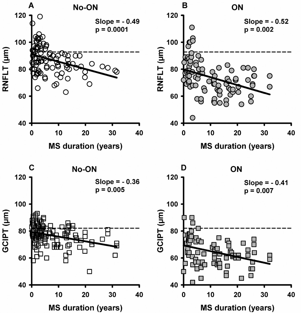
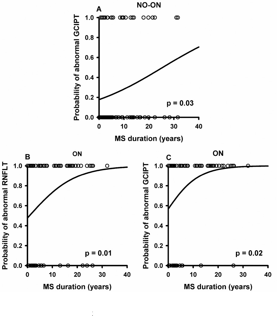
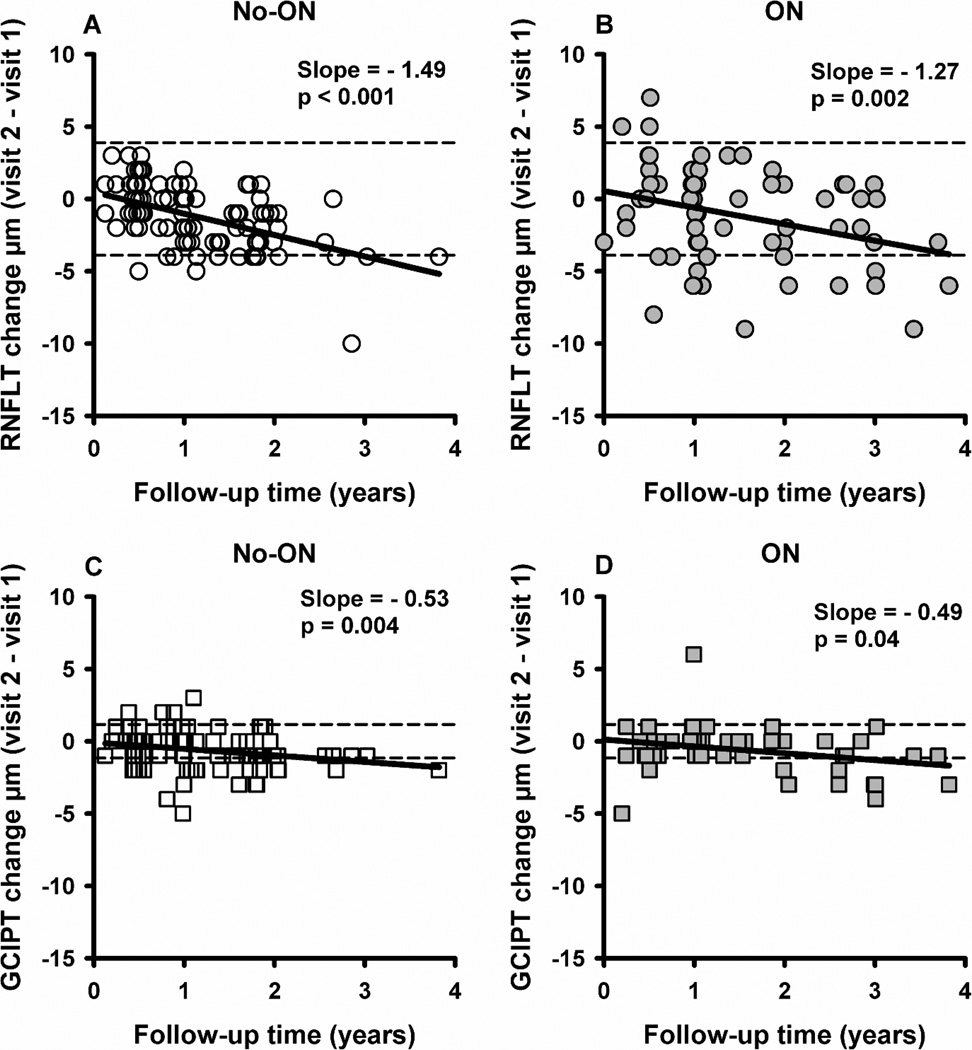
Results: GCIPT was abnormal in more eyes than RNFLT (27% vs 16% p = 0.004 in non-ON, 82% vs 72% p = 0.007 in ON). RNFLT and GCIPT decreased with MS duration by -0.49 µm/yr (p = 0.0001) and -0.36 (p = 0.005) for non-ON; -0.52 (p = 0.003) and -0.41 (p = 0.007) for ON. RNFLT and GCIPT decreased with follow-up time by -1.49 µm/yr (p < 0.0001) and -0.53 (p = 0.004) for non-ON, -1.27 (p = 0.002) and -0.49 (p = 0.04) for ON.
Conclusions: In RRMS eyes without clinically evident inflammation, progressive loss of RNFLT and GCIPT occurred, supporting the need for neuroprotection in addition to suppression of autoimmune responses and inflammation.
Keywords: Multiple sclerosis; ganglion cell inner plexiform layer; neurodegeneration; optic neuritis; optical coherence tomography; retinal nerve fiber layer.
© The Author(s) 2014.
Conflict of interest statement
The authors declare that they have no conflicts of interest.
Figures
Similar articles
-
Comparative diagnostic accuracy of ganglion cell-inner plexiform and retinal nerve fiber layer thickness measures by Cirrus and Spectralis optical coherence tomography in relapsing-remitting multiple sclerosis.Biomed Res Int. 2014;2014:128517. doi: 10.1155/2014/128517. Epub 2014 Sep 18. Biomed Res Int. 2014. PMID: 25313352 Free PMC article.
-
Optical coherence tomography segmentation analysis in relapsing remitting versus progressive multiple sclerosis.PLoS One. 2017 Feb 13;12(2):e0172120. doi: 10.1371/journal.pone.0172120. eCollection 2017. PLoS One. 2017. PMID: 28192539 Free PMC article.
-
The Effect of Glatiramer Acetate on Retinal Nerve Fiber Layer Thickness in Patients with Relapsing-Remitting Multiple Sclerosis: A Longitudinal Optical Coherence Tomography Study.CNS Drugs. 2018 Aug;32(8):763-770. doi: 10.1007/s40263-018-0521-9. CNS Drugs. 2018. PMID: 29767815
-
Optical coherence tomography as a prognostic tool for disability progression in MS: a systematic review.J Neurol. 2023 Feb;270(2):1178-1186. doi: 10.1007/s00415-022-11474-4. Epub 2022 Nov 13. J Neurol. 2023. PMID: 36372866
-
Optical coherence tomography detection of neurodegeneration in multiple sclerosis.CNS Neurol Disord Drug Targets. 2012 Aug;11(5):518-27. doi: 10.2174/187152712801661185. CNS Neurol Disord Drug Targets. 2012. PMID: 22583437 Review.
Cited by
-
Pharmacological Approaches to the Management of Secondary Progressive Multiple Sclerosis.Drugs. 2017 May;77(8):885-910. doi: 10.1007/s40265-017-0726-0. Drugs. 2017. PMID: 28429241 Review.
-
Optical coherence tomography (OCT) in neuro-ophthalmology.Eye (Lond). 2021 Jan;35(1):17-32. doi: 10.1038/s41433-020-01288-x. Epub 2020 Nov 25. Eye (Lond). 2021. PMID: 33239763 Free PMC article. Review.
-
Clinical trials in progressive multiple sclerosis: lessons learned and future perspectives.Lancet Neurol. 2015 Feb;14(2):208-23. doi: 10.1016/S1474-4422(14)70264-9. Lancet Neurol. 2015. PMID: 25772899 Free PMC article. Review.
-
Longitudinal evaluation of retinal neuroaxonal loss in epilepsy using optical coherence tomography.Epilepsia. 2024 Dec;65(12):3644-3654. doi: 10.1111/epi.18139. Epub 2024 Oct 9. Epilepsia. 2024. PMID: 39380535 Free PMC article.
-
Retinal ganglion cell analysis in multiple sclerosis and optic neuritis: a systematic review and meta-analysis.J Neurol. 2017 Sep;264(9):1837-1853. doi: 10.1007/s00415-017-8531-y. Epub 2017 May 31. J Neurol. 2017. PMID: 28567539
References
-
- Weinshenker BG, Bass B, Rice GP, et al. The natural history of multiple sclerosis: a geographically based study. 2. Predictive value of the early clinical course. Brain. 1989;112(Pt 6):1419–1428. - PubMed
-
- Noseworthy JH, Lucchinetti C, Rodriguez M, Weinshenker BG. Multiple sclerosis. N Engl J Med. 2000;343:938–952. - PubMed
-
- Rudick RA, Cutter GR, Baier M, et al. Estimating long-term effects of disease-modifying drug therapy in multiple sclerosis patients. Mult Scler. 2005;11:626–634. - PubMed
Publication types
MeSH terms
Grants and funding
LinkOut - more resources
Full Text Sources
Other Literature Sources
Miscellaneous
