. 2014 Jun 15;592(12):2605-23.
doi: 10.1113/jphysiol.2014.272468. Epub 2014 Mar 17.

Ponto-medullary nuclei involved in the generation of sequential pharyngeal swallowing and concomitant protective laryngeal adduction in situ

Affiliations

Ponto-medullary nuclei involved in the generation of sequential pharyngeal swallowing and concomitant protective laryngeal adduction in situ

Tara G Bautista et al. J Physiol. .

Abstract

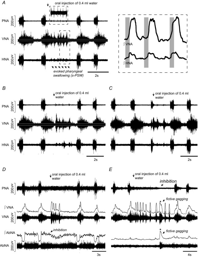
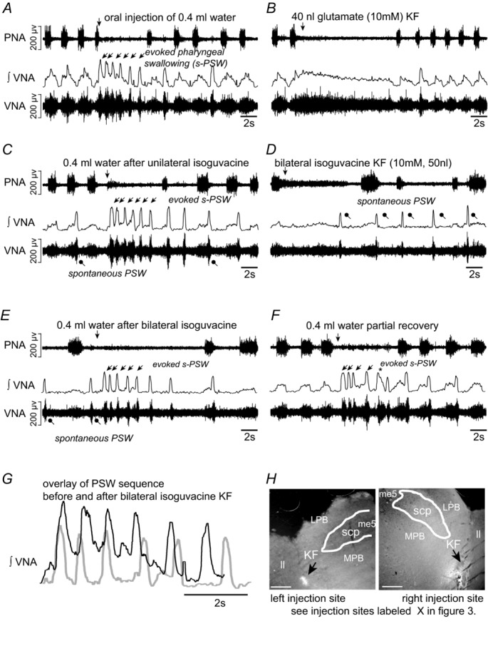
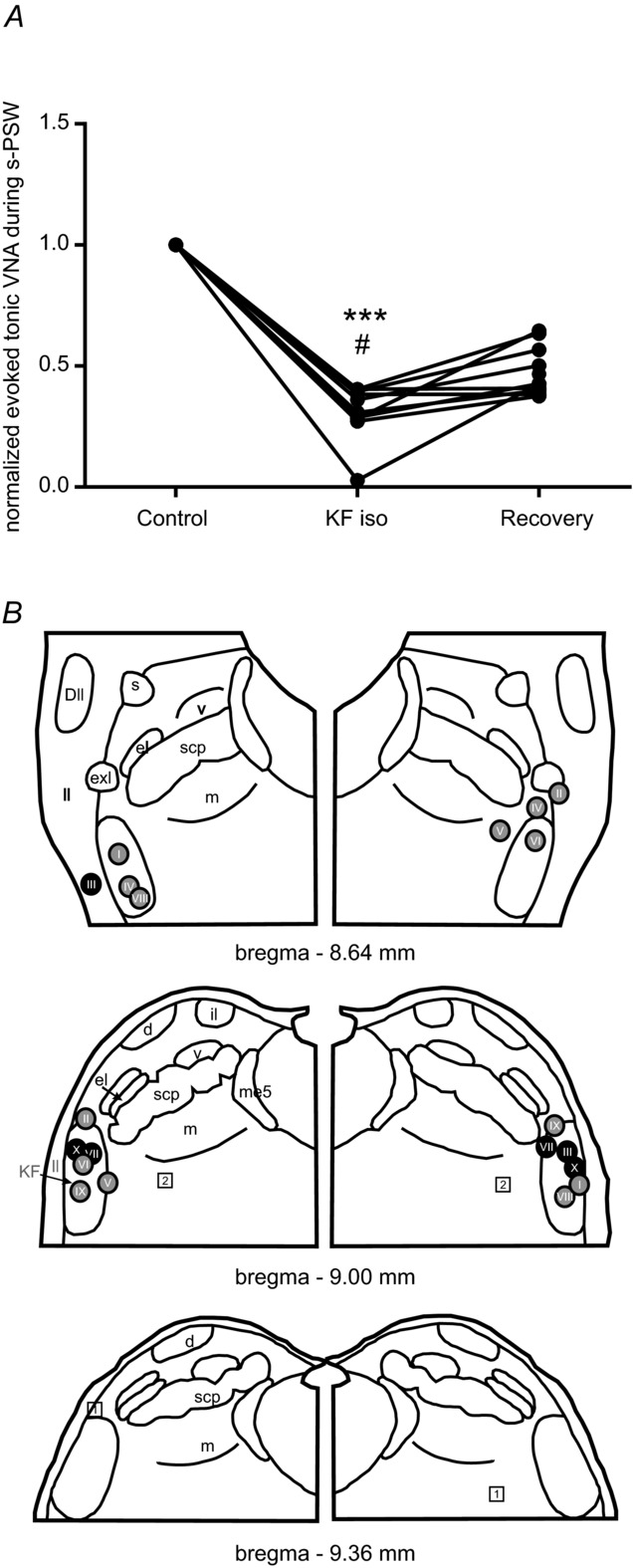
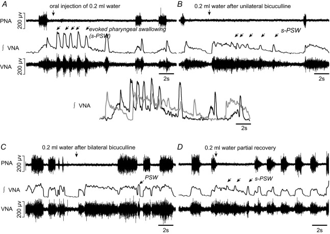
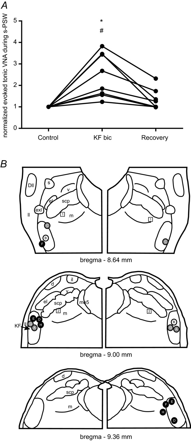
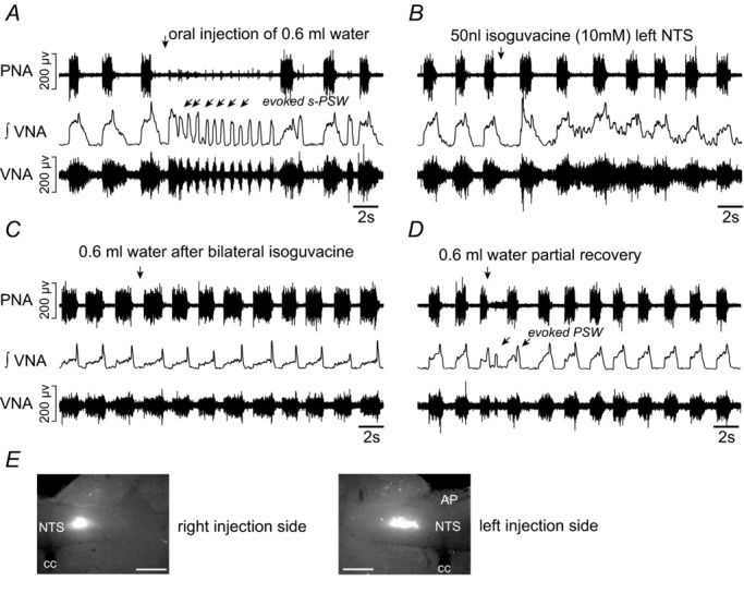
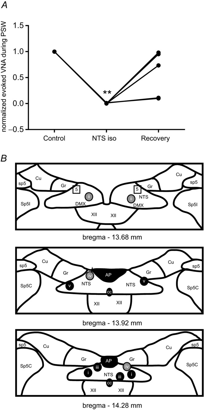
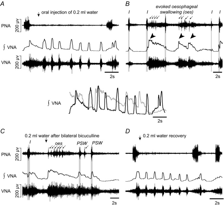
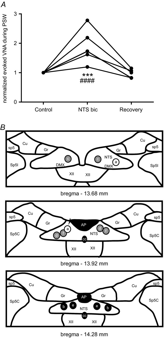
Both swallowing and respiration involve postinspiratory laryngeal adduction. Swallowing-related postinspiratory neurons are likely to be located in the nucleus of the solitary tract (NTS) and those involved in respiration are found in the Kölliker-Fuse nucleus (KF). The function of KF and NTS in the generation of swallowing and its coordination with respiration was investigated in perfused brainstem preparations of juvenile rats (n = 41). Orally injected water evoked sequential pharyngeal swallowing (s-PSW) seen as phasic, spindle-shaped bursting of vagal nerve activity (VNA) against tonic postinspiratory discharge. KF inhibition by microinjecting isoguvacine (GABAA receptor agonist) selectively attenuated tonic postinspiratory VNA (n = 10, P < 0.001) but had no effect on frequency or timing of s-PSW. KF disinhibition after bicuculline (GABAA receptor antagonist) microinjections caused an increase of the tonic VNA (n = 8, P < 0.01) resulting in obscured and delayed phasic s-PSW. Occurrence of spontaneous PSW significantly increased after KF inhibition (P < 0.0001) but not after KF disinhibition (P = 0.14). NTS isoguvacine microinjections attenuated the occurrence of all PSW (n = 5, P < 0.01). NTS bicuculline microinjections (n = 6) resulted in spontaneous activation of a disordered PSW pattern and long-lasting suppression of respiratory activity. Pharmacological manipulation of either KF or NTS also triggered profound changes in respiratory postinspiratory VNA. Our results indicate that the s-PSW comprises two functionally distinct components. While the primary s-PSW is generated within the NTS, a KF-mediated laryngeal adductor reflex safeguards the lower airways from aspiration. Synaptic interaction between KF and NTS is required for s-PSW coordination with respiration as well as for proper gating and timing of s-PSW.


Figures

Figure 1
Figure 1. Fictive swallow sequences elicited by water stimulation of the pharynx of the in situ perfused juvenile rat preparation
A, a typical example of a sequential pharyngeal swallowing (s-PSW) sequence elicited by oral injection of a small volume of distilled water during postinspiration. s-PSW corresponds with short, spindle-shaped bursts in cervical vagal nerve activity (VNA). Each s-PSW-related VNA burst is accompanied by a low amplitude burst in lateral hypoglossal nerve activity (HNA) (lower box; integrated VNA and HNA shown at higher magnification on the right). During the s-PSW sequence, inspiratory discharges in phrenic nerve activity (PNA) are inhibited, although a very low amplitude activity sometimes occurred in time with s-PSW-related VNA bursts (top box). Importantly, tonic lower amplitude activity is observed in VNA prior to and in between phasic swallow bursts. White lines indicate zero activity in VNA. B, prominent tonic s-PSW-related VNA still occurs if water injection is timed in late expiration. Note the medium-sized burst in HNA in the interval between the three fast swallows and the delayed fourth swallow. C, water stimulation timed during inspiration prematurely terminates this phase. Generally, fewer s-PSW bursts are elicited and the tonic activity between swallows is reduced. D, baseline activity from the iliohypogastric branch of abdominal nerve (AbNA) is inhibited during the evoked s-PSW. E, fictive gags (asterisk) are occasionally elicited by water stimulation. Gags are associated with longer bursts in VNA and with concurrent robust bursts in AbNA.
Figure 2
Figure 2. Swallowing activity after inhibition of the Kölliker–Fuse nucleus (KF)
A, evoked sequential pharyngeal swallowing (s-PSW, arrows) during baseline conditions. B, microinjection of glutamate into the intermediate KF region induces a postinspiratory apnoea. C, unilateral injection of GABAA receptor agonist isoguvacine (10 mm, 50 nl) into the left KF results in spontaneous swallows (dots). Tonic VNA during evoked s-PSW is slightly reduced. D, increased incidence of spontaneous swallows immediately after the second microinjection of isoguvacine into the right KF. E, bilateral microinjections of isoguvacine into the KF region causes apneusis. The tonic postinspiratory component of the evoked s-PSW is virtually abolished. F, partial recovery of the tonic s-PSW activity after 45 min. G, integrated VNA showing that the timing and duration of s-PSW remains comparable before (black) and after (grey) bilateral inhibition of KF. The decrease in amplitude of tonic VNA is the only obvious difference. H, microinjection sites marked with rhodamine beads. The KF is shown in relation to the lateral leminscus (ll), lateral parabrachial complex (LPB), medial parabrachial complex (MPB), superior cerebellar peduncle (scp) and mesencephalic trigeminal nucleus (Me5). Scale bar = 500 μm.
Figure 3
Figure 3. Summary of responses to water stimulation after inhibition of the Kölliker–Fuse nucleus
A, line graph showing a reduction of tonic VNA component during evoked s-PSW after inhibition of the KF region by bilateral microinjections of isoguvacine. Normalized values are shown. However, the raw mean value of tonic VNA during evoked s-PSW after bilateral isoguvacine microinjections into KF was significantly reduced compared to the mean at baseline (***P < 0.001) and during recovery (#P < 0.01). B, semi-schematic drawing of the pontine parabrachial complex to illustrate location of the microinjection sites (I–X). Colour-coding of the injection sites reflects the magnitude of the effect of isoguvacine on the tonic VNA during evoked s-PSW. Black circles reflect a reduction of tonic VNA of 70–100%. Grey circles reflect 30–70% reduction in tonic VNA. Squares reflect ineffective microinjection sites (0–20% reduction). Abbreviations: Dll, dorsal nucleus of the lateral lemniscus; me5, mesencephalic trigeminal neurons; scp, superior cerebellar peduncle. Nuclei of the parabrachial complex: d, dorsal nucleus; el, external lateral nucleus; exl, extreme lateral nucleus; il, internal lateral nucleus; KF, Kölliker–Fuse nucleus; m, medial nucleus; v, ventral nucleus.
Figure 4
Figure 4. Swallowing activity after disinhibition of the Kölliker–Fuse nucleus
A, evoked sequential pharyngeal swallowing (s-PSW, arrows) during baseline conditions. B, unilateral microinjection of GABAA receptor antagonist bicuculline (10 mm, 50 nl) increased postinspiratory activity in VNA. Amplitude of tonic VNA during evoked s-PSW was also increased. Below, the overlay of integrated evoked s-PSW-related VNA before (black) and after (grey) illustrates the relative increase in the tonic VNA. C, water stimulation after bilateral microinjections of bicuculline resulted in exaggerated tonic VNA without evidence of phasic s-PSW initially. A single delayed PSW was observed after an inspiratory burst in phrenic nerve. D, partial recovery of the s-PSW response to water stimulation after 90 min. Notice the reappearance of s-PSW-related VNA despite the elevated tonic VNA.
Figure 5
Figure 5. Summary of responses to water stimulation after disinhibition of the Kölliker–Fuse nucleus
A, line graph showing an increase in the tonic VNA component during evoked s-PSW after disinhibition of the KF region by bilateral microinjections of bicuculline. Normalized values are shown. However, the raw mean value of tonic VNA during evoked s-PSW after bilateral bicuculline microinjections into KF was significantly increased compared to the mean at baseline (*P < 0.01) and during recovery (#P < 0.01). B, semi-schematic drawing of the pontine parabrachial complex to illustrate location of the microinjection sites (A–H). Colour-coding of the injection sites reflects the magnitude of the effect of bicuculline on the tonic VNA during evoked s-PSW. Black circles reflect an increase in tonic VNA of >150%. Grey circles reflect 50–150% increase in tonic VNA. Squares reflect ineffective microinjection sites (increase <50%). For abbreviations see Fig. 3.
Figure 6
Figure 6. Swallowing activity after inhibition of the caudal nucleus of the solitary tract
A, evoked sequential pharyngeal swallowing (s-PSW, arrows) during baseline conditions. B, unilateral microinjection of isoguvacine (10 mm, 50 nl) into the caudal NTS induces immediate prolongation of inspiratory period and shortening of postinspiratory activity. C, all responses to water stimulation were abrogated after bilateral microinjections of isoguvacine into the NTS. D, partial recovery of evoked s-PSW after 45 min. E, microinjection sites marked by rhodamine beads were found in the caudal portions of the NTS. The NTS is shown in relation to the central canal (cc) and area postrema (AP). Scale bar = 200 μm.
Figure 7
Figure 7. Summary of responses to water stimulation after inhibition of the NTS
A, line graph showing a reduction of evoked VNA during s-PSW after isoguvacine injection into NTS. Normalized values are shown. However, the raw mean value of tonic VNA during evoked s-PSW after bilateral isoguvacine microinjections into NTS was significantly decreased compared to the mean at baseline (**P < 0.001) but not different compared to recovery. B, schematic diagrams illustrating bilateral injection (i–v) sites into the NTS. Colour-coding of the injection sites reflects the magnitude of the effect of isoguvacine on the tonic VNA during evoked s-PSW. Black circles reflect complete abolition of all spontaneous and evoked PSW. Grey circles reflect abolished spontaneous PSW and decreased VNA during evoked s-PSW (at least 25% decrease). Squares reflect ineffective injection site. Abbreviations: AP, area postrema; cc, central canal; Cu, cuneate nucleus; DMX, dorsal motor nucleus of the vagal nerve; Gr, gracile nucleus; NTS, nucleus of the solitary tract; sp5, spinal trigeminal tract; Sp5C, caudal nucleus of the spinal trigeminal tract; Sp5I, interpolar nucleus of the spinal trigeminal tract; XII, hypoglossal motor nucleus.
Figure 8
Figure 8. Responses to water stimulation after disinhibition of the NTS
A, evoked sequential pharyngeal swallowing (s-PSW, arrows) during baseline conditions. B, profound disruption to respiratory activity following bilateral microinjections of bicuculline (10 nm, 50 nl) into the caudal NTS caused by spontaneous cycles of large, decrementing s-PSW-related bursts in VNA (tilted arrowheads). The decrementing bursts in VNA were associated with tonic activity and small rhythmic bursts in PNA (arrows). Some relatively normal but shortened respiratory-related discharges in PNA and VNA (grey panelling; I-inspiration) were observed but were infrequent. C, the disordered response to water stimulation after bilateral microinjections of bicuculline into the NTS is very similar to the spontaneous cyclic activity in B. Some delayed bursts in VNA that resemble normal PSW-related bursts are shown in grey panelling. Above, an overlay of the response to water stimulation in integrated and smoothed VNA before (black) and after (grey) bilateral disinhibition of the NTS. D, recovery of the control response to swallow stimulation as well as normal respiratory discharges after 90 min. Note that both spontaneous (seen in B) and evoked (seen in C) PSW were always characterized by presumptive oesophageal swallowing (‘oes’) seen as the shortened bursts in the PNA recording.
Figure 9
Figure 9. Summary of responses to water stimulation after disinhibition of the NTS
A, line graph showing an increase of evoked VNA during PSW after bicuculline injection into NTS. Normalized values are shown. However, the raw mean value of evoked VNA during evoked PSW after bilateral bicuculline microinjections into NTS was significantly increased compared to the mean at baseline (***P < 0.0001) and during recovery (####P < 0.00001). B, schematic diagrams illustrating bilateral microinjection sites (a–f) into the NTS are shown. Colour-coding of the injection sites reflects the magnitude of the effect of bicuculline on VNA during evoked s-PSW. Black circles reflect an increase in evoked VNA >100%. Grey circles reflect 50–100% increase in evoked VNA. The white circle reflects an ineffective microinjection site. For abbreviations see Fig. 7.

References

    1. Abdala AP, Rybak IA, Smith JC, Paton JF. Abdominal expiratory activity in the rat brainstem-spinal cord in situ: patterns, origins and implications for respiratory rhythm generation. J Physiol. 2009;587:3539–3559. - PMC - PubMed
    1. Abdala APL, Dutschmann M, Bissonnette JM, Paton JFR. Correction of respiratory disorders in a mouse model of Rett syndrome. Proc Natl Acad Sci U S A. 2010;107:18208–18213. - PMC - PubMed
    1. Alagiakrishnan K, Bhanji RA, Kurian M. Evaluation and management of oropharyngeal dysphagia in different types of dementia: A systematic review. Arch Gerontol Geriatr. 2013;56:1–9. - PubMed
    1. Alheid GF, Milsom WK, McCrimmon DR. Pontine influences on breathing: an overview. Respir Physiol Neurobiol. 2004;143:105–114. - PubMed
    1. Aydogdu I, Tanriverdi Z, Ertekin C. Dysfunction of bulbar central pattern generator in ALS patients with dysphagia during sequential deglutition. Clin Neurophysiol. 2011;122:1219–1228. - PubMed

Publication types

LinkOut - more resources