. 2014 Apr 1;21(4):223-31.
doi: 10.1101/lm.034199.113.

Behavioral flexibility is increased by optogenetic inhibition of neurons in the nucleus accumbens shell during specific time segments

Affiliations

Behavioral flexibility is increased by optogenetic inhibition of neurons in the nucleus accumbens shell during specific time segments

Luca Aquili et al. Learn Mem. .

Abstract

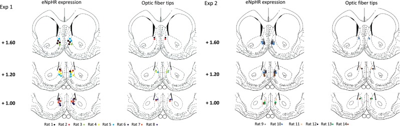
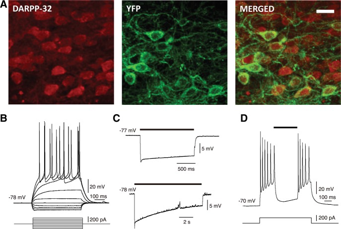
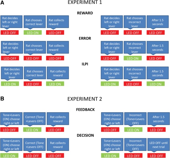
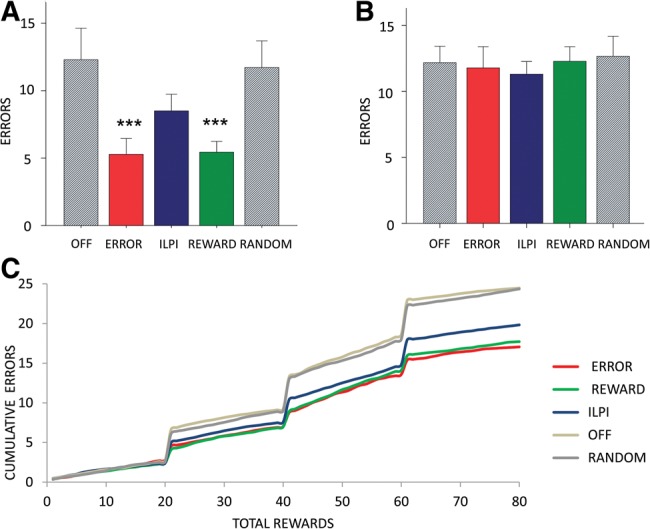
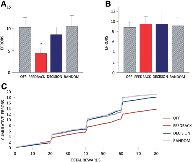
Behavioral flexibility is vital for survival in an environment of changing contingencies. The nucleus accumbens may play an important role in behavioral flexibility, representing learned stimulus-reward associations in neural activity during response selection and learning from results. To investigate the role of nucleus accumbens neural activity in behavioral flexibility, we used light-activated halorhodopsin to inhibit nucleus accumbens shell neurons during specific time segments of a bar-pressing task requiring a win-stay/lose-shift strategy. We found that optogenetic inhibition during action selection in the time segment preceding a lever press had no effect on performance. However, inhibition occurring in the time segment during feedback of results--whether rewards or nonrewards--reduced the errors that occurred after a change in contingency. Our results demonstrate critical time segments during which nucleus accumbens shell neurons integrate feedback into subsequent responses. Inhibiting nucleus accumbens shell neurons in these time segments, during reinforced performance or after a change in contingencies, increases lose-shift behavior. We propose that the activity of nucleus shell accumbens shell neurons in these time segments plays a key role in integrating knowledge of results into subsequent behavior, as well as in modulating lose-shift behavior when contingencies change.


Figures

Figure 1.
Figure 1.
Halorhodopsin expression and position of optical fibers in the NAcc shell. Location of maximal expression of halorhodopsin (eNpHR expression, left) and tips of optical fibers (right) are indicated by circles, color-coded by animal. Several animals showed halorhodopsin expression in multiple sections. Halorhodopsin expression extended ∼300 µm in the medial–lateral and 400 µm in the dorsal–ventral directions.
Figure 2.
Figure 2.
Cellular expression of halorhodopsin and electrophysiology of light-activated inhibition in medium spiny neurons. (A) Medium spiny neurons (DARPP-32, red), halorhodopsin (YFP, green), and their colocalization (MERGED) indicating halorhodopsin expression in medium spiny neurons (red + green). Scale bar, 20 µm. (B) Electrophysiological recording from YFP-positive neurons in NAcc shows the characteristic voltage response (above) of a medium spiny neuron to depolarizing and hyperpolarizing current pulses (below). (C) Optical stimulation of YFP-positive neurons induces hyperpolarization in medium spiny neurons on short and long timescales. Black bars indicate illumination time (upper trace, 1.5 sec; lower trace, 10 sec). (D) Illumination (black bar) blocked repetitive action potential firing induced by suprathreshold current injection.
Figure 3.
Figure 3.
Schematic representation of optogenetic conditions. (A) Experiment 1. REWARD: LED on after a correct lever press and off when the reward was collected. ERROR: LED on after an incorrect lever press and off after 1.5 sec. ILPI: LED on throughout but after a correct level press was turned off until reward collection and after an incorrect lever press was turned off for 1.5 sec. (B) Experiment 2. FEEDBACK: LED on for either REWARD or ERROR conditions as in Experiment 1. DECISION: LED on during tone and lever-out period until a correct or incorrect lever press.
Figure 4.
Figure 4.
Reversal errors are reduced by optogenetic inhibition during REWARD or ERROR epochs. (A) Mean total number of lever-pressing errors after reversal until first correct response summed over three reversals. There is a significant decrease in these errors in the ERROR and REWARD conditions. (B) Mean total number of lever-pressing errors excluding those shown in A. There is no difference between the conditions. (C) Learning curve showing cumulative errors over rewards acquired (80 rewards per session) for the three optogenetic conditions (ERROR, REWARD, ILPI) and two control conditions (OFF, RANDOM), confirming that the main effect is in the errors after reversal and before the first correct response.
Figure 5.
Figure 5.
Optogenetic stimulation has no effect on motivational variables. (A) There is no significant effect of condition on latency from correct lever press to reward collection. (B) There is no significant effect of condition on time to complete the task.
Figure 6.
Figure 6.
Optical stimulation has no effect on control rats with inactive halorhodopsin. (A) Total number of lever-pressing errors after reversal until first correct response summed over three reversals. There is no effect of condition. (B) Total number of lever-pressing errors excluding errors in the period after reversal until first correct response. There is no effect of condition.
Figure 7.
Figure 7.
Reversal errors are reduced by optogenetic inhibition during FEEDBACK. (A) Mean total number of lever-pressing errors after reversal until first correct response summed over three reversals. There is a significant decrease in these errors in the FEEDBACK condition but not in the DECISION, RANDOM, or OFF conditions. (B) Mean total number of lever-pressing errors excluding those shown in A. There is no difference between the conditions. (C) Learning curve showing cumulative errors over rewards acquired (80 rewards per session) for the two optogenetic conditions (FEEDBACK, DECISION) and the control conditions (OFF, RANDOM), confirming that the main effect occurs in the period between contingency switch and the first correct response.

References

    1. Aggarwal M, Hyland BI, Wickens JR 2012. Neural control of dopamine neurotransmission: implications for reinforcement learning. Eur J Neurosci 35: 1115–1123 - PubMed
    1. Ambroggi F, Ghazizadeh A, Nicola SM, Fields HL 2011. Roles of nucleus accumbens core and shell in incentive-cue responding and behavioral inhibition. J Neurosci 31: 6820–6830 - PMC - PubMed
    1. Aravanis AM, Wang LP, Zhang F, Meltzer LA, Mogri MZ, Schneider MB, Deisseroth K 2007. An optical neural interface: in vivo control of rodent motor cortex with integrated fiberoptic and optogenetic technology. J Neural Eng 4: S143–S156 - PubMed
    1. Arenkiel BR, Peca J, Davison IG, Feliciano C, Deisseroth K, Augustine GJ, Ehlers MD, Feng GP 2007. In vivo light-induced activation of neural circuitry in transgenic mice expressing channelrhodopsin-2. Neuron 54: 205–218 - PMC - PubMed
    1. Barto AG, Sutton RS 1982. Simulation of anticipatory responses in classical conditioning by a neuron-like adaptive element. Behav Brain Res 4: 221–235 - PubMed

MeSH terms

LinkOut - more resources