. 2014 Apr 1;111(13):4958-63.
doi: 10.1073/pnas.1403078111. Epub 2014 Mar 17.

Mycobacterial outer membrane is a lipid bilayer and the inner membrane is unusually rich in diacyl phosphatidylinositol dimannosides

Affiliations

Mycobacterial outer membrane is a lipid bilayer and the inner membrane is unusually rich in diacyl phosphatidylinositol dimannosides

Ritu Bansal-Mutalik et al. Proc Natl Acad Sci U S A. .

Abstract

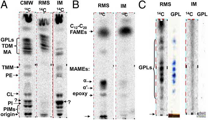
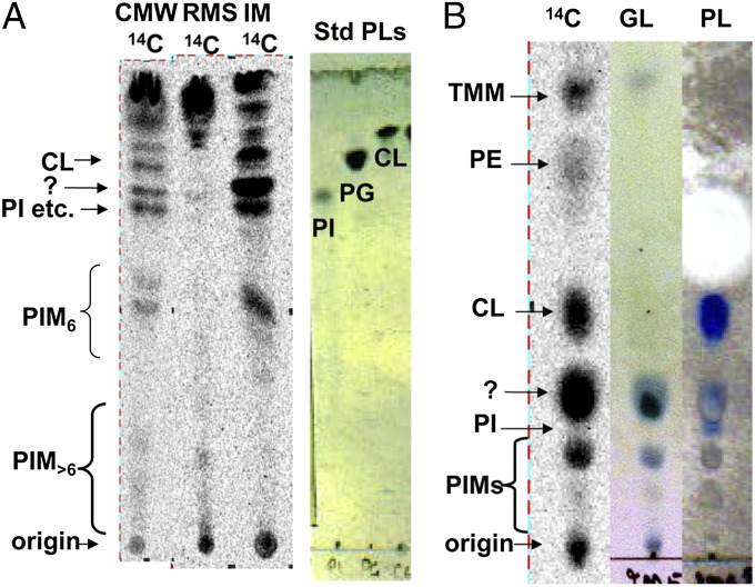
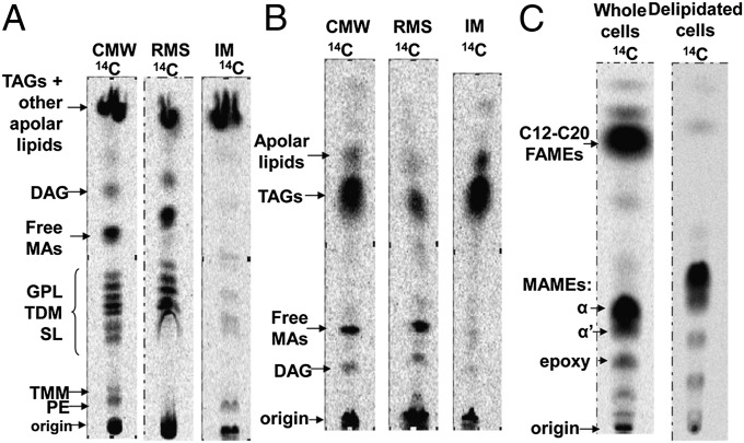
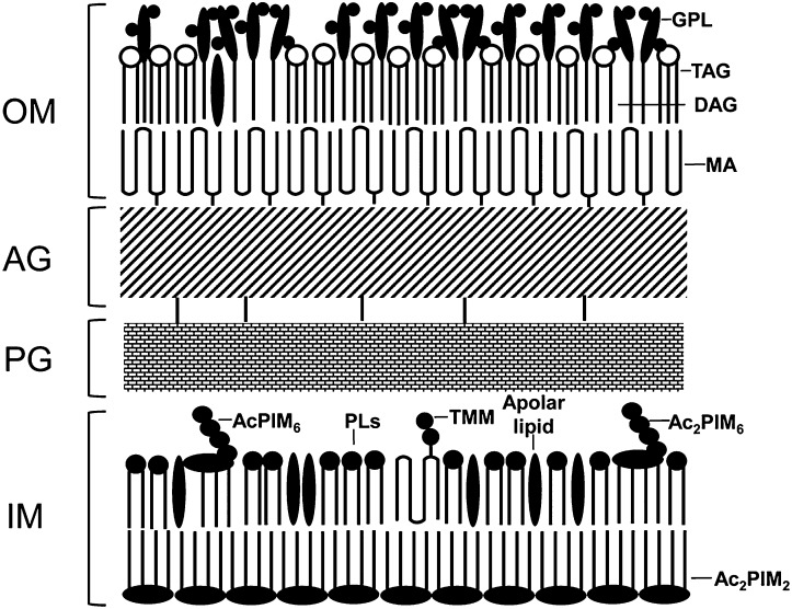
Mycobacterium species, including the human pathogen Mycobacterium tuberculosis, are unique among Gram-positive bacteria in producing a complex cell wall that contains unusual lipids and functions as a permeability barrier. Lipids in the cell wall were hypothesized to form a bilayer or outer membrane that would prevent the entry of chemotherapeutic agents, but this could not be tested because of the difficulty in extracting only the cell-wall lipids. We used reverse micellar extraction to achieve this goal and carried out a quantitative analysis of both the cell wall and the inner membrane lipids of Mycobacterium smegmatis. We found that the outer leaflet of the outer membrane contains a similar number of hydrocarbon chains as the inner leaflet composed of mycolic acids covalently linked to cell-wall arabinogalactan, thus validating the outer membrane model. Furthermore, we found that preliminary extraction with reverse micelles permitted the subsequent complete extraction of inner membrane lipids with chloroform-methanol-water, revealing that one-half of hydrocarbon chains in this membrane are contributed by an unusual lipid, diacyl phosphatidylinositol dimannoside. The inner leaflet of this membrane likely is composed nearly entirely of this lipid. Because it contains four fatty acyl chains within a single molecule, it may produce a bilayer environment of unusually low fluidity and may slow the influx of drugs, contributing to the general drug resistance phenotype of mycobacteria.

Keywords: Mycobacterium smegmatis; mycolic acid; phosphoinositides.


Conflict of interest statement

The authors declare no conflict of interest.

Figures

Fig. 1.
Fig. 1.
TLC profiles demonstrate the selective OM-solubilizing ability of RMS in M. smegmatis mc2 155. In all of the figures, CMW and RMS indicate CMW and RMS extracts of intact cells, whereas IM indicates CMW extract of residues of RMS-extracted cells. (A) TLC of M. smegmatis 14C-labeled lipids developed with CMW (30:8:1), showing that PLs (PE, CL, and PI) are abundant in IM but are totally absent in the RMS extract (OM lipids). (B) TLC of the FA esters obtained from 14C-labeled lipids of M. smegmatis, developed using petroleum ether:diethylether (85:15), detected by radioactivity. The material from the IM extract shows only traces of MAMEs. (C) TLC of alkali-treated lipids of M. smegmatis, developed with CM (9:1) and sprayed with anthrone reagent for visualization of GPLs.
Fig. 2.
Fig. 2.
TLC profiles of polar lipids from M. smegmatis. (A) TLC profiles of 14C-labeled lipids in different extracts, developed with chloroform–methanol–13 M ammonia–1 M ammonium acetate–water (180:140:9:9:23, vol/vol). Question mark indicates the suspected diacyl phosphatidylinositol dimannosides (Ac2PIM2). The band labeled “PI etc.” seems to contain not only PI but also AcPIM2, PIM2, and PIM4. For comparison, standard phospholipids (std PLs) were developed in the same solvent and visualized with phosphospray. (B) TLC profiles of IM lipids developed using chloroform–methanol–water (65:25:4, vol/vol) sprayed with anthrone for glycolipids (GLs) or phsophospray for PLs.
Fig. 3.
Fig. 3.
TLC of medium- and low-polarity lipids from M. smegmatis. (A) Development with CMW (100:14:0.8), showing that these lipids are confined to OM (RMS extract), except TAG. (B) Development with hexane–diethyl ether–acetic acid (70:30:1). (C) FA and mycolic acid methyl esters (FAMEs and MAMEs) obtained from whole cells or delipidated (CMW-extracted) cells, developed with petroleum ether: diethylether (85:15).
Fig. 4.
Fig. 4.
SDS/PAGE profile of lipoglycans in various aqueous and phenol water extracts of M. smegmatis. RMaq and IMaq denote aqueous washes following RMS extraction and following CMW extraction of RMS-treated cells, respectively. “PWE after IME” and “PWE after CME intact” indicate the phenol–water extracts of residues obtained after IM extraction and after CMW extraction of intact cells, respectively. The leftmost lane shows MW standards, the size indicated in kilodaltons. (A) Staining with basic fuschin. (B) Visualization with the CS-35 antibody probe specific for LAMs.
Fig. 5.
Fig. 5.
A probable model of mycobacterial cell envelope. Based on the lipid composition data obtained in this study, a model of the OM and IM is proposed. The ratios between various components reflect the quantitative composition data, but very roughly. In the OM, TDM is not shown because it is present in an amount corresponding to less than 1 in the scheme. Free MA is not shown because it is likely to exist largely as an extracellular material. The thickness of peptidoglycan (PG) and arabinogalactan (AG) layers is arbitrary.

References

    1. Minnikin DE. Lipids: Complex lipids, their chemistry, biosynthesis, and roles. In: Ratledge C, Stanford J, editors. The Biology of the Mycobacteria. New York: Academic; 1982. pp. 95–184.
    1. Brennan PJ, Nikaido H. The envelope of mycobacteria. Annu Rev Biochem. 1995;64:29–63. - PubMed
    1. Ortalo-Magné A, et al. Identification of the surface-exposed lipids on the cell envelopes of Mycobacterium tuberculosis and other mycobacterial species. J Bacteriol. 1996;178(2):456–461. - PMC - PubMed
    1. Bhamidi S, et al. Detailed structural and quantitative analysis reveals the spatial organization of the cell walls of in vivo grown Mycobacterium leprae and in vitro grown Mycobacterium tuberculosis. J Biol Chem. 2011;286(26):23168–23177. - PMC - PubMed
    1. Etienne G, et al. The cell envelope structure and properties of Mycobacterium smegmatis mc(2)155: Is there a clue for the unique transformability of the strain? Microbiology. 2005;151(Pt 6):2075–2086. - PubMed

Publication types