. 2014 Apr 1;111(13):E1291-9.
doi: 10.1073/pnas.1403244111. Epub 2014 Mar 17.

Cartography of neurexin alternative splicing mapped by single-molecule long-read mRNA sequencing

Affiliations

Cartography of neurexin alternative splicing mapped by single-molecule long-read mRNA sequencing

Barbara Treutlein et al. Proc Natl Acad Sci U S A. .

Abstract

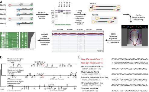
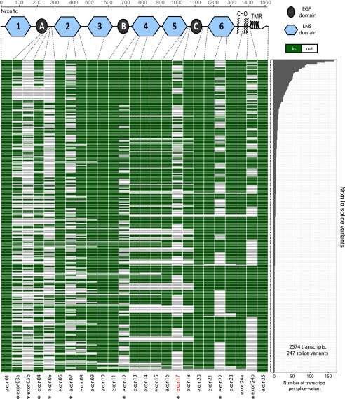
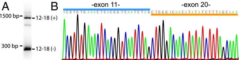
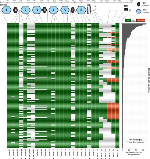
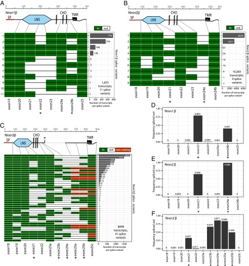
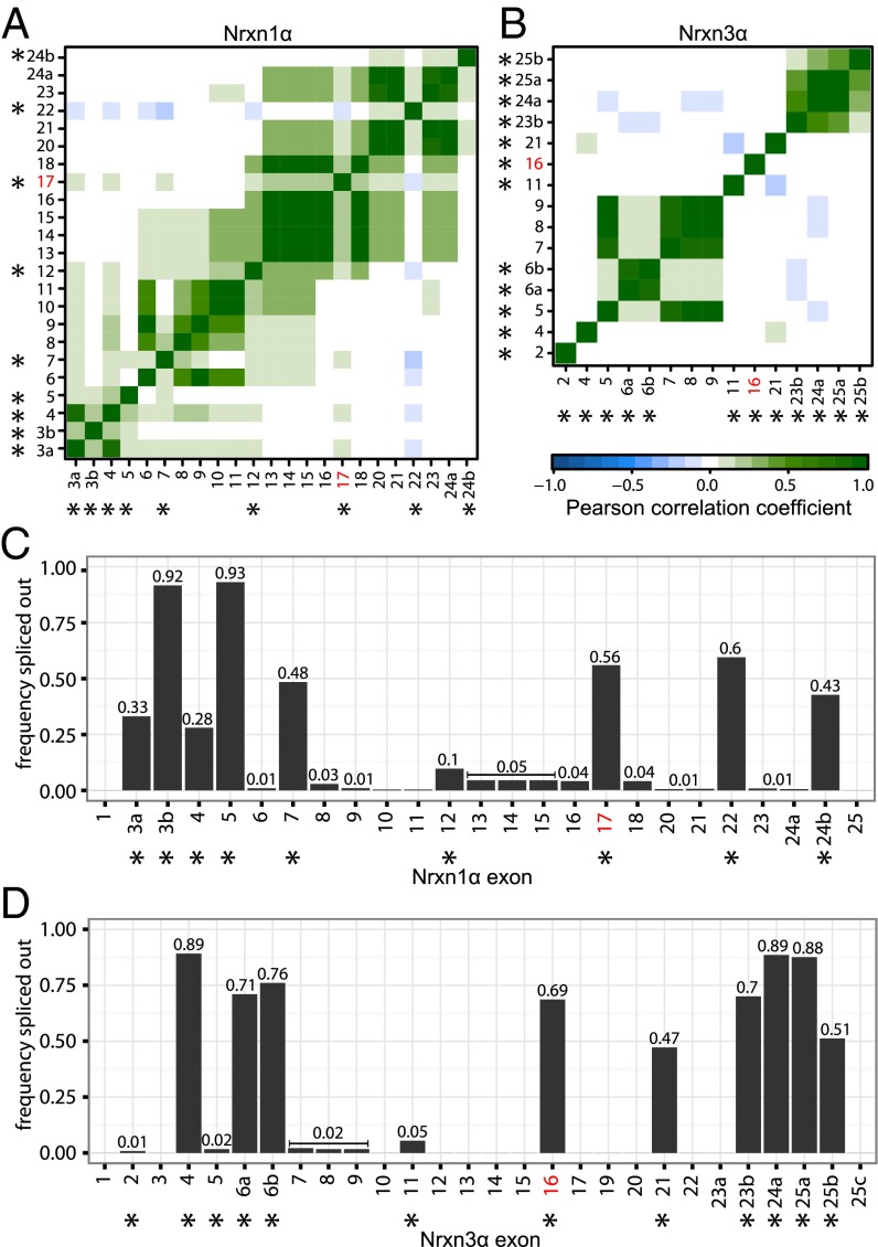
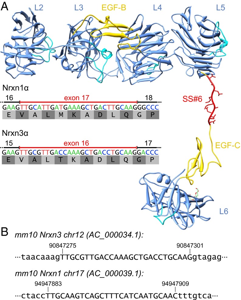
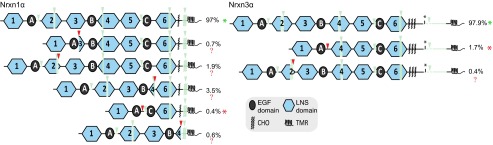
Neurexins are evolutionarily conserved presynaptic cell-adhesion molecules that are essential for normal synapse formation and synaptic transmission. Indirect evidence has indicated that extensive alternative splicing of neurexin mRNAs may produce hundreds if not thousands of neurexin isoforms, but no direct evidence for such diversity has been available. Here we use unbiased long-read sequencing of full-length neurexin (Nrxn)1α, Nrxn1β, Nrxn2β, Nrxn3α, and Nrxn3β mRNAs to systematically assess how many sites of alternative splicing are used in neurexins with a significant frequency, and whether alternative splicing events at these sites are independent of each other. In sequencing more than 25,000 full-length mRNAs, we identified a novel, abundantly used alternatively spliced exon of Nrxn1α and Nrxn3α (referred to as alternatively spliced sequence 6) that encodes a 9-residue insertion in the flexible hinge region between the fifth LNS (laminin-α, neurexin, sex hormone-binding globulin) domain and the third EGF-like sequence. In addition, we observed several larger-scale events of alternative splicing that deleted multiple domains and were much less frequent than the canonical six sites of alternative splicing in neurexins. All of the six canonical events of alternative splicing appear to be independent of each other, suggesting that neurexins may exhibit an even larger isoform diversity than previously envisioned and comprise thousands of variants. Our data are consistent with the notion that α-neurexins represent extracellular protein-interaction scaffolds in which different LNS and EGF domains mediate distinct interactions that affect diverse functions and are independently regulated by independent events of alternative splicing.

Keywords: LRRTM; autism; cerebellin; neuroligin; schizophrenia.


Conflict of interest statement

The authors declare no conflict of interest.

Figures

Fig. 1.
Fig. 1.
Single-molecule long-read sequencing of full-length neurexin mRNA transcripts. (A) Schema of the workflow of Pacific Biosciences sequencing of full-length neurexin transcripts for mapping neurexin diversity. Full-length transcripts of Nrxn1α, 1β, 2β, 3α, and 3β were separately reverse-transcribed and PCR-amplified (RT-PCR) from total RNA isolated from adult murine prefrontal cortex. PCR was performed with primer pairs specific to the first and last coding exons of each gene, with α- and β-specific the forward primers and identical reverse primers for the α- and β-versions of a given neurexin. The cDNA size distribution for each neurexin was examined by gel electrophoresis before preparation of Pacific Biosciences sequencing libraries by blunt-end ligation of hairpin adapters, annealing of the sequencing primer, and binding of biotinylated DNA polymerase. Subsequently, full-length transcripts were sequenced by single-molecule long-read sequencing (36), yielding an average read length of 3.6–5.8 kb. Reads containing at least two adapters (≥1 passes through circular DNA by the polymerase) were processed (quality filter, adapter removal) and then aligned to the mouse genome (mm10) using the STAR aligner (38) and used for further analysis. (B) Phylogenetic tree and structures of mouse neurexin genes. The diagrams depict the positions of exons and introns. Exons are numbered; asterisks mark exons subject to canonical alternative splicing. The positions of α- and β-neurexin promoters are indicated, and the position and size of the genes are shown above each gene diagram (see also Fig. S1). (C) Neurexin mRNAs from diverse species contain the newly identified SS#6. Sequences of murine Nrxn1α and Nrxn3α SS#6 that were identified in this study are colored in red.
Fig. 2.
Fig. 2.
Splice landscape of Nrxn1α. The transcript map visualizes the 247 unique alternatively spliced isoforms (rows) observed for the 2,574 full-length Nrxn1α mRNAs sequenced. Exons (columns) are colored in green if present and in white if absent, and are numbered at the bottom (asterisks, exons with canonical alternative splicing; for an explanation of the numbering, see Fig. S1). The domain structure of Nrxn1α is shown at the top (light blue hexagons, LNS domains; black ovals, EGF-like domains; CHO, O-linked sugar modifications; TMR, transmembrane region) and is connected to the exons that encode the respective domains by dotted gray lines. The abundance of each splice isoform is shown in the bar graph (Right).
Fig. 3.
Fig. 3.
Validation of newly identified alternatively spliced isoform of Nrxn1α lacking exons 12–18 by RT-PCR. (A) Agarose gel electrophoresis analysis of the PCR product obtained with primers specific to the end of exon 11 and beginning of exon 20 of Nrxn1α. Two major splice variants are identified: a long DNA fragment (∼1,500 bp) corresponding to transcripts containing exons 12–18 as well as a short DNA fragment (∼300 bp) corresponding to transcripts lacking exons 12–18. Note that exon 19 encodes the N terminus of Nrxn1β and is always missing from Nrxn1α mRNAs (12). (B) Sanger sequencing of the short PCR product as obtained in A.
Fig. 4.
Fig. 4.
Splice landscape of Nrxn3α. The transcript map visualizes the 138 unique alternatively spliced isoforms (rows) observed for the 934 full-length Nrxn3α mRNAs sequenced. Exons (columns) are colored in green for coding and red for exons with in-frame stop codons if present and in white if absent, and are numbered at the bottom (asterisks, exons with canonical alternative splicing; for an explanation of the numbering, see Fig. S1). The domain structure of Nrxn1α is shown at the top and is connected to the exons that encode the respective domains by dotted gray lines. In the domain structure, the asterisk denotes a stop codon that is present in some of the alternatively spliced exons and could produce secreted versions of neurexin-3. The abundance of each splice isoform is shown in the bar graph (Right).
Fig. 5.
Fig. 5.
Splice landscape of β-neurexins. (AC) Transcript maps visualizing unique splice isoforms (rows) observed for Nrxn1β (A; 1,653 transcripts, 11 splice variants), Nrxn2β (B; 10,283 transcripts, 9 splice variants), and Nrxn3β (C; 8,499 transcripts, 41 splice variants). Exons (columns) are colored in green if present and in white if absent, and are numbered at the bottom (asterisks, exons with canonical alternative splicing; for an explanation of the numbering, see Fig. S1). The domain structures of the respective β-neurexins are shown above the transcript maps and are connected to the exons that encode the respective domains by dotted gray lines. SP denotes the signal peptide; the asterisk in the domain structure of Nrxn3β indicates a stop codon encoded by one of the alternatively spliced exons. The abundance of each splice isoform is shown in the bar graph (Right). (DF) Bar graphs visualizing the relative frequency of exon skipping for the indicated exons in the Nrxn1β (D), Nrxn2β (E), and Nrxn3β mRNAs (F). Canonical alternatively spliced exons are marked by asterisks.
Fig. 6.
Fig. 6.
Independent and coordinated splicing of canonical and novel splice sites for Nrxn1α and Nrxn3α. (A and B) Correlograms showing the pairwise correlation in splice behavior between alternatively spliced exons of (A) Nrxn1α and (B) Nrxn3α. The color bar denotes the Pearson correlation coefficient from −1 (blue, anticorrelated splicing) through 0 (no correlation in splicing) to 1 (green, positively correlated splicing). Newly observed alternatively spliced exons tend to be spliced out in a coordinated way, whereas canonical events of alternative splicing appear to occur independent of each other. (C and D) Bar graphs visualizing the frequency of each exon to be spliced out for (C) Nrxn1α and (D) Nrxn3α. Canonical alternatively spliced exons (12) are marked by asterisks. Exons that are observed to be alternatively spliced in a coordinated way (A and B) show similar frequencies.
Fig. 7.
Fig. 7.
Sequence and structure of newly identified alternatively spliced exons for Nrxn1α and Nrxn3α. (A) mRNA sequence and translated 9-residue amino acid sequence of the newly identified alternatively spliced Nrxn1α exon 17 and Nrxn3α exon 16 (SS#6). A possible extended conformation of Nrxn1α exon 17 was modeled into the crystal structure of Nrxn1α [PDB ID 3QCW (39)] using Coot (42). SS#6 is located between the LNS5 and EGF-C domains, thereby extending the flexible hinge region between these structural features. The image of the Nrxn1α protein structure was created in Chimera (43). (B) Location of the newly identified exon of Nrxn1α and Nrxn3α in the mouse genome (mm10). Exonic nucleotides are shown in capital letters, whereas intronic nucleotides are shown in lowercase letters.
Fig. 8.
Fig. 8.
Protein domain structures of Nrxn1α and Nrxn3α splice isoforms. Protein domain structures corresponding to groups of splice isoforms are shown for Nrxn1α and Nrxn3α. Used splice sites are denoted by downward-pointing triangles colored in green for previously known canonical splice sites and red for novel splice sites. Protein parts that are spliced out only in a subset of splice variants of a given group are shown in semitransparent green. The percentage of total transcripts detected for each protein isoform is given. In the majority of detected transcripts only canonical splice sites are used, which are known to produce functional proteins (green asterisks). Most protein isoforms resulting from transcripts in which novel alternative splice sites are used appear to be nonfunctional (red question marks), because substantial parts of LNS domains are removed. However, a small subset of transcripts using novel alternative splice sites is suggested to produce functional, truncated proteins (red asterisks), because structural neurexin domains (LNS and EGF) are completely removed.

References

    1. Sperry RW. Chemoaffinity in the orderly growth of nerve fiber patterns and connections. Proc Natl Acad Sci USA. 1963;50(4):703–710. - PMC - PubMed
    1. Van Essen DC. Cartography and connectomes. Neuron. 2013;80(3):775–790. - PMC - PubMed
    1. Bargmann CI, Marder E. From the connectome to brain function. Nat Methods. 2013;10(6):483–490. - PubMed
    1. Meinertzhagen IA, Lee CH. The genetic analysis of functional connectomics in Drosophila. Adv Genet. 2012;80:99–151. - PMC - PubMed
    1. Kleinfeld D, et al. Large-scale automated histology in the pursuit of connectomes. J Neurosci. 2011;31(45):16125–16138. - PMC - PubMed

Publication types

MeSH terms

Associated data