. 2014 Apr 1;111(13):E1230-9.
doi: 10.1073/pnas.1320192111. Epub 2014 Mar 17.

Aβ-induced Golgi fragmentation in Alzheimer's disease enhances Aβ production

Affiliations

Aβ-induced Golgi fragmentation in Alzheimer's disease enhances Aβ production

Gunjan Joshi et al. Proc Natl Acad Sci U S A. .

Abstract

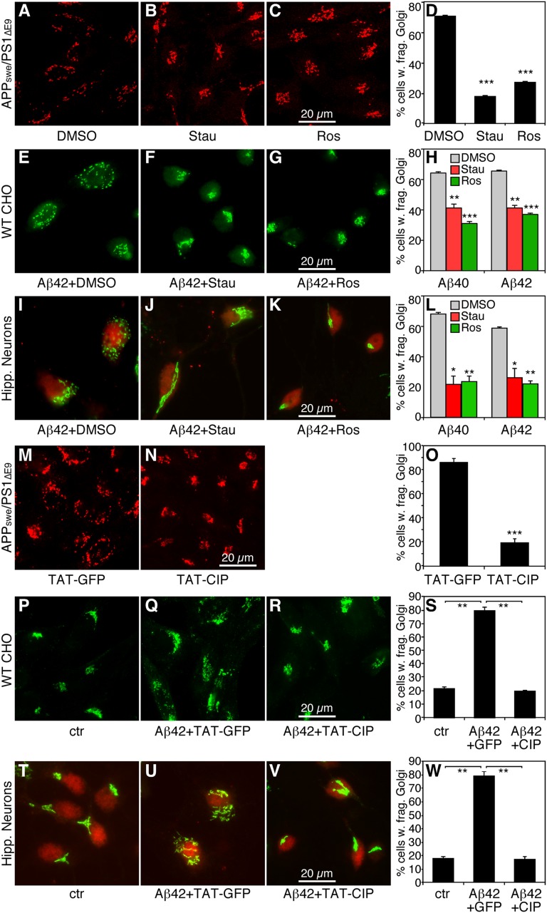
Golgi fragmentation occurs in neurons of patients with Alzheimer's disease (AD), but the underlying molecular mechanism causing the defects and the subsequent effects on disease development remain unknown. In this study, we examined the Golgi structure in APPswe/PS1E9 transgenic mouse and tissue culture models. Our results show that accumulation of amyloid beta peptides (Aβ) leads to Golgi fragmentation. Further biochemistry and cell biology studies revealed that Golgi fragmentation in AD is caused by phosphorylation of Golgi structural proteins, such as GRASP65, which is induced by Aβ-triggered cyclin-dependent kinase-5 activation. Significantly, both inhibition of cyclin-dependent kinase-5 and expression of nonphosphorylatable GRASP65 mutants rescued the Golgi structure and reduced Aβ secretion by elevating α-cleavage of the amyloid precursor protein. Our study demonstrates a molecular mechanism for Golgi fragmentation and its effects on amyloid precursor protein trafficking and processing in AD, suggesting Golgi as a potential drug target for AD treatment.

Keywords: APP processing; GRASP55; Golgi stacking; amyloidogenic.


Conflict of interest statement

The authors declare no conflict of interest.

Figures

Fig. 1.
Fig. 1.
Golgi is fragmented in APPswe/PS1∆E9 transgenic mice. (A and B) Fluorescence images of the Golgi in neurons of APPswe/PS1∆E9 transgenic mice. Cryostat sections of hippocampal tissues from 12-mo-old transgenic (B) and WT mice (A) were immunostained for TGN38. (C–F) EM micrographs of the Golgi region in neurons of APPswe/PS1∆E9 transgenic mice. Shown are EM images from ultrathin sections of hippocampal (C and D) and cortical (E and F) tissues from 12-mo-old transgenic mice (D and F) and WT mice (C and E).
Fig. 2.
Fig. 2.
APP and PS1 expression in tissue culture cells causes Golgi fragmentation. (A–C) CHO cells transfected with control vector (A), WT APP and PS1 (B), or APPswe/PS1∆E9 mutants (C) were immunostained for GRASP65. (D) Quantitation (mean ± SEM) of A–C from three independent experiments. (E–J) WT CHO cells and cells stably expressing APPswe/PS1∆E9 were immunostained for three different Golgi markers: GRASP65 (E and H, cis-Golgi), GRASP55 (F and I, medial-Golgi), and TGN38 (G and J, trans-Golgi). (K) Quantitation (mean ± SEM) of E–J. Note the significant Golgi fragmentation in APPswe/PS1∆E9-expressing cells using all three markers. (L–N) EM images of WT and APPswe/PS1∆E9-expressing cells with quantitation. Note that the APPswe/PS1∆E9-expressing cells have fewer cisternae per stack, shorter cisternae, and more vesicles. *P < 0.05; **P < 0.01; ***P < 0.001, Student t test.
Fig. 3.
Fig. 3.
Aβ accumulation causes Golgi fragmentation in CHO cells and primary hippocampal neurons. (A–D) Inhibition of Aβ production reduces Golgi fragmentation. APPswe/PS1∆E9-expressing CHO cells were cultured in growth medium containing DMSO (A), 20 µM α-secretase inhibitor TAPI (B), 1 µM β-secretase inhibitor BACEi (C), or 5 µM γ-secretase inhibitor DAPT (D) for 8 h and then analyzed by fluorescence microscopy for GRASP65. Note that inhibition of β- and γ-secretases, but not of α-secretase, reduced Golgi fragmentation. (E) Quantitation (mean ± SEM) of A–D. Statistical significance was assessed by comparison of secretase inhibitors to DMSO treatment. (F–H) Treatment with synthetic Aβ peptides leads to Golgi fragmentation in WT cells. WT CHO cells were treated without (control, F) or with 1 µM Aβ40 (G) or Aβ42 (H) for 6 h and immunostained for GRASP65. (I) Quantitation of F–H. Statistical significance was assessed by comparing Aβ-treated and PBS-treated cells. (J–L) EM images (J and K) and quantitation (L) of WT CHO cells treated with PBS or 1 µM Aβ40 as in F–G. (M–P) Cultured primary hippocampal neurons were treated with PBS (control, M), 1 µM Aβ40 (N), or Aβ42 (O) for 6 h and immunostained for GRASP65 in green and the neuronal marker NeuN in red. Fluorescence images are shown in A–C, and quantitation is shown in P. Statistical significance was assessed by comparing Aβ-treated and PBS-treated cells. (Q–S) EM images and quantitation of hippocampal neurons treated with PBS or with 1 µM Aβ42, as in M and O. **P < 0.01; ***P < 0.001, Student t test.
Fig. 4.
Fig. 4.
Kinase inhibitors reduce Aβ-induced Golgi fragmentation. (A–C) Kinase inhibitors reverse Golgi fragmentation in APPswe/PS1∆E9-expressing (AD) cells. Cells cultured overnight were treated with DMSO (A), 1 µM staurosporine (B), or 15 µM roscovitine (C) for 2 h. Chemicals were added directly to the tissue culture dishes without changing the media. Cells were immunostained for GRASP65. (D) Quantitation of A–C. (E–G) Kinase inhibitors reverse Golgi fragmentation in Aβ-treated WT CHO cells. WT CHO cells were treated with Aβ in the presence of DMSO (E), 1 µM staurosporine (F), or 8 µM roscovitine (G) for 2 h, and then stained for GRASP65. (H) Quantitation of E–G. (I–K) Kinase inhibitors reverse Aβ-induced Golgi fragmentation in hippocampal neurons. Primary hippocampal neurons were treated with Aβ peptides in the presence of DMSO (I), 1 µM staurosporine (J), or 8 µM roscovitine (K) for 2 h, and then stained for GRASP65. (L) Quantitation of I–K. (M and N) TAT-CIP treatment reverses Golgi fragmentation in AD cells. Cells cultured overnight were treated with 600 nM TAT-GFP (M) or TAT-CIP (N) (added directly into the medium) for 12 h, then immunostained for GRASP65. (O) Quantitation of M and N. (P–R) TAT-CIP inhibits Golgi fragmentation in Aβ-treated WT CHO cells. WT CHO cells were treated with 1 µm control peptide (P) or Aβ42 for 12 h in the presence of 600 nM TAT-GFP (Q) or TAT-CIP (R) added 30 min before Aβ42, then immunostained for GRASP65. (S) Quantitation of P–R. (T–W) TAT-CIP reverses Aβ-induced Golgi fragmentation in hippocampal neurons. Primary hippocampal neurons were treated with either control peptide (T) or 1 μM Aβ42 for 12 h in the presence of 600 nM TAT-GFP (U) or TAT-CIP (V) added 30 min before Aβ42, stained for GRASP65, and quantified (W). *P < 0.05; **P < 0.01; ***P < 0.001, Student t test.
Fig. 5.
Fig. 5.
Activation of cdk5 by Aβ accumulation phosphorylates GRASP65. (A) Cdk5 is activated by Aβ treatment. Hippocampal neurons in culture were treated with DMSO (lane 1) or 2 μM Aβ42 (lane 2) for 20 h. Lysates of these cells, of 12-mo-old control (lane 3), or of AD mouse brain (lane 4) were evaluated for p35/p25 and actin by Western blot analysis. Note the increased p25 levels in the Aβ-treated cells and AD mouse brain. (B) GRASP65 is phosphorylated in APP-expressing cells on Aβ accumulation. APPswe/PS1∆E9 cells stably expressing GFP-tagged GRASP65 WT protein (p65FL) were cultured overnight to accumulate Aβ in the media. The media for cells in lanes 1 and 4 were changed every hour for 4 h to remove Aβ from the medium, whereas cells in lanes 2, 3, and 5 were maintained in the old media. In lane 3, cells were treated with 15 μm roscovitine (Ros) in the old medium for another 2 h. After treatment, cells were lysed, immunoprecipitated using an anti-GRASP65 polyclonal antibody (lanes 1–3) or with control rabbit IgG (lanes 4 and 5), and evaluated by Western blot analysis for total GRASP65 (Lower) or phosphorylated GRASP65 (Upper). Note that GRASP65 is phosphorylated on Aβ accumulation (lane 2), and that this phosphorylation is inhibited by roscovitine (lane 3). (C) Aβ treatment-triggered GRASP65 phosphorylation depends on cdk5. Cells stably expressing GFP-tagged GRASP65 WT protein (p65FL) were treated with 4 µM control peptide (lane 1) or Aβ42 (lanes 2–4) for 12 h. During the last 2 h of incubation, 15 μM roscovitine (Ros) (lane 3) or 600 nM TAT-CIP (lane 4) was added directly into the medium. Cells were lysed and evaluated by Western blot analysis for phospho-GRASP65 and total GRASP65 as in B. (D) Activated cdk5 by p25 phosphorylates GRASP65. SH-SY5Y cells were transfected with cdk5 alone (lanes 2 and 3) or with cdk5+p25 (lanes 4–8). Cdk5 was immunoprecipitated and used to treat His-tagged recombinant GRASP65 in the presence or absence of roscovitine or TAT-CIP. The p25-activated cdk5 (lanes 4 and 5) phosphorylated the recombinant GRASP65 to a greater extent than the nonactivated cdk5 (lanes 2 and 3) or the nontransfected cdk5 (lane 1). Roscovitine (lane 6) and CIP (lanes 7 and 8) significantly reduced GRASP65 phosphorylation. (E–I) Activation of cdk5 by p25 causes Golgi fragmentation and enhances Aβ production. SH-SY5Y cells were transfected with cdk5 (F) and cdk5+p25 (G and H) for 16 h. Cells in H were subsequently treated with 600 nM TAT-CIP for 12 h. Nontransfected cells served as a negative control (E). Cells were stained for GRASP65, and images were quantified (I). (J and K) ELISA measurement of Aβ40 (J) and Aβ42 (K) secretion by the cells described in E–I. Shown are the average results from three independent experiments. *P < 0.05; **P < 0.01; ***P < 0.001, Student t test.
Fig. 6.
Fig. 6.
Golgi structure and function are rescued by expression of nonphosphorylatable GRASP mutants. (A–E) Expression of GRASP55, GRASP65, and their nonphosphorylatable mutants rescues the Golgi structure in AD cells. AD cells stably expressing APPswe/PS1∆E9 were transfected with GFP (A), GFP-tagged GRASP-domains p55GD (B) or p65GD (D), or full-length proteins p55FL (C) or p65FL (E) of GRASP55 or GRASP65. Shown are fluorescent images of GFP and GM130. Note that the Golgi is more compact in cells expressing GRASP55/65 and their mutants (positive cells indicated by asterisks). (F) Quantitation of A–E. (G) Western blot of the cells in A–E for indicated GFP and GFP-tagged proteins using a GFP antibody. Actin served as a loading control. *P < 0.05; **P < 0.01; ***P < 0.001, Student t test.
Fig. 7.
Fig. 7.
Rescue of the Golgi structure reduces Aβ production. (A–D) Expression of GRASPs and their nonphosphorylatable mutants reduces Aβ production. AD cells transfected with GFP, GFP-tagged GRASP domain (p55GD/p65GD), or the full-length protein (p55FL/p65FL) of GRASP55 or GRASP65 were incubated overnight, after which Aβ40 (A and C) and Aβ42 (B and D) were measured in the media by ELISA. Shown are the average results from three independent experiments. (E and F) Expression of nonphosphorylatable GRASP mutants reduces Aβ production. (E) AD cells transfected with indicated constructs were labeled by 35S-met/cys for 4 h at 37 °C, after which APP in the cell lysate and Aβ in the media were immunoprecipitated and analyzed by SDS/PAGE and autoradiography. (F) Quantitation of E from three independent experiments. *P < 0.05; **P < 0.01; ***P < 0.001, Student t test.
Fig. 8.
Fig. 8.
Rescue of the Golgi structure enhances APP α-cleavage. (A and B) Expression of nonphosphorylatable GRASP mutants results in APP accumulation in the Golgi. (A) APPswe/PS1∆E9-expressing AD cells expressing GFP, GFP-tagged GRASP domain (p65GD), or full-length GRASP65 (p65FL) were homogenized, fractionated by a sucrose equilibrium gradient, and evaluated by Western blot analysis for APP and syntaxin 5 (syn5) as a Golgi marker. (B) Quantitation of A for % APP in the Golgi fractions (fractions 3 and 4 of the sucrose gradient). Note that the relative amount of APP in Golgi-enriched fractions is increased in p65GD-expressing cells. (C) Expression of GRASP constructs increases sAPPα production. AD cells expressing indicated GRASP constructs were treated with CHX for the indicated times, after which sAPPα in the medium and APP in the cell lysate were evaluated by Western blot analysis. Note the increased sAPPα signal in the medium in GRASP-expressing cells (lanes 5, 6, 11, and 12) compared with GFP-expressing cells (lanes 4 and 10). (D) Expression of the GRASP domain of GRASP65 (p65GD) increases sAPPα production. APPswe/PS1∆E9-cells transfected with GFP or p65GD were pulse-labeled by 35S-met/cys for 15 min and then chased for the indicated times. APP from the cell lysate and sAPPα from the media were immunoprecipitated using the 6E10 antibody, followed by analysis with SDS/PAGE and autoradiography. Note the increased sAPPα signal in the p65GD cell medium (lane 6 vs. lane 5). (E) Working hypothesis. APP expression and processing cause Aβ accumulation (1), which induces Golgi fragmentation through modification of Golgi structural proteins (2), which in turn increases Aβ production by enhancing amyloidogenic cleavage (3). This deleterious feedback loop would impair the integrity of the secretory pathway and compromise neuronal function; thus, rescue of the Golgi structure and function may reduce Aβ production and thereby delay AD development (4).

References

    1. Thinakaran G, Teplow DB, Siman R, Greenberg B, Sisodia SS. Metabolism of the “Swedish” amyloid precursor protein variant in neuro2a (N2a) cells: Evidence that cleavage at the “beta-secretase” site occurs in the Golgi apparatus. J Biol Chem. 1996;271(16):9390–9397. - PubMed
    1. Choy RW, Cheng Z, Schekman R. Amyloid precursor protein (APP) traffics from the cell surface via endosomes for amyloid β (Aβ) production in the trans-Golgi network. Proc Natl Acad Sci USA. 2012;109(30):E2077–E2082. - PMC - PubMed
    1. Dries DR, Yu G. Assembly, maturation, and trafficking of the gamma-secretase complex in Alzheimer’s disease. Curr Alzheimer Res. 2008;5(2):132–146. - PMC - PubMed
    1. Prabhu Y, et al. Adaptor protein 2-mediated endocytosis of the β-secretase BACE1 is dispensable for amyloid precursor protein processing. Mol Biol Cell. 2012;23(12):2339–2351. - PMC - PubMed
    1. Schmidt V, et al. SorLA/LR11 regulates processing of amyloid precursor protein via interaction with adaptors GGA and PACS-1. J Biol Chem. 2007;282(45):32956–32964. - PubMed

Publication types

MeSH terms

LinkOut - more resources