. 2014 Mar 7:8:71.
doi: 10.3389/fncel.2014.00071. eCollection 2014.

Synaptic vesicle tethering and the CaV2.2 distal C-terminal

Affiliations

Synaptic vesicle tethering and the CaV2.2 distal C-terminal

Fiona K Wong et al. Front Cell Neurosci. .

Abstract

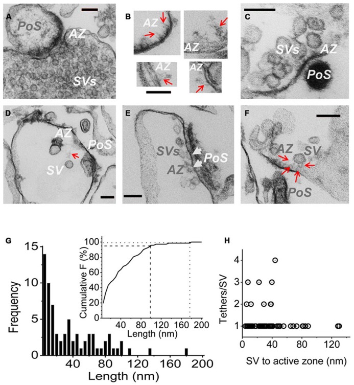
Evidence that synaptic vesicles (SVs) can be gated by a single voltage sensitive calcium channel (CaV2.2) predict a molecular linking mechanism or "tether" (Stanley, 1993). Recent studies have proposed that the SV binds to the distal C-terminal on the CaV2.2 calcium channel (Kaeser et al., 2011; Wong et al., 2013) while genetic analysis proposed a double tether mechanism via RIM: directly to the C terminus PDZ ligand domain or indirectly via a more proximal proline rich site (Kaeser et al., 2011). Using a novel in vitro SV pull down binding assay, we reported that SVs bind to a fusion protein comprising the C-terminal distal third (C3, aa 2137-2357; Wong et al., 2013). Here we limit the binding site further to the last 58 aa, beyond the proline rich site, by the absence of SV capture by a truncated C3 fusion protein (aa 2137-2299). To test PDZ-dependent binding we generated two C terminus-mutant C3 fusion proteins and a mimetic blocking peptide (H-WC, aa 2349-2357) and validated these by elimination of MINT-1 or RIM binding. Persistence of SV capture with all three fusion proteins or with the full length C3 protein but in the presence of blocking peptide, demonstrated that SVs can bind to the distal C-terminal via a PDZ-independent mechanism. These results were supported in situ by normal SV turnover in H-WC-loaded synaptosomes, as assayed by a novel peptide cryoloading method. Thus, SVs tether to the CaV2.2 C-terminal within a 49 aa region immediately prior to the terminus PDZ ligand domain. Long tethers that could reflect extended C termini were imaged by electron microscopy of synaptosome ghosts. To fully account for SV tethering we propose a model where SVs are initially captured, or "grabbed," from the cytoplasm by a binding site on the distal region of the channel C-terminal and are then retracted to be "locked" close to the channel by a second attachment mechanism in preparation for single channel domain gating.

Keywords: PDZ; RIM binding protein; SV-PD; calcium channel; cryoloading; presynaptic; synaptic vesicle; tether.


Figures

FIGURE 1
FIGURE 1
Fusion proteins. (A) Amino acid residues for native (cdB1) and C3 fusion protein constructs. Four constructs are shown: C3Strep, C3WildF, C3MutantF, and C3Prox with their common proximal region labeled C3. The proposed RBP P**P binding domain (blue), the terminal PDZ ligand domains (yellow) and the FLAG tag attached to C3WildF and C3MutantF (gray) are highlighted. In addition to the added FLAG tag, the DDWC PDZ ligand domain is mutated to DRYG in C3MutantF (dashed box). * any amino acid. (B) C3Strep, but not C3Prox, pulls down RIM from SSM membrane lysate. WB, Western blot; StrepV, strep vector alone; probing antibodies are indicated in brackets.
FIGURE 2
FIGURE 2
SV pull down requires the distal region of the C-terminal. (A) Bead-immobilized C3Strep was exposed to a suspension of purified SVs. SV capture was assayed by standard WB for key marker proteins (see text). (B) As in (A), but using immobilized C3Prox. SV capture failed, as indicated by the absence of integral protein markers.
FIGURE 3
FIGURE 3
Imaging SV tethers. (A–F) Each panel shows an electron micrograph (100 nm section) of a chick brain synaptosome AZ, comprising the presynaptic terminal with its attached postsynaptic “scab” (PoS) or a structure that corresponds to a “condensed” scab (e.g., C). (A) is an SSM fixed prior to osmotic rupture showing a presynaptic C-terminal with dense cytoplasm and clouds of synaptic vesicles while (B–F) are EMs of “SSM ghosts” in which the cytoplasm was discharged by osmotic rupture prior to fixation (buffer Ca2+ clamped at 0.1 μM). (B) Four examples of fibrous material extending from the AZ (red arrows). (C) AZ with a cluster of retained SVs showing short fibrous extensions in the AZ region. (D) AZ with a single remote SV and a faint but distinct fibrous connection. (E) Extensive AZ with tethered close SVs. SVs presumed to be docked are attached to the surface membrane and indicated by white arrow heads. (F) AZ with a single close (~30 nm) SV clearly showing multiple fibrous attachments. AZ, active zone; SV, synaptic vesicle. Scale bars are 100 nm. (G) Frequency histogram of tether lengths measured from the leading edge of the SV to the AZ membrane along the tether when visible, and directly where the SV was too close to the surface membrane to resolve tethers. N = 72. Inset. Cumulative frequency histogram of tether lengths with 95 (dashed line) and 99% (dotted line) confidence limits, corresponding to 98 and 176 nm, respectively, indicated. (H) Plot of number of tethers versus tether length for each SV. Note, up to ~45 nm the SVs are tethered by 1–4 visible links but more distant SVs only exhibit one tether.
FIGURE 4
FIGURE 4
Mutant PDZ ligand domain C-terminals and SV capture. (A) C3Strep, but not C3WildF or C3MutantF, pulls down MINT-1 from SSM lysate. (B) C3Strep, C3WildF or C3MutantF fail to pull down the integral SV proteins STG and VAMP from solubilized SV lysate while only C3Strep captures RIM. (C) C3Strep, C3WildF and C3MutantF capture SVs from a suspension in a detergent-free buffer (HB), as indicated by two proteins, SV2 and VAMP. The positive control of SV-PD with C3Strep is shown to the right. The use of different antibodies to identify the fusion proteins, anti-FLAG and anti-Strep, respectively, precluded quantitative comparison.
FIGURE 5
FIGURE 5
The C-terminal PDZ ligand domain blocking mimetic peptide, H-WC, does not block SV capture by the C-terminal. (A) SSM lysate was incubated with H-WC (HEADEDDWC) or HEADE (control, corresponding to the proximal, non-PDZ ligand domain, region of H-WC) peptides followed by pull-down with C3Strep fusion protein and Western blot analysis of captured MINT-1. In the absence of peptide or with HEADE, C3Strep successfully pulls down MINT-1 but this is markedly inhibited in the presence of H-WC. N = 4 comparing HEADE and H-WC (whole SSM or SSM membrane lysate). (B) RIM is pulled down from SV lysates by C3Strep alone or in the presence of control peptide (DDWA) but is markedly inhibited by H-WC. (C) SV-PD persists in the presence of H-WC, PDZ ligand domain-blocking peptide, as indicated by capture of STG and SV2. DDWA served as a control.
FIGURE 6
FIGURE 6
H-WC PDZ ligand domain-blocking peptide does not affect presynaptic styryl dye recycling. (A) SSMs were cryoloaded with H-WC peptide (1.2 mM) together with 3 kD Dextran-FITC 20 μM. The dextran marker (left panel) identifies SSMs (Nomarski bright field, right panel) that were cryoloaded with the peptide and was confirmed with an antibody raised against the C terminus (L4569; Khanna et al., 2006b; red, center panel). Images were taken with a fixed short shutter open time (300 ms) to avoid detection of intrinsic CaV2.2 channels. SSMs that were positive for both dextran and L4569 are indicated (yellow circles) while an SSM that failed to take up dextran was also negative for H-WC staining (blue circle). (B,C) The PDZ-ligand mimetic blocker H-WC does not block SV recycling in synaptosomes. SSMs were cryoloaded with the indicated test compounds as in (A). The defrosted terminals were depolarized with elevated K+ (40mM) in the presence of Ca2+ (1.2 mM) to trigger exocytosis and uptake of FM4-64 by membrane recycling. The fraction of FM-stained terminals was normalized to a paired dextran-only control experiment (see Nath et al., 2014). (B) Upper panel. SSMs loaded with H-WC alone (with carrier buffer as for Bot A-LC). Lower panel. SSMs loaded with H-WC, as in (A), together with Bot A-LC. Dashed circles indicate cryoloaded (dextran positive) SSMs that exhibit strong (yellow), moderate (orange) or no (blue) FM uptake. (C) Histograms of percent ± SE of dextran-positive, and hence cryoloaded terminals that were FM4-64 positive for three separate experiments. Cryoloaded compound(s), concentration and statistical test to the dextran-only Control were: DDWA (1.2 mM), p > 0.1; H-WC (1.2 mM), p > 0.1; P**P (1.2 mM), p > 0.1; P**P/H-WC (1.2 mM each), p > 0.1; HEADE/H-WC (1.2 mM each) p > 0.1; TxD/H-WC tetanus toxoid (200 nM and 1.2 mM, respectively) p > 0.1. BotA-LC/H-WC (200 nM and 1.2 mM, respectively) was significantly different from Control p < 0.05, H-WC (p < 0.01), SH3 + H-WC (p < 0.01), and TxD + HDWC (p < 0.05).
FIGURE 7
FIGURE 7
Working model of CaV tethering of SVs at the transmitter release site. Our suggest that the SV is initially tethered by binding to a site just proximal to the tip of the channel C-terminal. We hypothesize that this serves to grab, or “G-tether,” an SV from the cytoplasm in the AZ region and that an unknown mechanism then draws the SV into its docking site near the channel. However, since previous results suggest that the calcium sensor is within 25 nm of the channel mouth, we also hypothesize one or more additional CaV-SV links serves to lock or “L-tether” the SV within range of the calcium channel Ca2+ domain.

References

    1. Atlas D. (2001). Functional and physical coupling of voltage-sensitive calcium channels with exocytotic proteins: ramifications for the secretion mechanism. J. Neurochem. 77 972–985 10.1046/j.1471-4159.2001.00347.x - DOI - PubMed
    1. Atlas D. (2013). The voltage-gated calcium channel functions as the molecular switch of synaptic transmission. Annu. Rev. Biochem. 82 607–63510.1146/annurev-biochem-080411-121438 - DOI - PubMed
    1. Catterall W. A. (1999). Interactions of presynaptic Ca2+ channels and snare proteins in neurotransmitter release. Ann. N. Y. Acad. Sci. 868 144–15910.1111/j.1749-6632.1999.tb11284.x - DOI - PubMed
    1. Eggermann E., Bucurenciu I., Goswami S. P., Jonas P. (2011). Nanodomain coupling between Ca(2) channels and sensors of exocytosis at fast mammalian synapses. Nat. Rev. Neurosci. 13 7–2110.1038/nrn3125 - DOI - PMC - PubMed
    1. Fernandez-Busnadiego R., Asano S., Oprisoreanu A. M., Sakata E., Doengi M., Kochovski Z., et al. (2013). Cryo-electron tomography reveals a critical role of RIM1alpha in synaptic vesicle tethering. J. Cell Biol. 201 725–74010.1083/jcb.201206063 - DOI - PMC - PubMed

LinkOut - more resources