

The pink rim sign: location of pink as an indicator of melanoma in dermoscopic images
- PMID: 24639898
- PMCID: PMC3930131
- DOI: 10.1155/2014/719740
The pink rim sign: location of pink as an indicator of melanoma in dermoscopic images
Abstract
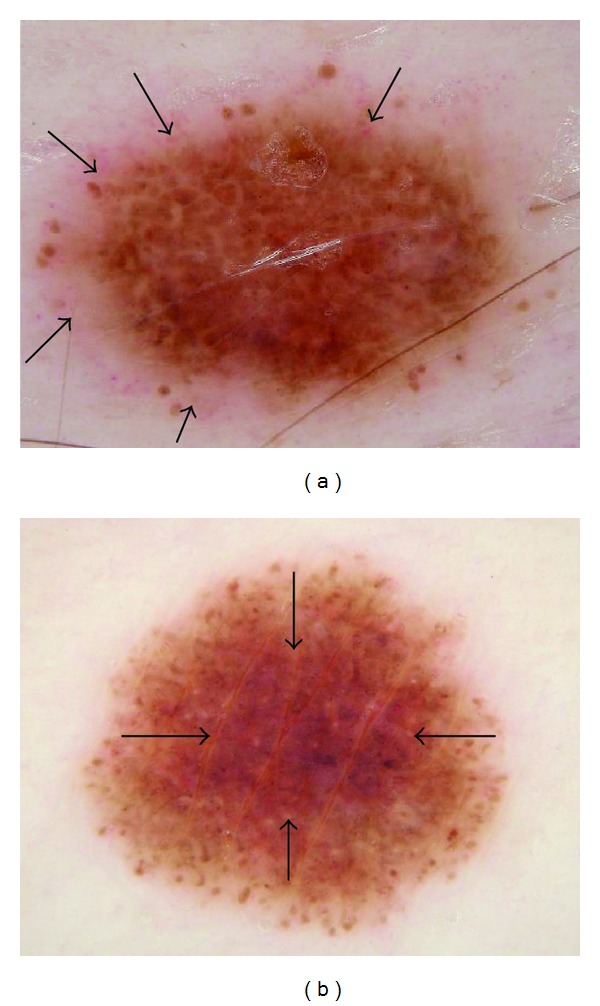
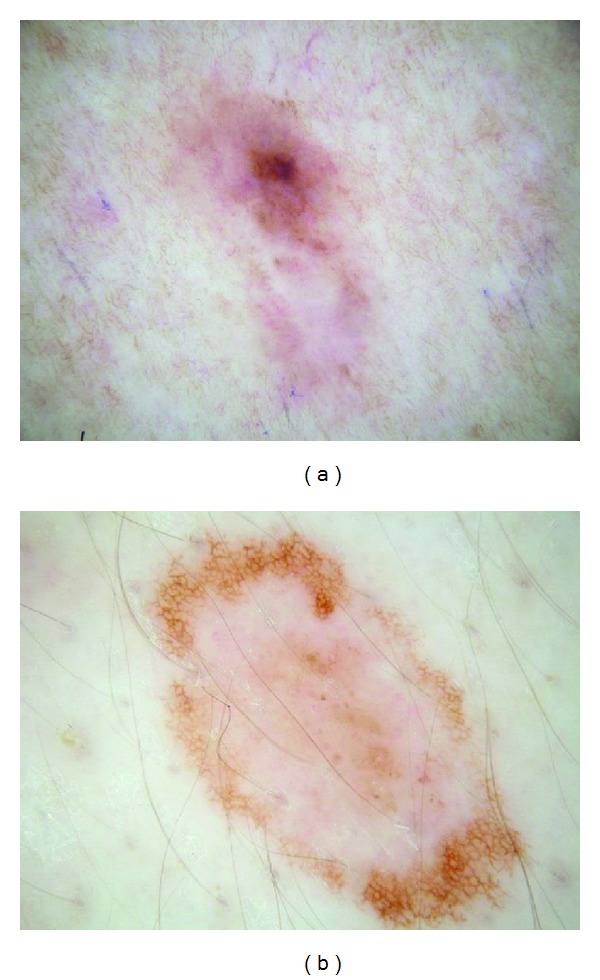
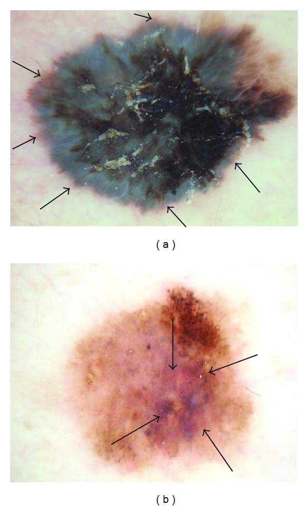
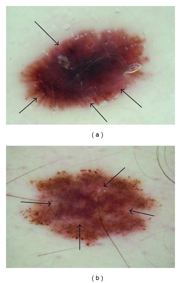
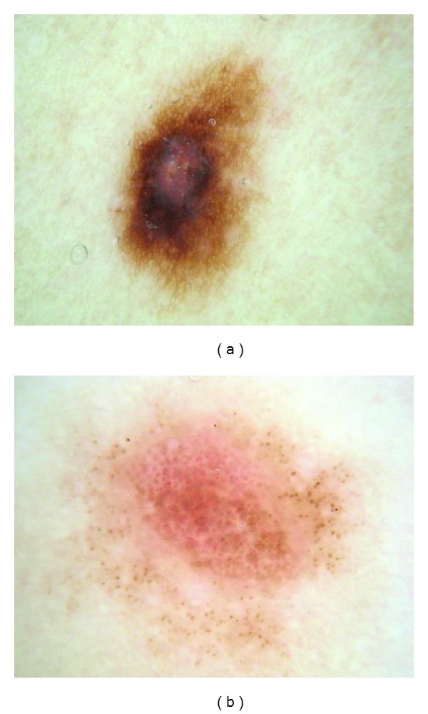
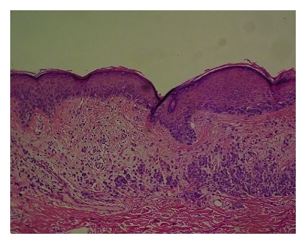
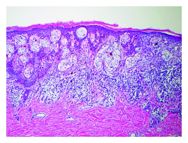
Background. In dermoscopic images, multiple shades of pink have been described in melanoma without specifying location of these areas within the lesion. Objective. The purpose of this study was to determine the statistics for the presence of centrally and peripherally located pink melanoma and benign melanocytic lesions. Methods. Three observers, untrained in dermoscopy, each retrospectively analyzed 1290 dermoscopic images (296 melanomas (170 in situ and 126 invasive), 994 benign melanocytic nevi) and assessed the presence of any shade of pink in the center and periphery of the lesion. Results. Pink was located in the peripheral region in 14.5% of melanomas and 6.3% of benign melanocytic lesions, yielding an odds ratio of 2.51 (95% CI: 1.7-3.8, P < 0.0001). Central pink was located in 12.8% of melanomas and 21.8% of benign lesions, yielding an odds ratio of 0.462 (95% CI: 0.67, P = 0.204). Pink in melanoma in situ tended to be present throughout the lesion (68% of pink lesions). Pink in invasive melanoma was present in 17% of cases, often presenting as a pink rim. Conclusions. The presence of pink in the periphery or rim of a dermoscopic melanocytic lesion image provides an indication of malignancy. We offer the "pink rim sign" as a clue to the dermoscopic diagnosis of invasive melanoma.
Figures


References
-
- Friedman RJ, Rigel DS, Kopf AW. Early detection of malignant melanoma: the role of physician examination and self-examination of the skin. Ca-A Cancer Journal for Clinicians. 1985;35(3):130–151. - PubMed
-
- Johr RH. Pink lesions. Clinics in Dermatology. 2002;20(3):289–296. - PubMed
-
- Argenziano G, Zalaudek I, Corona R, et al. Vascular structures in skin tumors: a dermoscopy study. Archives of Dermatology. 2004;140(12):1485–1489. - PubMed
-
- Pizzichetta MA, Talamini R, Stanganelli I, et al. Amelanotic/hypomelanotic melanoma: clinical and dermoscopic features. British Journal of Dermatology. 2004;150(6):1117–1124. - PubMed
-
- Menzies SW, Kreusch J, Byth K, et al. Dermoscopic evaluation of amelanotic and hypomelanotic melanoma. Archives of Dermatology. 2008;144(9):1120–1127. - PubMed
Grants and funding
LinkOut - more resources
Full Text Sources
Other Literature Sources
