. 2014 Jul;171(14):3448-62.
doi: 10.1111/bph.12693.

Suppression of cell membrane permeability by suramin: involvement of its inhibitory actions on connexin 43 hemichannels

Affiliations

Suppression of cell membrane permeability by suramin: involvement of its inhibitory actions on connexin 43 hemichannels

Yuan Chi et al. Br J Pharmacol. 2014 Jul.

Abstract

Background and purpose: Suramin is a clinically prescribed drug for treatment of human African trypanosomiasis, cancer and infection. It is also a well-known pharmacological antagonist of P2 purinoceptors. Despite its clinical use and use in research, the biological actions of this molecule are still incompletely understood. Here, we investigated the effects of suramin on membrane channels, as exemplified by its actions on non-junctional connexin43 (Cx43) hemichannels, pore-forming α-haemolysin and channels involved in ATP release under hypotonic conditions.

Experimental approach: Hemichannels were activated by removing extracellular Ca(2+) . The influences of suramin on hemichannel activities were evaluated by its effects on influx of fluorescent dyes and efflux of ATP. The membrane permeability and integrity were assessed through cellular retention of preloaded calcein and LDH release.

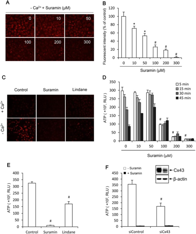
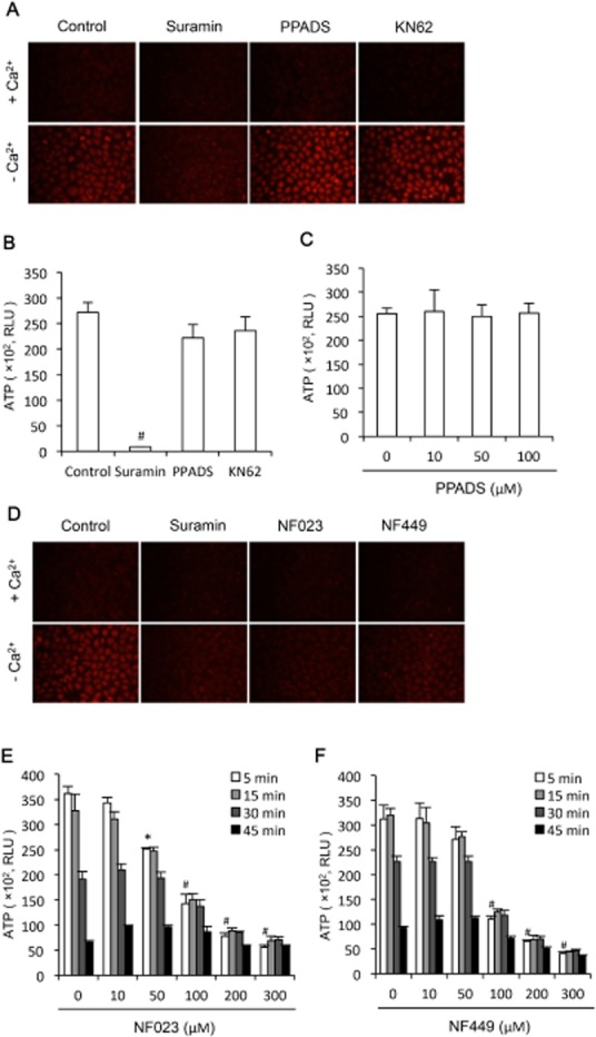
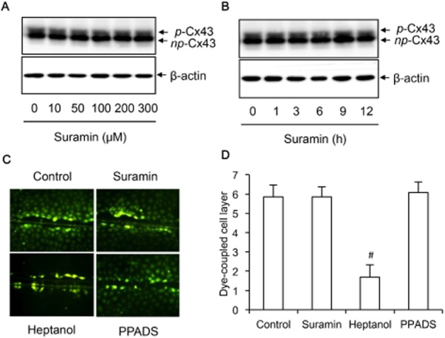
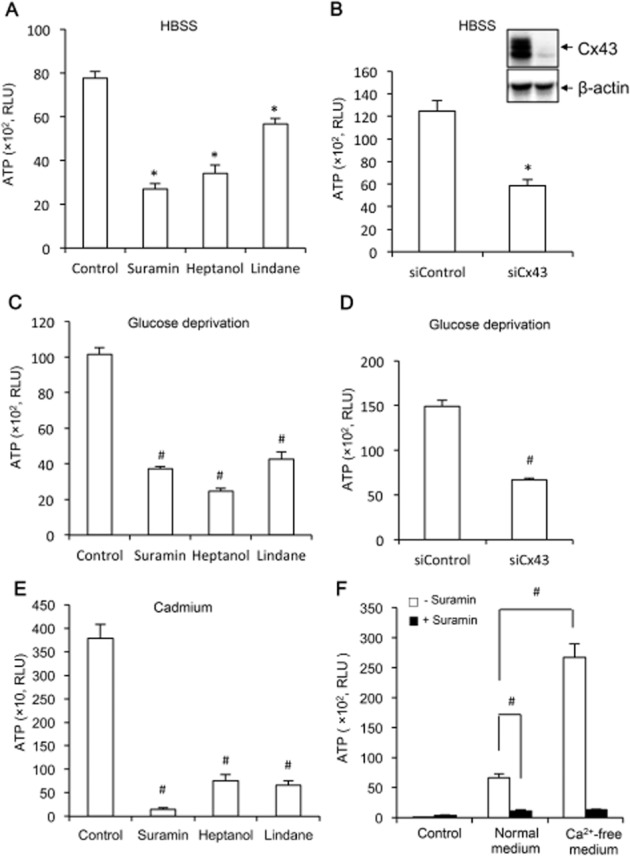
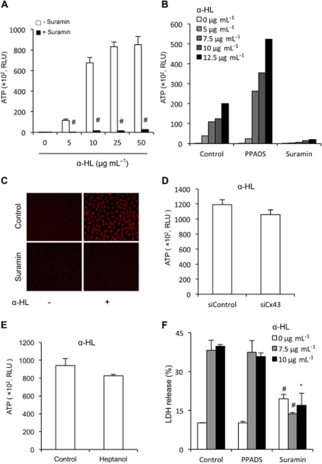
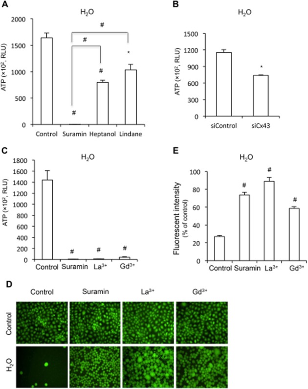
Key results: Suramin blocked Cx43 hemichannel permeability induced by removal of extracellular Ca(2+) without much effect on Cx43 expression and gap junctional intercellular communication. This action of suramin was mimicked by its analogue NF023 and NF449 but not by another P2 purinoceptor antagonist PPADS. Besides hemichannels, suramin also significantly blocked intracellular and extracellular exchanges of small molecules caused by α-haemolysin from Staphylococcus aureus and by exposure of cells to hypotonic solution. Furthermore, it prevented α-haemolysin- and hypotonic stress-elicited cell injury.

Conclusion and implications: Suramin blocked membrane channels and protected cells against toxin- and hypotonic stress-elicited injury. Our finding provides novel mechanistic insights into the pharmacological actions of suramin. Suramin might be therapeutically exploited to protect membrane integrity under certain pathological situations.

Keywords: channel permeability; connexin 43; hemichannels; hypotonic stress; suramin; α-haemolysin.


Figures

Figure 1
Figure 1
Effects of suramin on hemichannel permeability. (A) Effects of suramin on cellular uptake of EtBr following removal of extracellular Ca2+. NRK cells were pretreated with the indicated concentrations of suramin for 20 min. After that, they were exposed to either normal or Ca2+-free culture medium that contained 10 μM EtBr in the presence of the same concentrations of suramin for an additional 15 min. The cellular uptake of EtBr was photographed (magnification, ×320). (B) Quantitation of the cellular fluorescent intensity in (A). Results are expressed as means ± SEM (n = 10, *P < 0.05, #P < 0.01 vs. zero control). (C) Blockade of EtBr uptake by suramin and hemichannel inhibitor. Cells were treated the same as earlier in the presence of 300 μM suramin or 100 μM lindane (magnification, ×160). (D) Effects of suramin on Ca2+ depletion triggers ATP release. NRK cells were exposed to Ca2+-free culture medium in the presence of the indicated concentrations of suramin for the indicated time intervals. Cell supernatants were collected and quantitated for ATP concentration. Results are expressed as relative light unit (RLU; mean ± SE, n = 3). #P < 0.01 versus zero point control. (E) Effects of suramin and hemichannel inhibitors on ATP release. Cells were exposed to Ca2+-free medium in the presence or absence of 300 μM suramin or 100 μM lindane. (F) Effects of suramin and down-regulation of Cx43 with specific siRNA on ATP release. NRK cells were treated with either control siRNA or siRNA against Cx43 for 48 h. Thereafter, cells were exposed to Ca2+-free medium in the presence or absence of 300 μM suramin for 5 min. Cell supernatants were collected and quantitated for ATP concentration. Results are expressed as RLU (mean ± SE, n = 3). #P < 0.01 versus control. To verify the effectiveness of Cx43 siRNA in down-regulation of Cx43, the cellular lysates extracted from siControl and siCx43 were subjected to Western blot analysis of Cx43 (Figure 1F, insert). Note the obvious reduced level of Cx43 in Cx43 siRNA-treated cells (right lane).
Figure 2
Figure 2
Effects of other P2 purinoceptor antagonists on hemichannel permeability. (A) Effects of P2 purinoceptor antagonist PPADS on cellular uptake of EtBr following removal of extracellular Ca2+. NRK cells were pretreated with 300 μM suramin, 3 mM heptanol, 30 μM PPADS and 100 μM NK-62 for 20 min. After that, they were exposed to either normal or Ca2+-free culture medium that contained 10 μM EtBr in the presence of the same concentrations of the earlier agents for an additional 15 min. The cellular uptake of EtBr was photographed (magnification, ×200). (B) Effects of interception of P2 purinoceptor signalling pathway on Ca2+ depletion-triggers ATP release. NRK cells were exposed to Ca2+-free culture medium in the presence of 300 μM suramin, 30 μM PPADS or 100 μM KN-62 for 5 min. Cell supernatants were collected and quantitated for ATP concentration. Results are expressed as relative light unit (RLU; mean ± SE, n = 3). #P < 0.01 compared with control. (C) Effects of various concentrations of PPADS on ATP release. Cells were treated the same as earlier in the presence or absence of the indicated concentrations of PPADS. (D) Effect of suramin, NF-023 and NF-449 on cellular uptake of EtBr. NRK cells were pretreated with 300 μM suramin, NF-023 or NF-449 for 20 min. Thereafter, cells were exposed to either normal or Ca2+-free culture medium that contained 10 μM EtBr in the presence of the same concentrations of the previous agents for an additional 15 min. The cellular uptake of EtBr was photographed (magnification, ×200). (E,F) Effect of suramin analogue NF-023 and NF-449 on ATP release. NRK cells were exposed to Ca2+-free culture medium in the presence of the indicated concentrations of NF023 or NF449 for the indicated time intervals. Cell supernatants were collected and quantitated for ATP concentration. Results are expressed as RLU (mean ± SE, n = 3). #P < 0.01, *P < 0.05 as compared with zero point control.
Figure 3
Figure 3
Effects of suramin on Cx43 expression and function. (A,B) NRK cells were treated with the indicated concentrations of suramin for 30 min (A) or 300 μM suramin for the indicated time intervals (B). The cellular lysates were extracted and subjected to Western blot analysis of Cx43. The upper band represents phosphorylated Cx43 (p-Cx43), and the lower band indicates non-phosphorylated Cx43 (np-Cx43). (C,D) Effects of suramin on GJIC. NRK-E52 cells were treated with 300 μM suramin, 3 mM heptanol or 30 μM PPADS for 30 min. The micrographs of LY diffusion into cellular monolayer after scrape-loading were shown (magnification, ×200). (D) The distance of LY diffusion as shown in C. Results were expressed as cell layer of dye-coupled cells (mean ± SE, n = 13). #P < 0.01 versus control.
Figure 4
Figure 4
Effects of suramin on ATP release under several different conditions. (A,C,E) NRK (A,C) or EGFP-Cx43 LLC-PK1 cells (E) were pretreated with 300 μM suramin, 3 mM heptanol or 100 μM lindane for 20 min, and exposed to HBSS (A), glucose-deprived medium (C), or 50 μM Cd2+ (E) for an additional 30 min (A,B) or 3 h (E). Cell supernatants were collected and quantitated for ATP concentration. Results are expressed as relative light unit (RLU; mean ± SE, n = 3). *P < 0.05, #P < 0.01 compared with respective control. (B,D) Down-regulation of Cx43 with specific siRNA on HBSS and glucose deprivation-triggered ATP release. NRK cells were treated with either control siRNA or siRNA against Cx43 for 48 h. After that, they were exposed to HBSS or glucose-free medium for 30 min. The cellular lysates were also subjected to Western blot analysis of Cx43 to verify the effectiveness of Cx43 siRNA in down-regulation of Cx43 (Figure 4B, insert). Note the obvious reduced level of Cx43 in Cx43 siRNA-treated cells. Results are expressed as RLU (mean ± SE, n = 3). (F) Effect of suramin on basal ATP release triggered by medium exchange. NRK cells cultured in normal Ca2+ medium were incubated with or without 300 μM suramin for 20 min. Thereafter, culture medium were either left untouched (control) or changed to the same normal Ca2+ medium or Ca2+-free medium for additional 5 min. Cell supernatants were collected and quantitated for ATP concentration. Results are expressed as RLU (mean ± SE, n = 6). #P < 0.01.
Figure 5
Figure 5
Effects of suramin on α-HL-mediated permeability and cell injury. (A,B) Effect of suramin and PPADS on pore-mediated release of ATP. NRK cells were exposed to the indicated concentration of α-HL in the presence or absence of 300 μM suramin or 30 μM PPADS for 30 min. Cell supernatant were collected and quantitated for ATP activities. Results are expressed as relative light unit (RLU; mean ± SE, n = 4 in A and n = 1 in B). #P < 0.01 compared with control. (C) Effect of suramin on pore-mediated uptake of EtBr. NRK cells were treated with 10 μg·mL−1 α-HL in the presence or absence of 300 μM suramin for 30 min. The cells were exposed to 10 μM EtBr for 15 min. Cellular uptake of EtBr were photographed (magnification, ×160). (D,E) Effects of knockdown of Cx43 or blockade of hemichannels with heptanol on haemolysin-induced release of ATP. (D) NRK cells were treated with either control siRNA or siRNA against Cx43 for 48 h. Thereafter, cells were exposed to 10 μg·mL−1 alpha-haemolysin for 90 min. (E) NRK cells were exposed to 10 μg·mL−1 alpha-haemolysin in the presence or absence of 3 mM heptanol for 90 min. Cell supernatants were collected and quantitated for ATP activities. Results are expressed as RLU (mean ± SE, n = 6 for D and n = 4 for E). (F) Effect of suramin on α-HL-induced cell injury. NRK cells were exposed to the indicated concentration of α-HL in the presence of 300 μM suramin or 30 μM PPADS for 24 h. Cell supernatants were collected and assayed for LDH release. The results are expressed as % of total release (RLU; mean ± SE, n = 3). #P < 0.01, *P < 0.05 compared with respective control.
Figure 6
Figure 6
Effect of suramin on membrane permeability and integrity under hypotonic condition. (A,B) Effect of suramin, hemichannel inhibitor or Cx43 siRNA on ATP release under hypotonic condition. (A,B) NRK cells were pretreated with 300 μM suramin, 3 mM heptanol or 100 μM lindane for 20 min, or Cx43 siRNA for 48 h. After that, cells were exposed to distilled water in the presence or absence of the same amount of agents for 5 min. Cell supernatants were collected and quantitated for ATP concentration. Results are expressed as relative light unit (RLU; mean ± SE, n = 3). *P < 0.05, #P < 0.01 compared with untreated control. (C) Effect of suramin and non-specific channel blockers on hypotonicity-induced release of ATP. NRK cells were pretreated with 300 μM suramin, 500 μM La3+ or 500 μM Gd3+ for 20 min. Cells were exposed to distilled water in the presence or absence of the same amount of agents for 5 min. Cell supernatants were collected and quantitated for ATP concentration. Results are expressed as RLU (mean ± SE, n = 4). #P < 0.01 compared with untreated control. (D,E) Effect of suramin and non-specific channel blockers on membrane integrity under hypotonic stress. NRK cells were preincubated with 10 μM calcein for 1 h. After that, they were treated with 300 μM suramin, 500 μM La3+ or 500 μM Gd3+ and then exposed to distilled water in the presence or absence of the same amount of the earlier agents for an additional 45 min. The cell membrane integrity was evaluated by the retention of the preloaded calcein inside the cells under fluorescent microscopy (D; magnification, ×200) or the fluorescent intensity of the remaining cells under a fluorescent reader (E). Results in E are expressed as % of fluorescence relative to normal control (mean ± SE, n = 4). #P < 0.01 compared with H2O alone.

Similar articles

Cited by

References

    1. Alexander SPH, Benson HE, Faccenda E, Pawson AJ, Sharman JL, Catterall WA, et al. The Concise Guide to PHARMACOLOGY 2013/14: Ligand-Gated Ion Channels. Br J Pharmacol. 2013;170:1582–1606. - PMC - PubMed
    1. Anselmi F, Hernandez VH, Crispino G, Seydel A, Ortolano S, Roper SD, et al. ATP release through connexin hemichannels and gap junction transfer of second messengers propagate Ca2+ signals across the inner ear. Proc Natl Acad Sci U S A. 2008;105:18770–18775. - PMC - PubMed
    1. Bachmann A, Russ U, Quast U. Potent inhibition of the CFTR chloride channel by suramin. Naunyn Schmiedebergs Arch Pharmacol. 1999;360:473–476. - PubMed
    1. Baraldi PG, del Carmen Nuñez M, Morelli A, Falzoni S, Di Virgilio F, Romagnoli R. Synthesis and biological activity of N-arylpiperazine-modified analogues of KN-62, a potent antagonist of the purinergic P2X7 receptor. J Med Chem. 2003;46:1318–1329. - PubMed
    1. Baroja-Mazo A, Barbera-Cremades M, Pelegrin P. The participation of plasma membrane hemichannels to purinergic signaling. Biochim Biophys Acta. 2013;1828:79–93. - PubMed

Publication types