. 2014 Jun;16(6):502-11.
doi: 10.1016/j.micinf.2014.02.012. Epub 2014 Mar 15.

Extracellular traps are associated with human and mouse neutrophil and macrophage mediated killing of larval Strongyloides stercoralis

Affiliations

Extracellular traps are associated with human and mouse neutrophil and macrophage mediated killing of larval Strongyloides stercoralis

Sandra Bonne-Année et al. Microbes Infect. 2014 Jun.

Abstract

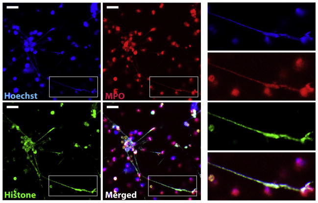
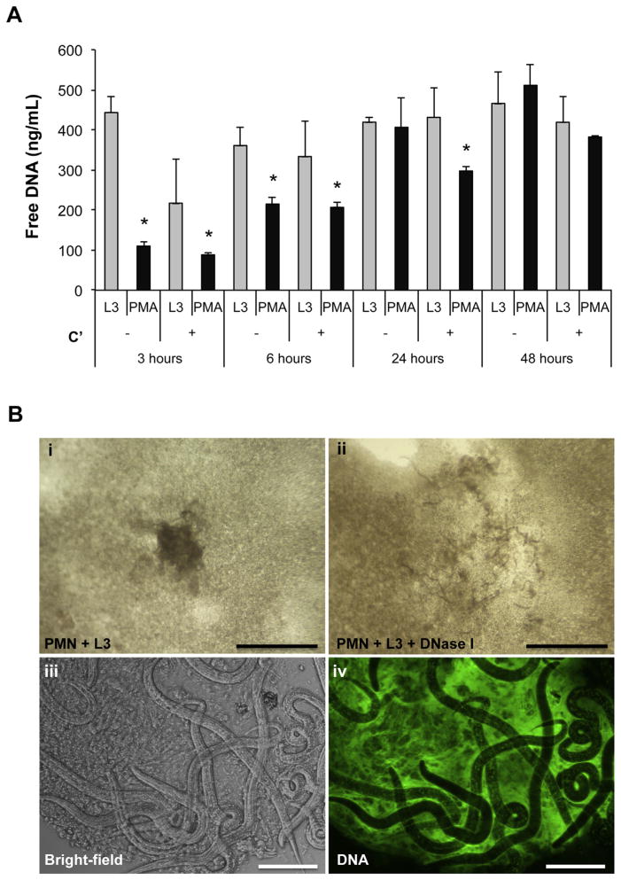
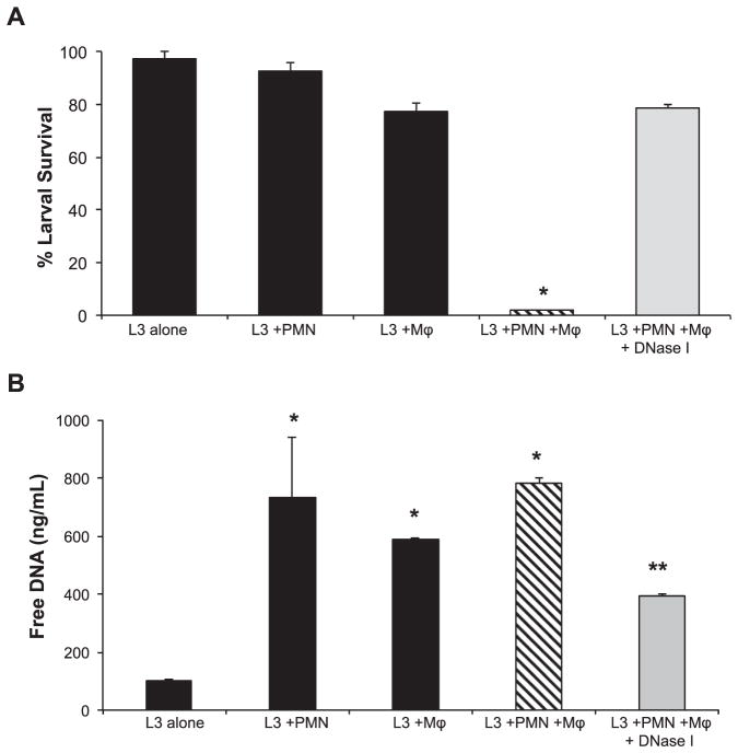
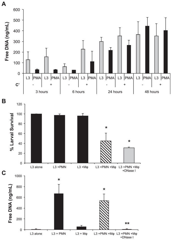
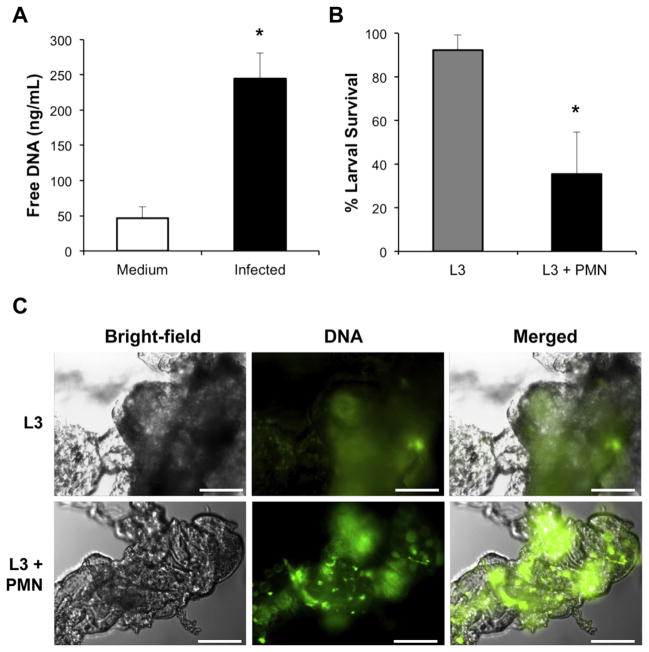
Neutrophils are multifaceted cells that are often the immune system's first line of defense. Human and murine cells release extracellular DNA traps (ETs) in response to several pathogens and diseases. Neutrophil extracellular trap (NET) formation is crucial to trapping and killing extracellular pathogens. Aside from neutrophils, macrophages and eosinophils also release ETs. We hypothesized that ETs serve as a mechanism of ensnaring the large and highly motile helminth parasite Strongyloides stercoralis thereby providing a static target for the immune response. We demonstrated that S. stercoralis larvae trigger the release of ETs by human neutrophils and macrophages. Analysis of NETs revealed that NETs trapped but did not kill larvae. Induction of NETs was essential for larval killing by human but not murine neutrophils and macrophages in vitro. In mice, extracellular traps were induced following infection with S. stercoralis larvae and were present in the microenvironment of worms being killed in vivo. These findings demonstrate that NETs ensnare the parasite facilitating larval killing by cells of the immune system.

Keywords: Extracellular traps; Human; Mice; NET; Neutrophils; Strongyloides stercoralis.


Figures

Fig. 1
Fig. 1
Human neutrophils release NETs following exposure to S. stercoralis larvae. Human neutrophils (2 × 10⁶) were exposed to 1000 dead larvae for 2 h at 37 °C. Cells were labeled with Hoechst (Blue) for DNA, anti-MPO (Red) and anti-histone (Green) to visualize NETs. Inserts on the right depict an enlarged NET co-labeled with Hoechst, anti-MPO, and anti-histone. Scale bar = 20 μm.
Fig. 2
Fig. 2
Human neutrophils release NETs that trap the larvae of S. stercoralis. A) Human neutrophils (1 × 10⁵) were stimulated ex vivo with 100 larvae (L3) or PMA in the presence of autologous human serum as a complement source (C′) or heat inactivated serum for 3, 6, 24 and 48 h. The culture supernatants were analyzed for extracellular DNA using the PicoGreen assay. Data are shown as mean ± SD. B) Human neutrophils, larvae and autologous human serum were cocultured for 24 h in vitro. Cultures revealed live larvae trapped within clots in wells containing only PMN and L3 (i, iii). The resulting clots were then extracted and stained with Sytox Green, a cell impermeable DNA dye, to visualize NET formation using a fluorescent microscope for Bright-field and fluorescent DNA (Green), (iv). Scale bars = 100 μm for (iii–iv). The larvae containing clots completely dissolved within 10 min of treatment with 100 U/ml of DNase I (ii). Scale bars = 6 mm for (i–ii).
Fig. 3
Fig. 3
NETs are required for larval killing by human neutrophils in vitro. A) Human macrophages (Mϕ) and neutrophils (PMN) cultured in vitro with 50 larvae (L3) for 48 h. The cultures were treated with 100 U/ml of DNase I to block NET formation and assessed for larval killing. Data are shown as mean ± SD. *, p < 0.001 when compared with all other groups. B) Supernatants from the in vitro assay were analyzed for extracellular DNA using the PicoGreen assay. Data are shown as mean ± SD. *, p < 0.001 compared to L3 alone group or **, p < 0.001 compared to the L3 + PMN + Mϕ group. Each group was performed in triplicate for at least two independent experiments.
Fig. 4
Fig. 4
Mouse NETs are not required for larval killing in vitro. A) Mouse neutrophils (1 × 10⁵) were stimulated ex vivo with 100 larvae (L3) or PMA in the presence of mouse serum as a complement source (C′) or heat inactivated serum for 3, 6, 24 and 48 h. The culture supernatants were analyzed for extracellular DNA using the PicoGreen assay. Data are shown as mean ± SD. B) Mouse bone marrow-derived macrophages (Mϕ) were cultured with neutrophils (PMN) and 50 larvae (L3) for 22 h. The cultures were treated with 100 U/ml of DNase I to degrade NETs and assessed for larval killing. Data are shown as mean ± SD. *, p < 0.01 when compared with wells containing L3 alone. C) Supernatants from the in vitro assay were analyzed for extracellular DNA using the PicoGreen assay. Data are shown as mean ± SD. *, p < 0.001 compared to L3 alone group or **, p < 0.001 when compared to wells with Mϕ and PMN but not treated with DNase I. Each group was performed in duplicate for at least two independent experiments.
Fig. 5
Fig. 5
Mice release both ET and NETs following exposure to S. stercoralis larvae in vivo. A) Naïve C57BL/6J mice were injected with medium or 10,000 larvae in the peritoneal cavity (infected) and subjected to peritoneal lavages after 3 h. The peritoneal lavage fluid was measured for extracellular DNA using the PicoGreen assay. Data displayed as mean ± SD, where n = 3 mice per group, representative of at least three independent experiments. Data are shown as mean ± SD. *, p < 0.04 when compared to mice injected with medium. B) Neutrophils (PMN) (2 × 10⁶) were isolated from naïve C57BL/6J mice and 50 larvae (L3) were placed in cell impermeable diffusion chambers, with a 0.1 um pore-size membrane. The chambers were then implanted into naive WT mice for 3 days at which time larval survival was assessed. Data are shown as mean ± SD. *, p ≤ 0.001 when compared to L3 alone control. C) Fluorescent images of Sytox Green stained clots taken from diffusion chambers containing L3 or L3 + PMN. Scale bars = 40 μm.

References

    1. Brinkmann V, Reichard U, Goosmann C, Fauler B, Uhlemann Y, Weiss DS, et al. Neutrophil extracellular traps kill bacteria. Science. 2004;303:1532–5. - PubMed
    1. Yousefi S, Gold JA, Andina N, Lee JJ, Kelly AM, Kozlowski E, et al. Catapult-like release of mitochondrial DNA by eosinophils contributes to antibacterial defense. Nat Med. 2008;14:949–53. - PubMed
    1. von Kockritz-Blickwede M, Goldmann O, Thulin P, Heinemann K, Norrby-Teglund A, Rohde M, et al. Phagocytosis-independent antimicrobial activity of mast cells by means of extracellular trap formation. Blood. 2008;111:3070–80. - PubMed
    1. Chow OA, von Kockritz-Blickwede M, Bright AT, Hensler ME, Zinkernagel AS, Cogen AL, et al. Statins enhance formation of phagocyte extracellular traps. Cell Host Microbe. 2010;8:445–54. - PMC - PubMed
    1. Steinberg BE, Grinstein S. Unconventional roles of the NADPH oxidase: signaling, ion homeostasis, and cell death. Sci STKE. 2007;2007:pe11. - PubMed

Publication types

LinkOut - more resources