Review
. 2014 Jun 15;159(3-4):192-201.
doi: 10.1016/j.vetimm.2014.02.016. Epub 2014 Feb 24.

Canine lymphoma as a comparative model for human non-Hodgkin lymphoma: recent progress and applications

Affiliations
Review

Canine lymphoma as a comparative model for human non-Hodgkin lymphoma: recent progress and applications

Daisuke Ito et al. Vet Immunol Immunopathol. .

Abstract

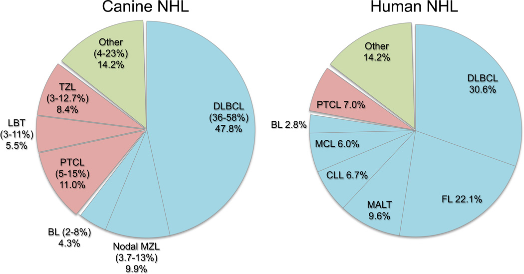
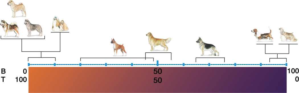
The term "lymphoma" describes a heterogeneous group of disorders involving monoclonal proliferation of malignant lymphocytes. As a group, lymphomas are among the most common tumors of dogs. Yet our enumeration and understanding of the many subtypes of lymphoma have been relatively slow, perhaps in part because for many years lymphoma was treated as a singular entity rather than a group of distinct diseases. The recognition that the full spectrum of lymphoid malignancies seen in humans also occurs in dogs, and that these tumors retain not only morphologic similarities and biological behavior but also synonymous driver molecular abnormalities, sets an ideal stage for dual-purpose research that can accelerate progress for these diseases in both species. Specifically, dogs represent exceptional models for defining causality, understanding progression, and developing new treatments for lymphoma in comparatively brief windows of time. Unique advantages of canine models include (1) spontaneous disease occurring without an isogenic background or genetic engineering; (2) chronology of disease adapted to lifespan, (3) shared environment and societal status that allows dogs to be treated as "patients," while at the same time being able to ethically explore translational innovations that are not possible in human subjects; and (4) organization of dogs into breeds with relatively homogeneous genetic backgrounds and distinct predisposition for lymphomas. Here, we will review recent studies describing intrinsic and extrinsic factors that contribute to the pathogenesis of canine and human lymphomas, as well as newly developed tools that will enhance the fidelity of these models to improve diagnosis and develop new treatments.


Figures

Figure 1
Figure 1
Relative distribution of common subtypes of canine lymphoma (Frantz et al., 2012; Ponce et al., 2010; Thomas et al., 2011; Valli et al., 2011) and human NHL (Anon, 1997). DLBCL; diffuse large B-cell lymphoma, MZL; marginal zone lymphoma, BL; Burkitt lymphoma, PTCL; peripheral T cell lymphoma not otherwise specified, TZL; nodal T-zone lymphoma, LBT; lymphoblastic T-cell lymphoma, FL; follicular lymphoma, MALT; mucosa associated lymphoid tissue lymphoma (extranodal MZL), CLL; chronic lymphocytic leukemia, MCL; mantle cell lymphoma.
Figure 2
Figure 2
Breed-specific distribution of B-cell and T-cell lymphomas in dogs. The prevalence of B-cell-derived and T-cell derived tumors in all dogs (considered as a single group) are ~65% and ~35%, respectively (Modiano et al., 2005). The incidence of B-cell tumors and T-cell tumors in most dogs and mixed breed dogs falls within these ranges (shown by German Shepherd Dog in this figure). In contrast, a prevalence of excess B-cell tumors and T-cell tumors has been shown in certain breeds. Reproduced with permission from (Modiano et al., 2006).
Figure 3
Figure 3
Conserved cytogenetic rearrangement in canine Burkitt lymphoma. (A) Interphase tumor cell from a dog with BL showing heterozygous co-localization of a canine BAC clone containing the MYC gene (white spots indicated by yellow arrows) and a BAC clone that maps to the same cytogenetic band as the IgH locus (red spots, identified by red arrows). Scale bar = 5 µm. (B) Myc expression in non-stimulated normal canine peripheral blood lymphocytes (PBL) (0 h), mitogen-stimulated PBL (55 h), or lymph node cells from dogs with anaplastic large cell lymphoma (ALCL), BL, DLBCL (1–9) or nodal MZL. Beta-actin was used as a loading control. Reproduced with permission from (Breen and Modiano, 2008).
Figure 4
Figure 4
Statistically significant genes define molecular subtypes of canine lymphoma. Genes differentially expressed with >3-fold average change and P values <0.001 were identified for the comparison of groups composed of (A) B-cell and T-cell lymphomas (n = 624), (B) high-grade and low-grade T-cell lymphomas (n = 389), and (C) high-grade and low-grade B-cell lymphomas (n = 25) using t test statistics. The second panel of (A–C) is an independent “validation” set (6 samples, right inset) of the results obtained in the initial set (29 samples, left panel). (D) Venn diagram showing the number of unique and overlapping genes for each 2-group test. Reproduced with permission from (Frantz et al., 2012).
Figure 5
Figure 5
Tumor engraftment of primary canine B-cell lymphoma in NSG mice. (A) Photomicrograph showing splenomegaly in an NSG recipient at autopsy. (B) Photomicrograph of diffuse infiltration of tumor cells in spleen. (C and D) Immunostaining of the donor (dog) cells for expression of canine CD20 (C) and CD79a (D). (E) Spleen cells of a secondary NSG recipient are virtually all canine B-cells, expressing CD21, CD22, and CD45, analyzed by multi-parameter flow cytometry. A small population of hematopoietic progenitor antigens CD34 and KIT positive cells is found within the CD22+ B-cell tumor population. Reproduced with permission from (Ito et al., 2011).
Figure 6
Figure 6
Putative TICs in primary canine B-cell lymphomas and human B-cell ALLs. (A) Existence of a putative Ly-IC population (CD22+CD34/KIT/CD133+; blue dots) in primary canine B-cell lymphoma samples (n = 24) and a putative TIC population (CD22+KIT+; red dots) in primary human B-cell ALL samples (n = 2) were shown using multi-parameter flow cytometry. Reproduced with permission from (Ito et al., 2011). (B) Heat map showing 44 differentially expressed transcripts between enriched Ly-ICs and BTCs (greater than 2-fold change by a two group T-test, p <0.05). Heat map colors represent median-centered fold change expression in log-space. Up-regulated genes are shown in red and down-regulated genes are shown in green.

References

    1. Alizadeh AA, Eisen MB, Davis RE, Ma C, Lossos IS, Rosenwald A, Boldrick JC, Sabet H, Tran T, Yu X, Powell JI, Yang L, Marti GE, Moore T, Hudson J, Lu L, Lewis DB, Tibshirani R, Sherlock G, Chan WC, Greiner TC, Weisenburger DD, Armitage JO, Warnke R, Levy R, Wilson W, Grever MR, Byrd JC, Botstein D, Brown PO, Staudt LM. Distinct types of diffuse large B-cell lymphoma identified by gene expression profiling. Nature. 2000;403:503–511. - PubMed
    1. Alvarez CE, Akey JM. Copy number variation in the domestic dog. Mammalian Genome. 2012;23:144–163. - PubMed
    1. Andersen NS, Larsen JK, Christiansen J, Pedersen LB, Christophersen NS, Geisler CH, Jurlander J. Soluble CD40 ligand induces selective proliferation of lymphoma cells in primary mantle cell lymphoma cell cultures. Blood. 2000;96:2219–2225. - PubMed
    1. Anon A clinical evaluation of the International Lymphoma Study Group classification of non-Hodgkin's lymphoma. The non-Hodgkin's lymphoma classification project. Blood. 1997;89:3909–3918. - PubMed
    1. Bernt KM, Armstrong SA. Leukemia stem cells and human acute lymphoblastic leukemia. Semin Hematol. 2009;46:33–38. - PMC - PubMed

Publication types

Substances