. 2014 Mar 18;9(3):e92263.
doi: 10.1371/journal.pone.0092263. eCollection 2014.

A non-invasive method of quantifying pancreatic volume in mice using micro-MRI

Affiliations

A non-invasive method of quantifying pancreatic volume in mice using micro-MRI

Jose L Paredes et al. PLoS One. .

Abstract

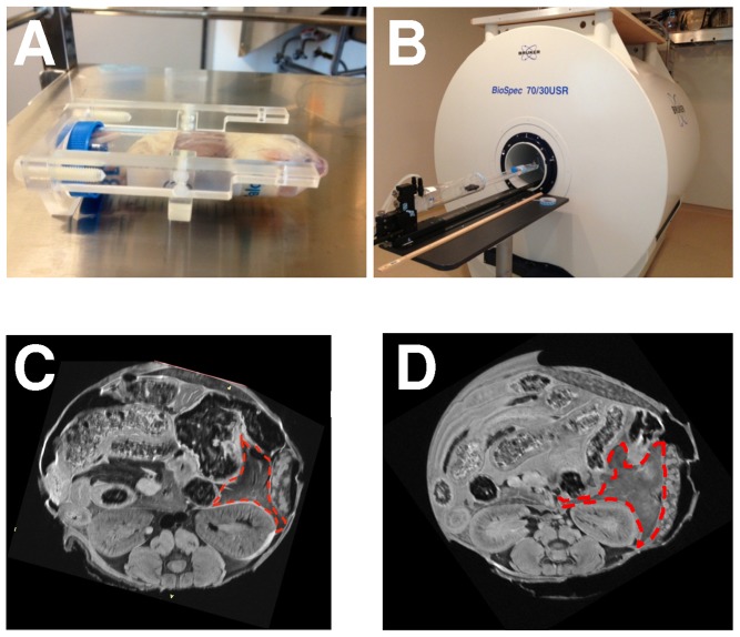
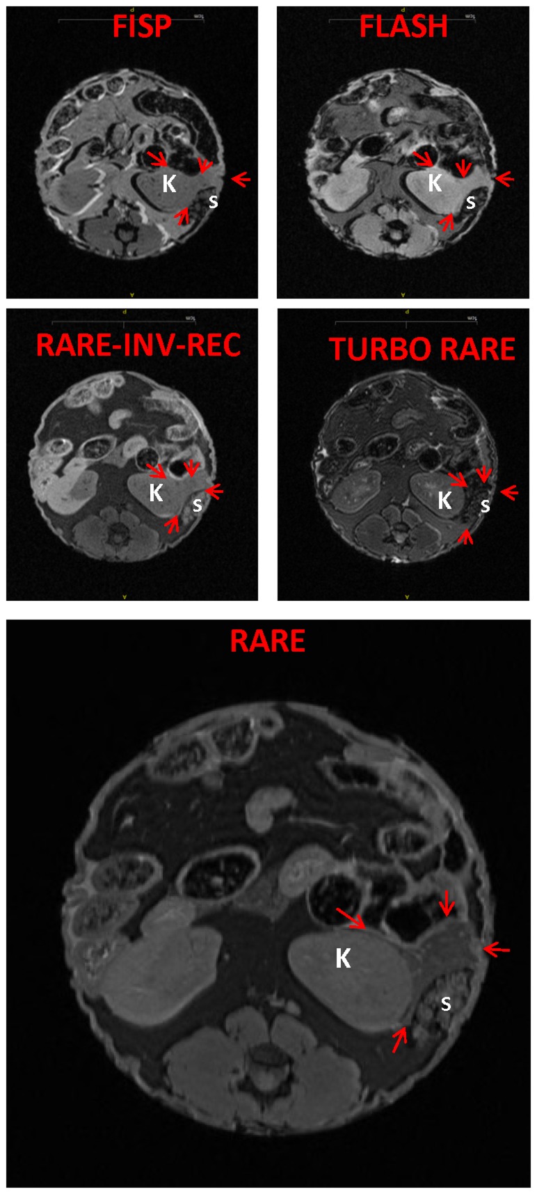
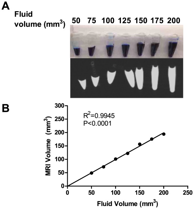
In experimental models of pancreatic growth and recovery, changes in pancreatic size are assessed by euthanizing a large cohort of animals at varying time points and measuring organ mass. However, to ascertain this information in clinical practice, patients with pancreatic disorders routinely undergo non-invasive cross-sectional imaging of the pancreas using magnetic resonance imaging (MRI) or computed tomography (CT). The aim of the current study was to develop a thin-sliced, optimized sequence protocol using a high field MRI to accurately calculate pancreatic volumes in the most common experimental animal, the mouse. Using a 7 Telsa Bruker micro-MRI system, we performed abdominal imaging in whole-fixed mice in three standard planes: axial, sagittal, and coronal. The contour of the pancreas was traced using Vitrea software and then transformed into a 3-dimensional (3D) reconstruction, from which volumetric measurements were calculated. Images were optimized using heart perfusion-fixation, T1 sequence analysis, and 0.2 to 0.4 mm thick slices. As proof of principle, increases in pancreatic volume among mice of different ages correlated tightly with increasing body weight. In summary, this is the first study to measure pancreatic volumes in mice, using a high field 7 Tesla micro-MRI and a thin-sliced, optimized sequence protocol. We anticipate that micro-MRI will improve the ability to non-invasively quantify changes in pancreatic size and will dramatically reduce the number of animals required to serially assess pancreatic growth and recovery.


Conflict of interest statement

Competing Interests: The authors have declared that no competing interests exist.

Figures

Figure 1
Figure 1. Preparation of the mouse for MRI.
In these studies, whole-fixed mice were (A) placed in a conical tube and (B) inserted into a Bruker 7 Tesla micro-MRI. (C) Compared to immersion fixation, (D) in vivo perfusion fixation yielded a more homogenous pancreatic MRI signal (red outline).
Figure 2
Figure 2. RARE is superior to other sequence formats in delineating the pancreas.
Representative slices of the various sequence protocols demonstrate that RARE sequence provides the best delineation of the pancreas from adjacent organs. The arrows point to the pancreas. S, spleen; K, kidney.
Figure 3
Figure 3. Method for generating a 3D reconstruction of the mouse pancreas.
(A) Gross dissection of the pancreas with its adjoining organs. The pancreas was traced in each (B) axial image and, for further delineation, tracings were cross-checked, as necessary, using (C) sagittal and (D) coronal planes. (E) Three representative 3D reconstructions of the pancreas, generated using these tracings, are shown.
Figure 4
Figure 4. Thin cross-sectional slices are necessary to obtain accurate mouse pancreatic volume measurements.
First, pancreatic volumes were calculated from the thinnest cross-sectional slice (0.2 mm; as described in the Methods). Next, volume calculations were also performed by skipping slices. (A) Volume calculations from 3 individual mice with increasing slice thickness. (B) Mean volumes ±1 standard deviation (for the 3 mice) demonstrate that volumes calculated from slices greater than 0.4 mm were substantially higher than the 0.2 mm “gold standard” and had higher variance.
Figure 5
Figure 5. Micro-MRI can accurately measure small volumes in phantom tubes.
(A) Known volumes of gadolinium contrast, ranging from 50 to 200 mm3, with 25 mm3 increments, were loaded into small conical tubes (top row) and imaged using an optimized MRI protocol (bottom row). (B) There was a tight correlation between known and MRI-measured volumes.
Figure 6
Figure 6. Micro-MRI can accurately measure increases in pancreatic volume with advancing age.
(A) Changes in pancreatic volume (in red) correlate with body weight (in blue). (n = 3 mice per age group). *, p<0.05, using a one way ANOVA. (B) Scatter plot demonstrating that pancreatic volume correlates tightly with body weight.

References

    1. Criscimanna A, Speicher JA, Houshmand G, Shiota C, Prasadan K, et al. (2011) Duct cells contribute to regeneration of endocrine and acinar cells following pancreatic damage in adult mice. Gastroenterology 141: 1451–1462. - PMC - PubMed
    1. Williams JA (2010) Regulation of acinar cell function in the pancreas. Curr Opin Gastroenterol 26: 478–483. - PMC - PubMed
    1. Gurda GT, Crozier SJ, Ji B, Ernst SA, Logsdon CD, et al... (2010) Regulator of calcineurin 1 controls growth plasticity of adult pancreas. Gastroenterology 139: 609–619, 619 e601–606. - PMC - PubMed
    1. Stanger BZ, Tanaka AJ, Melton DA (2007) Organ size is limited by the number of embryonic progenitor cells in the pancreas but not the liver. Nature 445: 886–891. - PubMed
    1. Bonner-Weir S, Baxter LA, Schuppin GT, Smith FE (1993) A second pathway for regeneration of adult exocrine and endocrine pancreas. A possible recapitulation of embryonic development. Diabetes 42: 1715–1720. - PubMed

Publication types