. 2014;51(2):118-31.
doi: 10.1159/000358920. Epub 2014 Mar 7.

Increased calcification in osteoprotegerin-deficient smooth muscle cells: Dependence on receptor activator of NF-κB ligand and interleukin 6

Affiliations

Increased calcification in osteoprotegerin-deficient smooth muscle cells: Dependence on receptor activator of NF-κB ligand and interleukin 6

Andrea Callegari et al. J Vasc Res. 2014.

Abstract

Objective: Vascular calcification is highly correlated with cardiovascular disease morbidity and mortality. Osteoprotegerin (OPG) is a secreted decoy receptor for receptor activator of NF-κB ligand (RANKL). Inactivation of OPG in apolipoprotein E-deficient (ApoE-/-) mice increases lesion size and calcification. The mechanism(s) by which OPG is atheroprotective and anticalcific have not been entirely determined. We investigated whether OPG-deficient vascular smooth muscle cells (VSMCs) are more susceptible to mineralization and whether RANKL mediates this process.

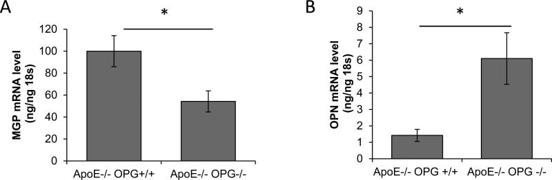
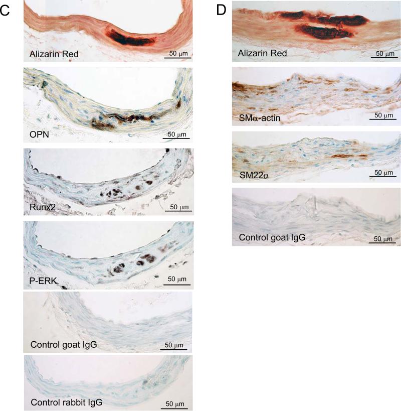
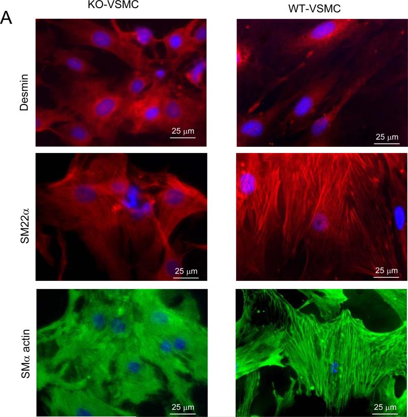
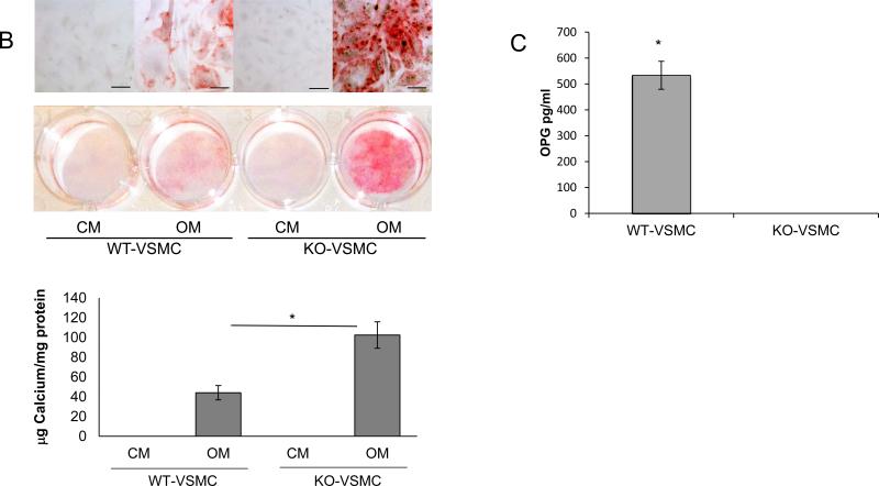
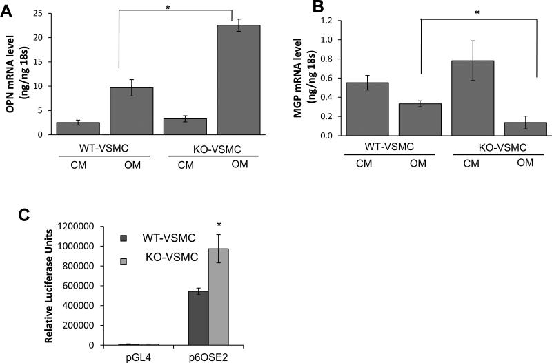
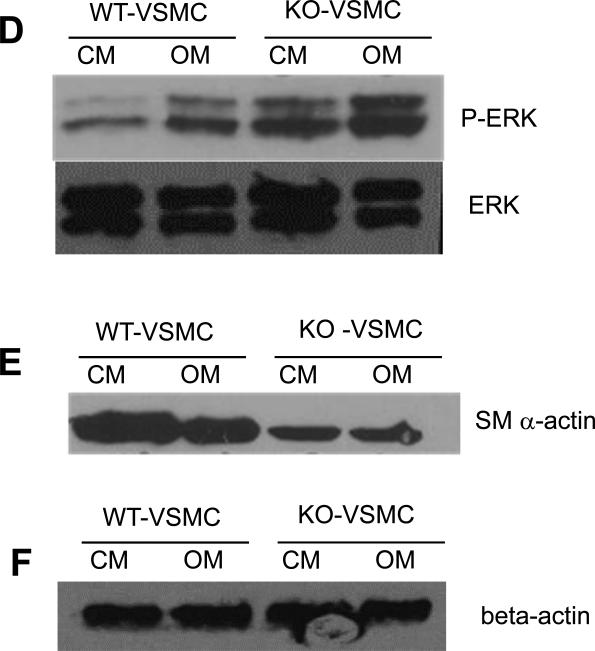
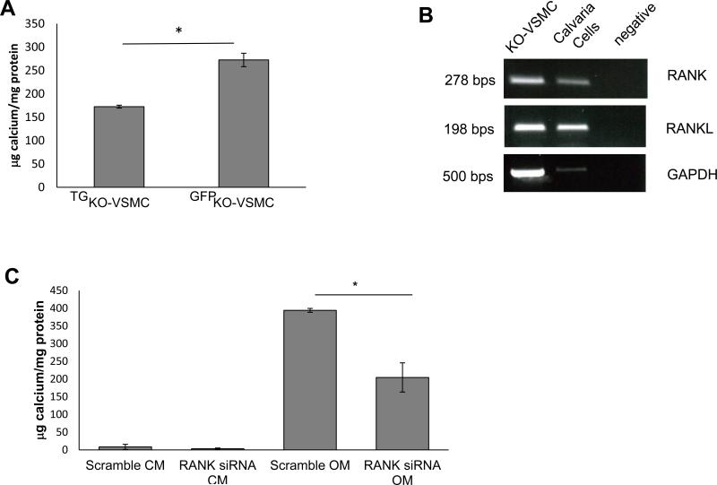
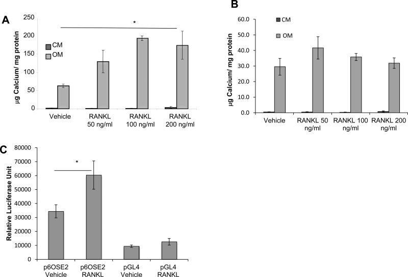
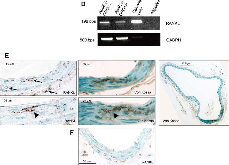
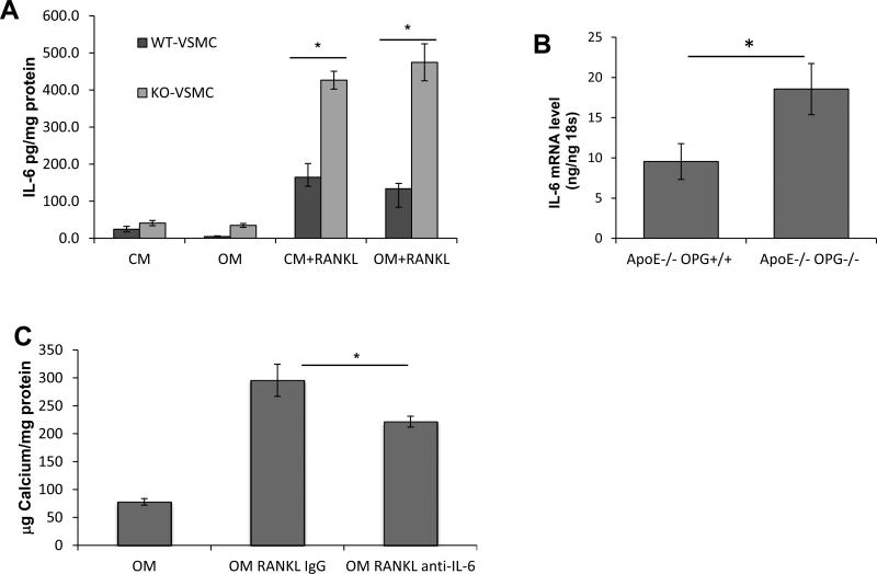
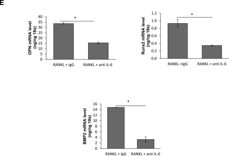
Results: Lesion-free aortas from 12-week-old ApoE-/-OPG-/- mice had spotty calcification, an appearance of osteochondrogenic factors and a decrease of smooth muscle markers when compared to ApoE-/-OPG+/+ aortas. In osteogenic conditions, VSMCs isolated from ApoE-/-OPG-/- (KO-VSMC) mice deposited more calcium than VSMCs isolated from ApoE-/-OPG+/+ (WT-VSMC) mice. Gene expression and biochemical analysis indicated accelerated osteochondrogenic differentiation. Ablation of RANKL signaling in KO-VSMCs rescued the accelerated calcification. While WT-VSMCs did not respond to RANKL treatment, KO-VSMCs responded with enhanced calcification and the upregulation of osteochondrogenic genes. RANKL strongly induced interleukin 6 (IL-6), which partially mediated RANKL-dependent calcification and gene expression in KO-VSMCs.

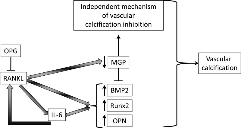
Conclusions: OPG inhibits vascular calcification by regulating the procalcific effects of RANKL on VSMCs and is thus a possible target for therapeutic intervention.


Figures

Figure 1
Figure 1
Altered expression of calcification regulatory factors in mouse aortas. (A-B) qPCR of RNA from aortic tissue (ascending, arch, thoracic) isolated from ApoE−/−OPG−/− mice (n=12) and ApoE−/−OPG+/+ (n=6) at 12 weeks of age. (A) MGP, (B) OPN *p<0.05. (C and D) Immunohistochemistry of ApoE−/−OPG−/− innominate artery serial sections for phosphorylated ERK, Runx2, OPN, SM22α SMα-actin, control goat rabbit and mouse IgG, and for Alizarin Red.
Figure 1
Figure 1
Altered expression of calcification regulatory factors in mouse aortas. (A-B) qPCR of RNA from aortic tissue (ascending, arch, thoracic) isolated from ApoE−/−OPG−/− mice (n=12) and ApoE−/−OPG+/+ (n=6) at 12 weeks of age. (A) MGP, (B) OPN *p<0.05. (C and D) Immunohistochemistry of ApoE−/−OPG−/− innominate artery serial sections for phosphorylated ERK, Runx2, OPN, SM22α SMα-actin, control goat rabbit and mouse IgG, and for Alizarin Red.
Figure 2
Figure 2
Characterization of freshly isolated KO-VSMCs and WT-VSMCs. (A) Immunofluorescence for Desmin, SM22α, SMα-actin in KO- and WT-VSMCs one passage after isolation. (B) Calcification assay with WT- and KO-VSMCs. Cells were cultured in control medium (CM) and osteogenic medium(OM) for 7 days. Calcium deposition was measured as described in the methods. *p<0.05. Alizarin Red S staining was also performed directly in the plate wells (low, bottom, and high, top, magnifications) scale bar=50 μm. (C) ELISA for OPG on supernatant of WT- and KO-VSMCs. *p<0.05.
Figure 2
Figure 2
Characterization of freshly isolated KO-VSMCs and WT-VSMCs. (A) Immunofluorescence for Desmin, SM22α, SMα-actin in KO- and WT-VSMCs one passage after isolation. (B) Calcification assay with WT- and KO-VSMCs. Cells were cultured in control medium (CM) and osteogenic medium(OM) for 7 days. Calcium deposition was measured as described in the methods. *p<0.05. Alizarin Red S staining was also performed directly in the plate wells (low, bottom, and high, top, magnifications) scale bar=50 μm. (C) ELISA for OPG on supernatant of WT- and KO-VSMCs. *p<0.05.
Figure 3
Figure 3
Differential expression of regulatory factors in KO-VSMCs versus WT-VSMCs. (A-B) Gene expression analysis between WT- and KO-VSMCs after 4 days treatment with CM and OM (A) showed higher level of OPN and (B) lower level of MGP in KO-VSMCs compared to WT-VSMCs in OM. Data are collected from 3 independent experiments. *p<0.05. (C) Dual luciferase reporter assay indicating that Runx2 activity is higher in KO-VSMCs compared to control WT-VSMC. Cells were transduced with p6OSE2 and pGL4 as describe in the methods and cultured 72 hours before harvesting for Luciferase measurement. *<0.05 versus p6OSE2-WT-VSMC. (D) Western blot for phosphorylated-ERK (P-ERK, top panel) and ERK (bottom panel) showing increased ERK activity in KO-VSMCs and increased ERK activity in OM regardless of cell genotype. (E) KO-VSMCs express less SMα-actin than WT-VSMCs. (F) beta-actin was used as loading control.
Figure 3
Figure 3
Differential expression of regulatory factors in KO-VSMCs versus WT-VSMCs. (A-B) Gene expression analysis between WT- and KO-VSMCs after 4 days treatment with CM and OM (A) showed higher level of OPN and (B) lower level of MGP in KO-VSMCs compared to WT-VSMCs in OM. Data are collected from 3 independent experiments. *p<0.05. (C) Dual luciferase reporter assay indicating that Runx2 activity is higher in KO-VSMCs compared to control WT-VSMC. Cells were transduced with p6OSE2 and pGL4 as describe in the methods and cultured 72 hours before harvesting for Luciferase measurement. *<0.05 versus p6OSE2-WT-VSMC. (D) Western blot for phosphorylated-ERK (P-ERK, top panel) and ERK (bottom panel) showing increased ERK activity in KO-VSMCs and increased ERK activity in OM regardless of cell genotype. (E) KO-VSMCs express less SMα-actin than WT-VSMCs. (F) beta-actin was used as loading control.
Figure 4
Figure 4
RANKL signaling inhibition in KO-VSMCs rescues increased calcification. (A) Calcification assay with TGKO-VSMCs, transgenic KO-VSMCs, in which OPG was retrovirally reintroduced. TGKO-VSMCs calcified and more than vector control KO-VSMCs (GPFKO-VSMCs), which were retrovirally infected with the control vector expressing GFP. TGKO-VSMCs and GFPKO-VSMCs were treated with OM for 7 days. *p<0.05. (B) RT-PCR expression of RANK and RANKL in KO-VSMCs and positive control calvaria cells. (C) Calcification assay with scrambled transduced (Scramble) or RANK siRNA transduced (RANK siRNA) KO-VSMCs. Cells were treated with CM or OM or 7 days. *p<0.05.
Figure 5
Figure 5
RANKL enhance calcification of KO-VSMCs but not WT-VSMCs. KO-VSMCs (A) and WT-VSMCs (B) were treated with RANKL at 50, 100, 200 ng/ml for 7 days. *p<0.05 RANKL all concentrations versus vehicle. (C) Dual luciferase reporter assay showing Runx2 activity increased in KO-VSMCs after treatment with RANKL for 72 hours. Cells were transduced with p6OSE2 and pGL4 as described in the methods and cultured with Vehicle or 100 ng/ml of RANKL before harvesting for Luciferase measurement.*p<0.05 p6OSE RANKL versus p6OSE2. (D) RT-PCR for RANKL expression in freshly isolated aortas from 12 week old ApoE−/−OPG−/−and ApoE−/−OPG+/+ mice. Calvaria cells were used as positive control. (E) Staining for RANKL and Von Kossa of 12 week old ApoE−/−OPG−/− innominate artery. Left top panel, wide view showing RANKL expression in medial cells closely associated with areas of calcification, in medial cells further from the calcified area, and in few adventitial cells (arrows). Left bottom panel shows higher magnification of RANKL staining associated with calcified area (arrowhead). Middle top panel, wide view showing VonKossa staining in the medial layer. Middle bottom panel, higher magnification VonKossa staining. Arrowhead, indicate VonKossa staining intimately associated with RANKL positive area. Right panel, wide whole vessel view of VonKossa staining. (F) RANKL staining on 12 week old ApoE−/−OPG+/+ innominate artery.
Figure 5
Figure 5
RANKL enhance calcification of KO-VSMCs but not WT-VSMCs. KO-VSMCs (A) and WT-VSMCs (B) were treated with RANKL at 50, 100, 200 ng/ml for 7 days. *p<0.05 RANKL all concentrations versus vehicle. (C) Dual luciferase reporter assay showing Runx2 activity increased in KO-VSMCs after treatment with RANKL for 72 hours. Cells were transduced with p6OSE2 and pGL4 as described in the methods and cultured with Vehicle or 100 ng/ml of RANKL before harvesting for Luciferase measurement.*p<0.05 p6OSE RANKL versus p6OSE2. (D) RT-PCR for RANKL expression in freshly isolated aortas from 12 week old ApoE−/−OPG−/−and ApoE−/−OPG+/+ mice. Calvaria cells were used as positive control. (E) Staining for RANKL and Von Kossa of 12 week old ApoE−/−OPG−/− innominate artery. Left top panel, wide view showing RANKL expression in medial cells closely associated with areas of calcification, in medial cells further from the calcified area, and in few adventitial cells (arrows). Left bottom panel shows higher magnification of RANKL staining associated with calcified area (arrowhead). Middle top panel, wide view showing VonKossa staining in the medial layer. Middle bottom panel, higher magnification VonKossa staining. Arrowhead, indicate VonKossa staining intimately associated with RANKL positive area. Right panel, wide whole vessel view of VonKossa staining. (F) RANKL staining on 12 week old ApoE−/−OPG+/+ innominate artery.
Figure 6
Figure 6
RANKL induces up-regulation of IL-6. (A) ELISA with cell supernatant from WT- and KO-VSMCs treated with RANKL in CM and OM conditions *p<0.05 KO-VSMC RANKL versus WT-VSMC RANKL. (B) qPCR for IL-6 expression in aortas freshly isolated from 12 week old ApoE−/−OPG−/− and ApoE−/−OPG+/+ mice. (C) Calcification assay with KO-VSMCs cultured in OM, treated RANKL and isotype control antibody (OM RANKL IgG), and treated RANKL and anti-IL-6 neutralizing antibody (OM RANKL anti-IL-6) for 7 days. *p<0.05 OM RANKL IgG versus OM RANKL anti-IL-6. (D) qPCR for OPN, Runx2 and BMP2 of KO-VSMCs treated with RANKL or RANKL and anti-IL-6 antibody for four days. *p<0.05.
Figure 6
Figure 6
RANKL induces up-regulation of IL-6. (A) ELISA with cell supernatant from WT- and KO-VSMCs treated with RANKL in CM and OM conditions *p<0.05 KO-VSMC RANKL versus WT-VSMC RANKL. (B) qPCR for IL-6 expression in aortas freshly isolated from 12 week old ApoE−/−OPG−/− and ApoE−/−OPG+/+ mice. (C) Calcification assay with KO-VSMCs cultured in OM, treated RANKL and isotype control antibody (OM RANKL IgG), and treated RANKL and anti-IL-6 neutralizing antibody (OM RANKL anti-IL-6) for 7 days. *p<0.05 OM RANKL IgG versus OM RANKL anti-IL-6. (D) qPCR for OPN, Runx2 and BMP2 of KO-VSMCs treated with RANKL or RANKL and anti-IL-6 antibody for four days. *p<0.05.
Figure 7
Figure 7
Proposed model for RANKL-mediated vascular calcification. In absence of OPG, RANKL in an IL-6 dependent and independent manner up-regulates a subset of osteochondrogenic genes (BMP2, Runx2, and OPN) and in an IL-6-independent manner and down-regulates MGP. The combination of these effects leads to increased vascular calcification. Further, IL-6 induces RANKL, and OPG possibly suggesting a feed-forward process.

Similar articles

Cited by

References

    1. Sage AP, Tintut Y, Demer LL. Regulatory mechanisms in vascular calcification. Nat Rev Cardiol. 2010;7:528–536. - PMC - PubMed
    1. Moe SM, Reslerova M, Ketteler M, O'neill K, Duan D, Koczman J, Westenfeld R, Jahnen-Dechent W, Chen NX. Role of calcification inhibitors in the pathogenesis of vascular calcification in chronic kidney disease (ckd). Kidney Int. 2005;67:2295–2304. - PubMed
    1. Shao JS, Cheng SL, Sadhu J, Towler DA. Inflammation and the osteogenic regulation of vascular calcification: A review and perspective. Hypertension. 2010;55:579–592. - PMC - PubMed
    1. Abedin M, Tintut Y, Demer LL. Vascular calcification: Mechanisms and clinical ramifications. Arterioscler Thromb Vasc Biol. 2004;24:1161–1170. - PubMed
    1. Rattazzi M, Bennett BJ, Bea F, Kirk EA, Ricks JL, Speer M, Schwartz SM, Giachelli CM, Rosenfeld ME. Calcification of advanced atherosclerotic lesions in the innominate arteries of apoe-deficient mice: Potential role of chondrocyte-like cells. Arterioscler Thromb Vasc Biol. 2005;25:1420–1425. - PubMed

Publication types

MeSH terms