

A rare case of diffuse pulmonary lymphangiomatosis in a middle-aged woman
- PMID: 24642766
- PMCID: PMC3955798
- DOI: 10.3348/kjr.2014.15.2.295
A rare case of diffuse pulmonary lymphangiomatosis in a middle-aged woman
Abstract
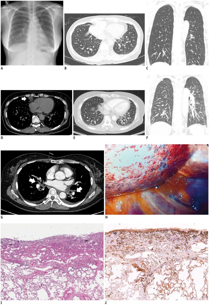
Diffuse pulmonary lymphangiomatosis (DPL) is a rare lymphatic disorder characterized by lymphatic channel proliferation. It is mostly reported in children and young adults. Here, we report a case involving a 52-year-old asymptomatic woman who presented with increased interstitial markings, as seen on a chest radiograph. Diffuse interstitial septal thickening was found on a serial follow-up chest computed tomography scan, and lymphangitic metastasis was the primary radiologic differential diagnosis. However, histologic sections of wedge resected lung revealed diffuse pleural and interlobular septal lymphatic proliferation characteristic of DPL.
Keywords: Computed tomography; Interstitial; Lung; Lymphangiomatosis.
Figures
References
-
- Swensen SJ, Hartman TE, Mayo JR, Colby TV, Tazelaar HD, Müller NL. Diffuse pulmonary lymphangiomatosis: CT findings. J Comput Assist Tomogr. 1995;19:348–352. - PubMed
-
- Yoo SH, Song JS, Lee JJ, Lee M, Hwang HS, Jang SJ. Diffuse pulmonary lymphangiomatosis: Pulmonary lymphatic disorder in an adult. Basic Appl Pathol. 2012;5:63–66.
-
- El Hajj L, Mazières J, Rouquette I, Mittaine M, Bolduc JP, Didier A, et al. Diagnostic value of bronchoscopy, CT and transbronchial biopsies in diffuse pulmonary lymphangiomatosis: case report and review of the literature. Clin Radiol. 2005;60:921–925. - PubMed
-
- Boland JM, Tazelaar HD, Colby TV, Leslie KO, Hartman TE, Yi ES. Diffuse pulmonary lymphatic disease presenting as interstitial lung disease in adulthood: report of 3 cases. Am J Surg Pathol. 2012;36:1548–1554. - PubMed
-
- Du MH, Ye RJ, Sun KK, Li JF, Shen DH, Wang J, et al. Diffuse pulmonary lymphangiomatosis: a case report with literature review. Chin Med J (Engl) 2011;124:797–800. - PubMed
Publication types
MeSH terms
LinkOut - more resources
Full Text Sources
Other Literature Sources
Medical
