. 2014 Mar 18;9(3):e92003.
doi: 10.1371/journal.pone.0092003. eCollection 2014.

cAMP signaling prevents podocyte apoptosis via activation of protein kinase A and mitochondrial fusion

Affiliations

cAMP signaling prevents podocyte apoptosis via activation of protein kinase A and mitochondrial fusion

Xiaoying Li et al. PLoS One. .

Abstract

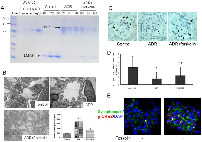
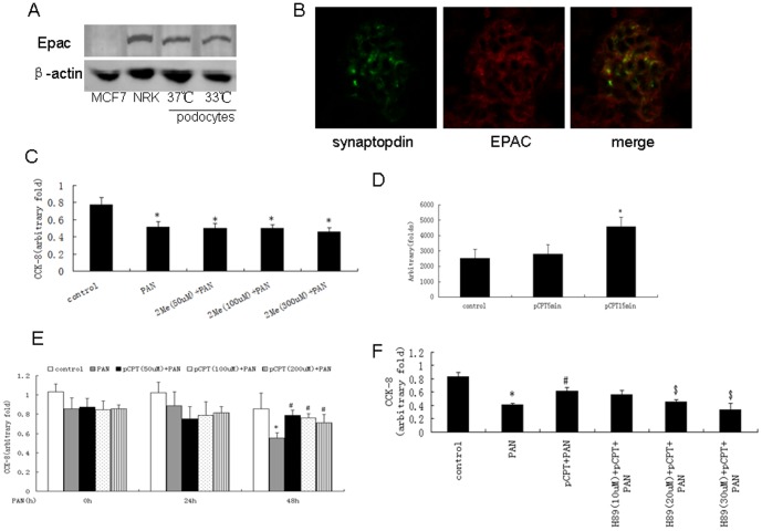
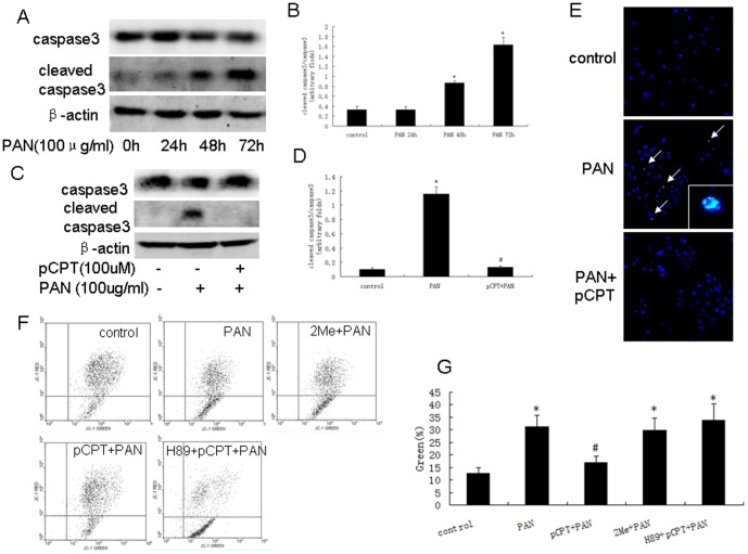
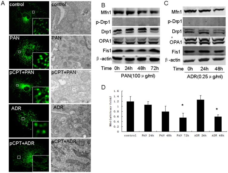
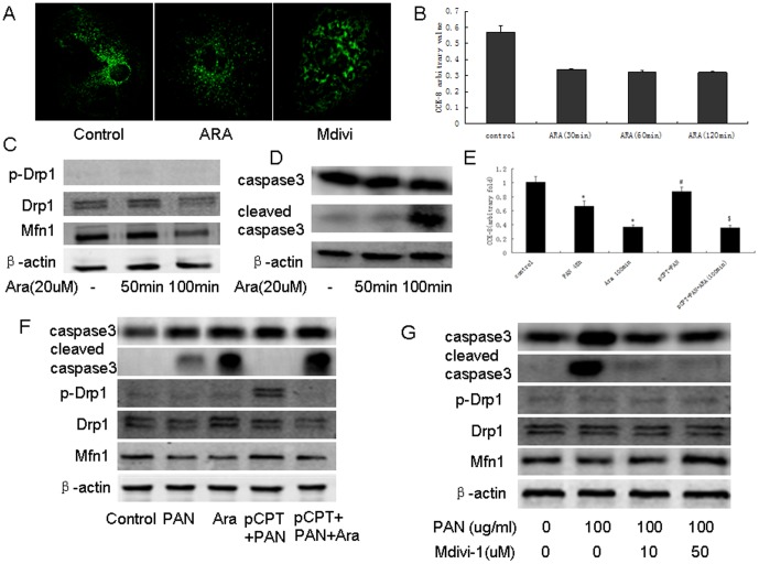
Our previous in vitro studies suggested that cyclic AMP (cAMP) signaling prevents adriamycin (ADR) and puromycin aminonucleoside (PAN)-induced apoptosis in podocytes. As cAMP is an important second messenger and plays a key role in cell proliferation, differentiation and cytoskeleton formation via protein kinase A (PKA) or exchange protein directly activated by cAMP (Epac) pathways, we sought to determine the role of PKA or Epac signaling in cAMP-mediated protection of podocytes. In the ADR nephrosis model, we found that forskolin, a selective activator of adenylate cyclase, attenuated albuminuria and improved the expression of podocyte marker WT-1. When podocytes were treated with pCPT-cAMP (a selective cAMP/PKA activator), PKA activation was increased in a time-dependent manner and prevented PAN-induced podocyte loss and caspase 3 activation, as well as a reduction in mitochondrial membrane potential. We found that PAN and ADR resulted in a decrease in Mfn1 expression and mitochondrial fission in podocytes. pCPT-cAMP restored Mfn1 expression in puromycin or ADR-treated podocytes and induced Drp1 phosphorylation, as well as mitochondrial fusion. Treating podocytes with arachidonic acid resulted in mitochondrial fission, podocyte loss and cleaved caspase 3 production. Arachidonic acid abolished the protective effects of pCPT-cAMP on PAN-treated podocytes. Mdivi, a mitochondrial division inhibitor, prevented PAN-induced cleaved caspase 3 production in podocytes. We conclude that activation of cAMP alleviated murine podocyte caused by ADR. PKA signaling resulted in mitochondrial fusion in podocytes, which at least partially mediated the effects of cAMP.


Conflict of interest statement

Competing Interests: The authors have declared that no competing interests exist.

Figures

Figure 1
Figure 1. Forskolin attenuated ADR nephrosis.
A: Coomassie blue staining of urine proteins resolved by gel electrophoresis. Creatinine concentration indicated to variable dilutions among urine samples. LMWP: low molecular weight protein; B: Electron microscopy of a capillary loop (×13500). C: Immunohistochemical staining was performed to detect WT-1 positive cells in glomeruli (×200). WT-1 positive cell numbers were counted by one renal pathologist using a blinded method. At least 50 glomeruli per kidney were calculated. Black arrow: WT-1 positive cells. D: A bar graph of the data expressed as average number per glomerulus. E: Immunofluorescence staining of kidney from mice treated with or without forskolin (×200). White arrow: p-CREB positive cells. *: P<0.05 compared with control group, #: P<0.05 compared with ADR group.
Figure 2
Figure 2. PKA-mediated cAMP protection against podocyte injury.
A: Western blot of MCF7 cells, NRK cells, differentiated and undifferentiated podocytes. B: Immunofluorescence staining of kidney from control mice (×200). C: CCK-8 test was performed using podocytes treated with PAN in the presence or absence of 2Me-cAMP. Data were obtained from five independent studies. D: PKA activity was detected using podocytes treated with pCPT-cAMP for the time indicated. Data were obtained from five independent studies. E: CCK-8 test was performed using podocytes treated with PAN in the presence or absence of pCPT-cAMP. Data were obtained from five independent studies. F: CCK-8 results from podocytes treated with PAN in the presence or absence of pCPT-cAMP and H89. Data were obtained from five independent studies. *: P<0.05 compared with control group, #: P<0.05 compared with PAN group, $: P<0.05 compared with pCPT+PAN group. 2Me: 2Me-cAMP, pCPT: pCPT-cAMP.
Figure 3
Figure 3. PKA signaling prevented PAN-induced podocyte apoptosis.
A and B: Western blot of podocytes treated with PAN for the indicated time. Bar graph of data obtained from five independent experiments. C and D: Western blot for podocytes treated with PAN in the presence or absence of pCPT-cAMP. Bar graph of data obtained from five independent experiments. E: TUNEL staining results (×100). White arrows indicate the TUNEL positive cells. F and G: JC-1 staining and bar graph of data obtained from four to five independent experiments. *: P<0.05 compared with control group, #: P<0.05 compared with PAN group. 2Me: 2Me-cAMP, pCPT: pCPT-cAMP.
Figure 4
Figure 4. Mitochondria fission in podocytes was induced by PAN or ADR treatment.
A: Mitochondria staining (×200) and electron microscopy (×17500) of podocytes treated with PAN in the presence or absence of pCPT-cAMP. B: Western blot of podocytes treated with PAN for the indicated time. C: Bar graph of data from five independent experiments. D: Western blot of podocytes treated with PAN for the indicated time. *: P<0.05 compared with control group. pCPT: pCPT-cAMP.
Figure 5
Figure 5. PKA signaling promoted mitochondria fusion in podocytes.
A and B: Western blot of podocytes treated with pCPT-cAMP for the indicated time. Bar graph of data obtained from five independent experiments. C and D: Western blot results from podocytes treated with pCPT-cAMP for 30 and 60 minutes. Bar graph of data obtained from at least five independent experiments. E: Western blot performed for podocytes treated with pCPT-cAMP for the indicated time. F and G: Western blot performed for podocytes treated with PAN or ADR in the presence or absence of pCPT-cAMP. H: Bar graph of data from five independent experiments. I: Western blot performed for podocytes treated with PAN or ADR in the presence or absence of pCPT-cAMP. J: Immunofluorescence and immunohistochemical staining of kidney from the control, ADR and forskolin+ADR groups (×200). *: P<0.05 compared with control group, #: P<0.05 compared with PAN group. pCPT: pCPT-cAMP.
Figure 6
Figure 6. Mitochondria fusion/fission was involved in podocyte apoptosis.
A: Mitochondria staining of podocytes treated with ARA or Mdivi. B: CCK-8 results from podocytes treated with ARA for the indicated time. Data were obtained from five independent experiments. C: Western blot of podocytes treated with ARA for the indicated time. D: CCKD-8 was performed using podocytes incubated with the indicated reagents. Data from five independent experiments are shown. E: Western blot of podocytes treated with the indicated reagents. F: Western blot of podocytes treated with PAN in the presence or absence of Mdivi-1. *: P<0.05 compared with control group, #: P<0.05 compared with PAN group, $: P<0.05 compared with pCPT+PAN group. pCPT: pCPT-cAMP.

Similar articles

Cited by

References

    1. Johnstone DB, Holzman LB (2006) Clinical impact of research on the podocyte slit diaphragm. Nat Clin Pract Nephrol 2: 271–282. - PubMed
    1. Pavenstadt H, Kriz W, Kretzler M (2003) Cell biology of the glomerular podocyte. Physiol Rev 83: 253–307. - PubMed
    1. Kriz W, Gretz N, Lemley KV (1998) Progression of glomerular diseases: is the podocyte the culprit? Kidney Int 54: 687–697. - PubMed
    1. Kriz W, Hosser H, Hahnel B, Gretz N, Provoost AP (1998) From segmental glomerulosclerosis to total nephron degeneration and interstitial fibrosis: a histopathological study in rat models and human glomerulopathies. Nephrol Dial Transplant 13: 2781–2798. - PubMed
    1. Shankland SJ (2006) The podocyte's response to injury: Role in proteinuria and glomerulosclerosis. Kidney Int 69: 2131–2147. - PubMed

Publication types

MeSH terms