. 2014 Mar 19:4:4271.
doi: 10.1038/srep04271.

Hybrid hydrogels containing vertically aligned carbon nanotubes with anisotropic electrical conductivity for muscle myofiber fabrication

Affiliations

Hybrid hydrogels containing vertically aligned carbon nanotubes with anisotropic electrical conductivity for muscle myofiber fabrication

Samad Ahadian et al. Sci Rep. .

Abstract

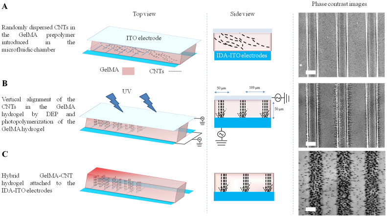
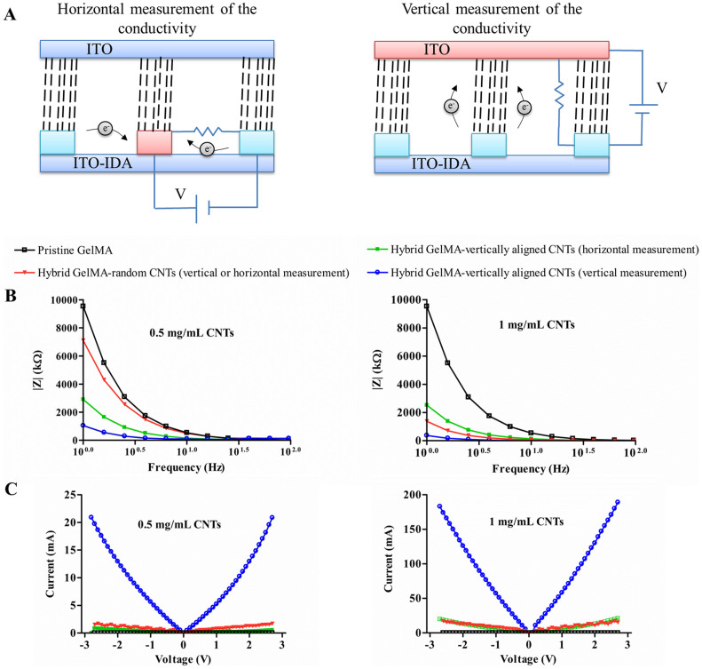
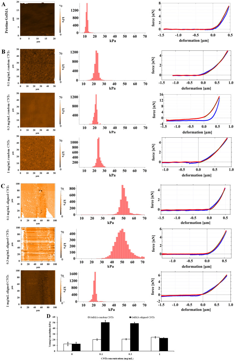
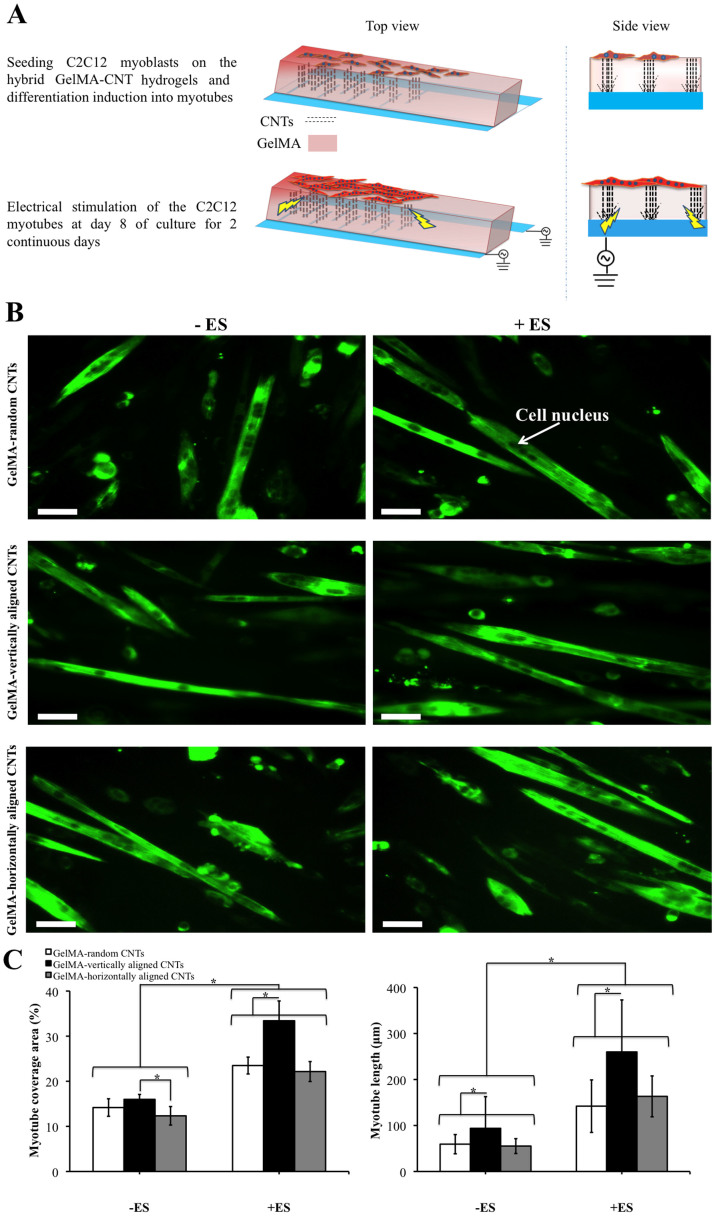
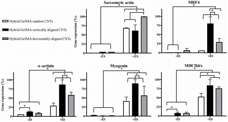
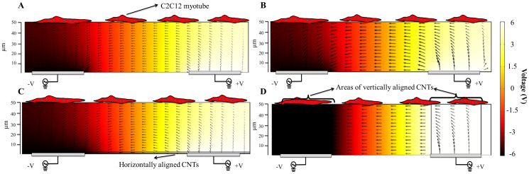
Biological scaffolds with tunable electrical and mechanical properties are of great interest in many different fields, such as regenerative medicine, biorobotics, and biosensing. In this study, dielectrophoresis (DEP) was used to vertically align carbon nanotubes (CNTs) within methacrylated gelatin (GelMA) hydrogels in a robust, simple, and rapid manner. GelMA-aligned CNT hydrogels showed anisotropic electrical conductivity and superior mechanical properties compared with pristine GelMA hydrogels and GelMA hydrogels containing randomly distributed CNTs. Skeletal muscle cells grown on vertically aligned CNTs in GelMA hydrogels yielded a higher number of functional myofibers than cells that were cultured on hydrogels with randomly distributed CNTs and horizontally aligned CNTs, as confirmed by the expression of myogenic genes and proteins. In addition, the myogenic gene and protein expression increased more profoundly after applying electrical stimulation along the direction of the aligned CNTs due to the anisotropic conductivity of the hybrid GelMA-vertically aligned CNT hydrogels. We believe that platform could attract great attention in other biomedical applications, such as biosensing, bioelectronics, and creating functional biomedical devices.


Figures

Figure 1
Figure 1. DEP mediated vertical alignment of CNTs within GelMA hydrogels.
(A) Randomly dispersed CNTs within GelMA prepolymer were introduced into a 50-μm-tall microfabricated chamber. (B) The CNTs in the GelMA prepolymer were vertically aligned on the IDA-ITO electrodes using DEP (voltage 20 V and frequency 2 MHz). The GelMA was then crosslinked by applying UV light for 150 s. (C) The GelMA gel containing vertically aligned CNTs was detached from the glass slide and retained on the IDA-ITO electrodes. Scale bars, 50 μm.
Figure 2
Figure 2. Anisotropic conductivity of pristine GelMA and hybrid GelMA-CNT gels containing different concentrations of CNTs.
(A) Schematic of the systems used for horizontal and vertical measurements of electrical conductivity for hybrid GelMA-vertically aligned CNT hydrogels. (B) Impedance measurements of pristine GelMA and hybrid gels loaded with 0.5 and 1 mg/mL CNTs. The perturbation amplitude was 25 mV. (C) I-V curves for pristine GelMA and GelMA hydrogels containing vertically aligned and random CNTs.
Figure 3
Figure 3. Mechanical properties of pristine GelMA and hybrid GelMA-CNT hydrogels containing different concentrations of CNTs (0.1, 0.3, and 1 mg/mL), as measured by AFM.
(A) Young's modulus map, Young's modulus distribution, and force deformation curves for pristine 5% GelMA hydrogels (left to right, respectively). The red curve represents the force deformation curve as the AFM cantilever approached the surface, and the blue curve is the force deformation curve produced when the cantilever left the surface. The green line was calculated according to the DMT theory. (B) Young's modulus map, Young's modulus distribution, and force deformation curves for hybrid GelMA-random CNT hydrogels (left to right, respectively). (C) Young's modulus map, Young's modulus distribution, and force deformation curves for hybrid GelMA-vertically aligned CNT hydrogels (left to right, respectively). Data in part (D) are presented as mean ± standard deviation obtained from at least 5 AFM pictures with 4096 independent data points in each.
Figure 4
Figure 4. Differentiation of C2C12 myoblasts on GelMA-0.3 mg/mL CNT hydrogels and characterization of the C2C12 myotubes obtained under ES.
(A) Schematic representation of the procedure used to produce and electrically stimulate C2C12 myotubes. (B) Immunostaining of the fast skeletal myosin heavy chain in the C2C12 myotubes fabricated on hybrid GelMA-random CNT, GelMA-vertically aligned CNT, and GelMA-horizontally aligned CNT hydrogels with and without ES application (indicated as +ES and −ES, respectively) on day 10 of culture. Cell nuclei within the C2C12 myotubes were obvious after the staining procedure. The ES parameters were as follows: a voltage of 8 V, a frequency of 1 Hz, and a duration of 10 ms. The scale bars represent 50 μm. (C) Quantification of the myotube coverage area and myotube length of the C2C12 myotubes fabricated on hybrid GelMA-random CNT, GelMA-vertically aligned CNT, and GelMA-horizontally aligned CNT hydrogels with and without ES on day 10 of culture. Data in part (C) are presented as mean ± standard deviation obtained from at least 40 myotubes of 2 independent experiments. Asterisks indicate significant differences between samples (*p < 0.05).
Figure 5
Figure 5. Gene expression analysis of C2C12 myotubes fabricated on GelMA-0.3 mg/mL CNT hydrogels subjected to ES.
Expression levels of sarcomeric actin, MRF4, α-actinin, myogenin, and MHCIId/x in fabricated muscle myofibers grown on GelMA hydrogels containing random CNTs, vertically aligned CNTs, and horizontally aligned CNTs. The ES was applied on day 8 of culture with a voltage of 8 V, a frequency of 1 Hz, and a duration of 10 ms continuously for 2 consecutive days. The expression levels of the genes were normalized based on the level of the internal reference gene GAPDH. Data are presented as mean ± standard deviation obtained from at least 8 measurements of 2 independent experiments. Asterisks indicate significant differences between samples (*p < 0.05).
Figure 6
Figure 6. Simulation of applied electric fields to muscle cells cultured on hydrogels.
Cross-sectional views of the numerically calculated electric fields (V/m) applied to muscle cells cultured on (A) pristine GelMA, (B) GelMA-random CNT, (C) GelMA-horizontally aligned CNT, and (D) GelMA-vertically aligned CNT hydrogels. Arrows show the current density (A/m2). Bottom grey bars show the ITO electrodes.

References

    1. Peppas N. A., Hilt J. Z., Khademhosseini A. & Langer R. Hydrogels in biology and medicine: From molecular principles to bionanotechnology. Adv. Mater. 18, 1345–60 (2006).
    1. Slaughter B. V., Khurshid S. S., Fisher O. Z., Khademhosseini A. & Peppas N. A. Hydrogels in regenerative medicine. Adv. Mater. 21, 3307–29 (2009). - PMC - PubMed
    1. Dvir T. et al. Nanowired three-dimensional cardiac patches. Nat. Nano. 6, 720–5 (2011). - PMC - PubMed
    1. Tian B. et al. Macroporous nanowire nanoelectronic scaffolds for synthetic tissues. Nat. Mater. 11, 986–94 (2012). - PMC - PubMed
    1. Xu L. et al. Design and synthesis of diverse functional kinked nanowire structures for nanoelectronic bioprobes. Nano Lett. 13, 746–51 (2013). - PMC - PubMed

Publication types

MeSH terms