. 2014 Mar;26(3):1360-76.
doi: 10.1105/tpc.114.123588. Epub 2014 Mar 18.

N-glycosylation of effector proteins by an α-1,3-mannosyltransferase is required for the rice blast fungus to evade host innate immunity

Affiliations

N-glycosylation of effector proteins by an α-1,3-mannosyltransferase is required for the rice blast fungus to evade host innate immunity

Xiao-Lin Chen et al. Plant Cell. 2014 Mar.

Abstract

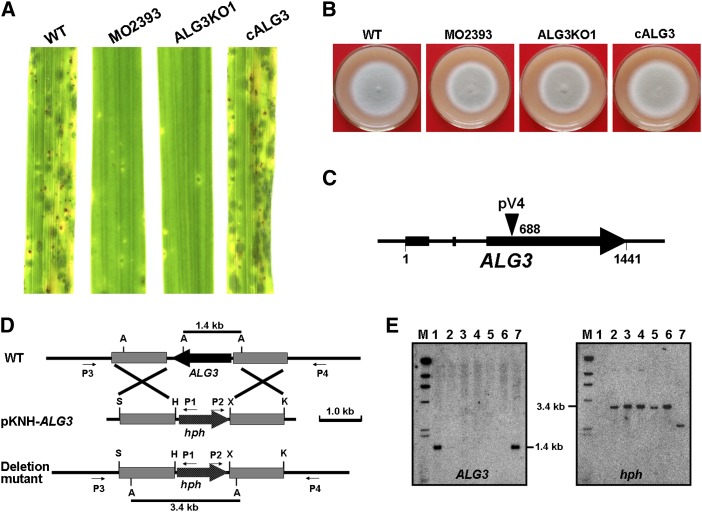
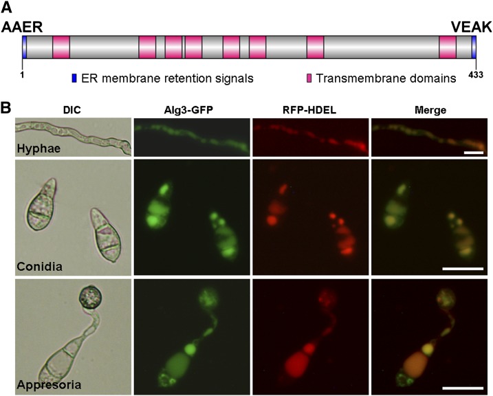
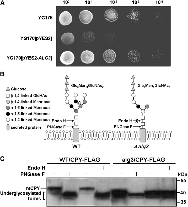
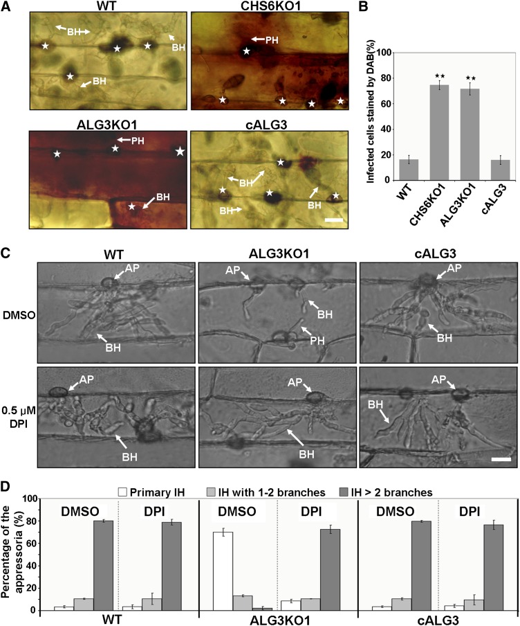
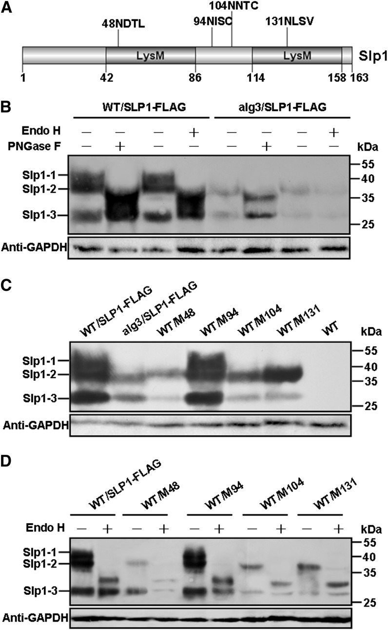
Plant pathogenic fungi deploy secreted effectors to suppress plant immunity responses. These effectors operate either in the apoplast or within host cells, so they are putatively glycosylated, but the posttranslational regulation of their activities has not been explored. In this study, the ASPARAGINE-LINKED GLYCOSYLATION3 (ALG3)-mediated N-glycosylation of the effector, Secreted LysM Protein1 (Slp1), was found to be essential for its activity in the rice blast fungus Magnaporthe oryzae. ALG3 encodes an α-1,3-mannosyltransferase for protein N-glycosylation. Deletion of ALG3 resulted in the arrest of secondary infection hyphae and a significant reduction in virulence. We observed that Δalg3 mutants induced massive production of reactive oxygen species in host cells, in a similar manner to Δslp1 mutants, which is a key factor responsible for arresting infection hyphae of the mutants. Slp1 sequesters chitin oligosaccharides to avoid their recognition by the rice (Oryza sativa) chitin elicitor binding protein CEBiP and the induction of innate immune responses, including reactive oxygen species production. We demonstrate that Slp1 has three N-glycosylation sites and that simultaneous Alg3-mediated N-glycosylation of each site is required to maintain protein stability and the chitin binding activity of Slp1, which are essential for its effector function. These results indicate that Alg3-mediated N-glycosylation of Slp1 is required to evade host innate immunity.


Figures

Figure 1.
Figure 1.
ALG3 Is Important for Virulence and Vegetative Growth of M. oryzae. (A) Rice leaves were sprayed with conidium suspensions (1 × 10⁵ spores/mL) of the wild-type P131, REMI mutant MO2393, ALG3 deletion mutant ALG3KO1, and complemented transformant cALG3. The inoculated leaves were photographed at 5 d post inoculation. (B) Five-day-old oatmeal agar cultures of P131, MO2393, ALG3KO1, and cALG3. (C) Schematic diagram of the ALG3 gene and the integration site of pV4 in the REMI mutant MO2393. (D) Schematic diagram of the ALG3 deletion strategy. Letters A, S, H, K, and X mark the recognition sites of ApaI, SpeI, HindIII, KpnI, and XhoI, respectively. hph, hygromycin phosphotransferase gene; P1 to P4, PCR primers. (E) DNA gel blot analysis of the ALG3 deletion mutants. ApaI-digested genomic DNAs were hybridized with a deleted fragment of ALG3 (left) and hph (right). Lane M, HindIII-digested fragments of λDNA; lane 1, wild-type strain P131; lanes 2 to 6, ALG3 deletion mutants ALG3KO1, ALG3KO2, ALG3KO3, ALG3KO4, and ALG3KO5; lane 7, ectopic transformant 08010-E1.
Figure 2.
Figure 2.
ALG3 Deletion Mutants Are Defective in Invasive Growth. Barley leaves were inoculated with conidium suspensions of the wild-type P131, ALG3 deletion mutant ALG3KO1, and complemented transformant cALG3. (A) Penetration and infectious hyphae were examined at 18, 24, and 30 hpi. AP, PH, and BH indicate appressoria, primary IH, and branched IH, respectively. Bar = 20 μm. (B) Percentages of appressoria with distinct types of IH, primary IH, IH with one or two branches, and IH with more than two branches, at 18, 24, and 30 hpi.
Figure 3.
Figure 3.
Alg3 Localizes to the ER in M. oryzae. (A) Predicted ER membrane retaining signals and transmembrane domains of Alg3 are indicated by blue and red boxes, respectively. AAER and VEAK motifs are predicted ER retaining signals. (B) Dual-color imaging by confocal laser scanning microscopy of transformant ALG3DW-1 expressing both Alg3-GFP and RFP-HDEL. Top row, vegetative hyphae; middle row, conidia; bottom row, appressoria. DIC, differential interference contrast. Bars = 20 μm.
Figure 4.
Figure 4.
ALG3 Encodes an α-1,3-Mannosyltransferase Required for Protein N-Glycosylation. (A) Complementation of the S. cerevisiae alg3 stt3 mutant by ALG3. Cells of YG176 (stt3) and transformants of YG170 (alg3 stt3) carrying pYES2 or pYES2-ALG3 were spotted in 10-fold dilutions on SC-Gal plates and incubated at 30°C for 2 d. (B) Lipid-linked or secreted protein–linked oligosaccharide structures of the wild-type and alg3 strains of S. cerevisiae. The digestion sites of Endo H and PNGase F are labeled with arrows. The functional site of Alg3 is indicated by the star. (C) Assays for CPY glycosylation by immunoblot analysis. Total proteins isolated from the transformants P131/CPY-FLAG and alg3/CPY-FLAG were treated with or without Endo H or PNGase F. The fully glycosylated form (mCPY) and underglycosylated forms of CPY were detected with an anti-FLAG antibody.
Figure 5.
Figure 5.
A Δalg3 Mutant Induces ROS Generation and Arrest of Invasive Growth during Plant Infection. (A) DAB staining of penetrated plant cells. Barley leaves inoculated with conidium suspensions (1 × 10⁵ spores/mL) of the wild-type P131, CHS6 deletion mutant CHS6KO1, ALG3 deletion mutant ALG3KO1, and complementation strain cALG3 were stained with DAB at 30 hpi. CHS6KO1 was used as a positive control strain (Kong et al., 2012). Stars and arrows indicate appressoria and IH, respectively. BH, branched IH; PH, primary IH. Bar = 20 μm. (B) Percentages of the DAB-stained cells. Means and se were calculated from three independent replicates. Significant differences are indicated by stars (P = 0.01; n > 100). (C) Invasive growth of IH restored by DPI treatment. Barley leaves were treated with or without DPI (0.5 μM) dissolved in DMSO. Invasive growth was observed at 30 hpi. AP, appressoria. Bar = 20 μm. (D) Percentages of appressoria with distinct types of IH, primary IH, IH with one or two branches, and IH with more than two branches, formed at 30 hpi by P131, ALG3KO1, and cALG3 in barley leaves treated with or without DPI. DMSO treatment was used as a control. Means and se were calculated from three independent replicates.
Figure 6.
Figure 6.
Alg3 Is Required for N-Glycosylation of the Effector Protein Slp1 in M. oryzae. (A) The Slp1 effector has two LysM domains (dark boxes) and four predicted N-glycosylation sites. (B) Assays for N-glycosylation of Slp1 in the wild type and the ALG3 deletion mutant. Total proteins isolated from the transformants WT/SLP1-FLAG and alg3/SLP1-FLAG were treated with or without PNGase F or Endo H and detected with an anti-FLAG antibody by immunoblot analysis. (C) Assays for the effects of point mutations at individual N-glycosylation sites of Slp1. Immunoblots of total proteins isolated from labeled strains were detected with an anti-FLAG antibody. Transformants WT/M48, WT/M94, WT/M104, and WT/M131 contain SLP1-3xFLAG fusion constructs carrying N48G, N94G, N104G, and N131G mutations, respectively. WT/SLP1-FLAG and alg3/SLP1-FLAG were used as controls. (D) Assays for N-glycosylation in WT/M48, WT/M94, WT/M104, and WT/M131 strains. Total proteins were digested by Endo H, and immunoblot analysis was performed as in (C). Three Slp1 forms (Slp1-1, Slp1-2, and Slp1-3) were detected and are marked on the left. GAPDH (glyceraldehyde-3-phosphate dehydrogenase) levels were used as a loading control.
Figure 7.
Figure 7.
N-Glycosylation of Slp1 Is Required for Virulence and Evasion of Chitin-Triggered Immunity. (A) Barley leaves sprayed with the wild-type strain P131, SLP1 deletion mutant SLP1KO1, and complement strains cSLP1, M48/104/131, M48, M94, M104, and M131 were photographed at 5 d post inoculation. M48, M94, M104, and M131 contain the SLP1-3xFLAG fusion construct carrying the N48G, N94G, N104G, and N131G mutations, respectively. M48/104/131 contains the SLP1-3xFLAG fusion construct with the N48G, N104G, and N131G triple mutation. (B) Lesion numbers on barley leaves caused by the same set of strains described in (A). (C) Average lesion size of barley leaves inoculated with the same set of strains described in (A). Lesion size was measured as reported by Mentlak et al. (2012). For (B) and (C), means and se were calculated from three independent replicates. Significant differences are indicated by stars (P = 0.05). (D) DAB staining assays of penetrated plant cells. Barley leaves were sprayed with the same set of strains described in (A), and the epidermis was stained with DAB at 30 hpi and observed with a Nikon microscope. AP, appressoria. Bar = 20 μm. (E) Percentages of DAB-stained cells. Means and se were calculated from three independent replicates. Significant differences are indicated by double stars (P = 0.01; n > 100).
Figure 8.
Figure 8.
N-Glycosylation of Slp1 Is Critical for Its Chitin Binding Activity. (A) The N-glycosylated and underglycosylated Slp1 proteins were similarly located at the plant–fungus interface. slp1/Slp1RFP, the Δslp1 mutant transformed with the SLP1-RFP fusion construct; M48/104/131-RFP, the Δslp1 mutant transformed with the SLP1N48G N104G N131G-RFP fusion construct; alg3/Slp1RFP, the Δalg3 mutant transformed with the SLP1-RFP fusion construct. Stars indicate appressoria, and arrows point to the plant–fungus interface. Bars = 20 μm. (B) Affinity precipitation of ySlp1, ySlp1M48/104/131, and bSlp1 with chitin beads detected by immunoblot analysis with an anti-His antibody. Most ySlp1 coprecipitated with chitin beads, but only trace amounts of ySlp1M48/104/131 and bSlp1 coprecipitated with chitin beads. P, pellet; S, supernatant. (C) Effect of exogenous addition of ySlp1, ySlp1M48/104/131, and bSlp1 proteins on the infection hyphal growth of Δslp1. Purified ySlp1, ySlp1M48/104/131, and bSlp1 proteins were added to the inoculation sites at 16 hpi, and the growth of IH was photographed at 28 hpi. Appressoria are indicated by stars. Purified 6xHis peptide was used as a control. Bars = 20 μm. (D) Percentages of appressoria that formed primary IH, IH with one to three branches, and IH with more than three branches at 28 hpi in (C). Means and se were calculated from three independent replicates, and double stars indicate statistically significant differences (P < 0.01). For (B) to (D), ySlp1 and bSlp1 were 6xHis-tagged Slp1 expressed in P. pastoris and E. coli, respectively. ySlp1M48/104/131 was 6xHis-tagged Slp1N48G N104G N131G expressed in P. pastoris.

Similar articles

Cited by

References

    1. Adam T., Bouhidel K., Der C., Robert F., Najid A., Simon-Plas F., Leborgne-Castel N. (2012). Constitutive expression of clathrin hub hinders elicitor-induced clathrin-mediated endocytosis and defense gene expression in plant cells. FEBS Lett. 586: 3293–3298. - PubMed
    1. Aebi M., Gassenhuber J., Domdey H., te Heesen S. (1996). Cloning and characterization of the ALG3 gene of Saccharomyces cerevisiae. Glycobiology 6: 439–444. - PubMed
    1. Anelli T., Sitia R. (2008). Protein quality control in the early secretory pathway. EMBO J. 27: 315–327. - PMC - PubMed
    1. Bourett T.M., Sweigard J.A., Czymmek K.J., Carroll A., Howard R.J. (2002). Reef coral fluorescent proteins for visualizing fungal pathogens. Fungal Genet. Biol. 37: 211–220. - PubMed
    1. Bowman S.M., Free S.J. (2006). The structure and synthesis of the fungal cell wall. Bioessays 28: 799–808. - PubMed

Publication types

Associated data