. 2014 Aug 1;23(15):4064-76.
doi: 10.1093/hmg/ddu120. Epub 2014 Mar 18.

iRHOM2-dependent regulation of ADAM17 in cutaneous disease and epidermal barrier function

Affiliations

iRHOM2-dependent regulation of ADAM17 in cutaneous disease and epidermal barrier function

Matthew A Brooke et al. Hum Mol Genet. .

Abstract

iRHOM2 is a highly conserved, catalytically inactive member of the Rhomboid family, which has recently been shown to regulate the maturation of the multi-substrate ectodomain sheddase enzyme ADAM17 (TACE) in macrophages. Dominant iRHOM2 mutations are the cause of the inherited cutaneous and oesophageal cancer-susceptibility syndrome tylosis with oesophageal cancer (TOC), suggesting a role for this protein in epithelial cells. Here, using tissues derived from TOC patients, we demonstrate that TOC-associated mutations in iRHOM2 cause an increase in the maturation and activity of ADAM17 in epidermal keratinocytes, resulting in significantly upregulated shedding of ADAM17 substrates, including EGF-family growth factors and pro-inflammatory cytokines. This activity is accompanied by increased EGFR activity, increased desmosome processing and the presence of immature epidermal desmosomes, upregulated epidermal transglutaminase activity and heightened resistance to Staphylococcal infection in TOC keratinocytes. Many of these features are consistent with the presence of a constitutive wound-healing-like phenotype in TOC epidermis, which may shed light on a novel pathway in skin repair, regeneration and inflammation.


Figures

Figure 1.
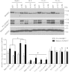
Figure 1.
ADAM17 and iRHOM2 expression in control and TOC keratinocyte cell lines. Expression of iRHOM2 and ADAM17 is shown in two control (Neb1 and K17) and two TOC (TYLK1 and 2) keratinocyte cell lines in whole cell lysates collected 24 h after transfection with NTP siRNA, or siRNA against either ADAM17 (si-A17) or iRHOM2 (si-iR2). The graph shown represents results from three separate experiments. *signifies that expression of mature ADAM17 is individually significantly higher in both TOC cell lines than both control cell lines (P < 0.01, in all cases); #signifies that expression of both pro- and mature ADAM17 is significantly reduced in all keratinocytes treated with si-A17, compared with those treated with NTP (P < 0.01, in all cases); §symbols signify that expression of mature ADAM17 is significantly reduced in both TOC cell lines treated with si-iR2 compared with NTP, but not in either of the control cell lines (P < 0.05, in each case).
Figure 2.
Figure 2.
ADAM17 expression in TOC. (A) ADAM17 is highly expressed at the cell surface of TOC keratinocytes, and intracellularly, in compartments that co-localize with the Golgi marker GM130, higher levels of ADAM17 are observed at the cell surface of TOC cells compared with controls (left panel, and detail) (B) western blotting of the ADAM17 pro-domain (released upon ADAM17 maturation) also shows much higher levels of this free pro-domain in TOC. (C) 3D cultures of TOC and control keratinocytes yield fully differentiated skin equivalent models (see also Supplementary Material, Fig. S2). (D) Western blotting of lysates from 3D cultures reveals the same pattern of increased mature ADAM17 and free ADAM17 pro-domain as is seen in keratinocyte monolayers. These data represent results from a single experiment. (E): 3D skin equivalent cultures also display much higher levels of ADAM17 at the cell surface of TOC keratinocytes compared with controls (quantified in graph 2F). These data represent results from three separate experiments. Scale bars on all histology and immunofluorescence images (A, C and E) represent 100 µm.
Figure 3.
Figure 3.
Shedding of ADAM17 substrates and EGFR activation. (A) Shedding of ADAM17's best known substrate TNFα was significantly higher in both TOC keratinocytes and PBMCs compared with controls, following stimulation with 100 ng/ml PMA. (nd, not detected) (B) TOC keratinocytes constitutively shed significantly higher levels of the EGFR ligands amphiregulin, TGFα and HB-EGF than control keratinocytes. This increase in shedding was ADAM17 dependent and could be significantly reduced by siRNA knockdown of ADAM17. (C) Amphiregulin (and other growth factors) secretion from 3D culture models was significantly higher in TOC cells than controls. (D) Secretion of IL-6 and IL-8 was also found to be significantly higher from TOC keratinocytes, with this level of secretion also sensitive to siRNA knockdown of ADAM17. (E) Phosphorylation of the EGFR at three separate tyrosine residues was noticeably higher in TOC 3D culture models than the control. (F) mRNA expression of ADAM17 substrates and secreted cytokines was variable, but was not affected by ADAM17 knockdown, illustrating that siRNA-mediated reductions in substrate shedding were dependent on loss of shedding by ADAM17. ADAM17 siRNA did, however, reduce ADAM17 mRNA levels, as expected. Shedding of all substrates was analysed by ELISA. Graphs in A, B and D represent results from experiments in which each cell line (K17, Neb1, TYLK1 and TYLK2) was tested on three separate occasions, with the data from the two control cell lines and two tylosis cell lines subsequently pooled. To calculate significance results were analysed by two-way ANOVA followed by Bonferroni's post-test. The graph in C represents results from ELISA tests on supernatants from three 3D cultures of each cell line, which were again pooled and were analysed by student's T-test. Ns, not significant, *P < 0.05, **P < 0.01, ***P < 0.001.
Figure 4.
Figure 4.
Desmosomes in TOC. (A) Analysis of desmosomes in TOC skin compared with controls showed the presence in TOC skin of desmosomes lacking the electron-dense midline (white arrows) found in mature, calcium-independent desmosomes of control skin. (B) levels of the ADAM17 substrate desmoglein 2 (DSG2) were reduced in TOC keratinocytes. This effect is EGFR-activation dependent and was most noticeable when keratinocytes were cultured in the absence of exogenous EGFR ligands. DSG2 expression levels were not significantly different between any of the four cell lines in the presence of EGF (analysed in three separate experiments, using two-way ANOVA followed by Bonferroni's post-test) but were significantly higher in both control cell lines than both TOC cell lines in the absence of EGF (P < 0.05, in all cases). (C) Expression of a DSG2 cleavage product is also shown when keratinocytes are cultured in the presence and absence of EGF. Expression of this cleavage product is comparable when cells are in the presence of EGF, but notably reduced in control cells in the absence of EGF, whereas expression in TOC keratinocytes in the absence of EGF remains robust.
Figure 5.
Figure 5.
Barrier function and infection resistance in TOC. (A) Analysis of transglutaminase 1 activity in the granular layer of the epidermis of TOC and control skin by the Biot-MDC assay revealed significantly greater activity in TOC skin compared with controls (white arrows). (B) This pattern was replicated in 3D cultures of TOC skin. Scale bars in Panels A and B represent 100 µm. (C and D) These increases were quantified, with activity shown to be significantly higher in TOC skin and 3D cultures of TOC keratinocytes. Data in C represent results from experiments on three sections each from two control and two tylosis patients, whilst data in D represent results from six sections of each of the organotypic cultures. Significance in both cases was analysed using student's t-test. (E) TOC keratinocytes displayed significantly increased resistance to both adhesion and invasion by S. aureus, with this resistance dependent on ADAM17 and vulnerable to siRNA knockdown of ADAM17. (F) Secretion of IL-6 and IL-8 was measured by ELISA during S. aureus infection, found to be significantly higher from TOC cells compared with controls and could be significantly reduced by siRNA-mediated knockdown of ADAM17. Data in E and F were analysed by two-way ANOVA, followed by Bonferroni's post-test; *P < 0.05, **P < 0.01, ***P < 0.001.

Similar articles

Cited by

References

    1. Etheridge S.L., Brooke M.A., Kelsell D.P., Blaydon D.C. Rhomboid proteins: a role in keratinocyte proliferation and cancer. Cell Tissue Res. 2013;351:301–307. doi: 10.1007/s00441-012-1542-1. - DOI - PubMed
    1. Freeman M. Rhomboid proteases and their biological functions. Ann. Rev. Genet. 2008;42:191–210. doi: 10.1146/annurev.genet.42.110807.091628. - DOI - PubMed
    1. Zettl M., Adrain C., Strisovsky K., Lastun V., Freeman M. Rhomboid family pseudoproteases use the ER quality control machinery to regulate intercellular signaling. Cell. 2011;145:79–91. doi: 10.1016/j.cell.2011.02.047. - DOI - PMC - PubMed
    1. Adrain C., Zettl M., Christova Y., Taylor N., Freeman M. Tumor necrosis factor signaling requires iRhom2 to promote trafficking and activation of TACE. Science. 2012;335:225–228. doi: 10.1126/science.1214400. - DOI - PMC - PubMed
    1. McIlwain D.R., Lang P.A., Maretzky T., Hamada K., Ohishi K., Maney S.K., Berger T., Murthy A., Duncan G., Xu H.C., et al. iRhom2 regulation of TACE controls TNF-mediated protection against Listeria and responses to LPS. Science. 2012;335:229–232. doi: 10.1126/science.1214448. - DOI - PMC - PubMed

Publication types

MeSH terms

Supplementary concepts