. 2014 Jun;75(6):851-63.
doi: 10.1002/ana.24145. Epub 2014 May 28.

Homocysteine exacerbates β-amyloid pathology, tau pathology, and cognitive deficit in a mouse model of Alzheimer disease with plaques and tangles

Affiliations

Homocysteine exacerbates β-amyloid pathology, tau pathology, and cognitive deficit in a mouse model of Alzheimer disease with plaques and tangles

Jian-Guo Li et al. Ann Neurol. 2014 Jun.

Abstract

Objective: High level of homocysteine (Hcy) is a recognized risk factor for developing Alzheimer disease (AD). However, the mechanisms involved are unknown. Previously, it was shown that high Hcy increases brain β-amyloid (Aβ) levels in amyloid precursor protein transgenic mice, but no data are available on the effect that it may have on the other main pathologic features of AD such as tau.

Methods: 3xTg mice with diet-induced high Hcy were compared with mice having normal Hcy. Neuronal cells were incubated with and without Hcy.

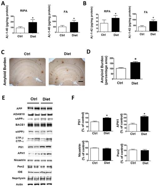
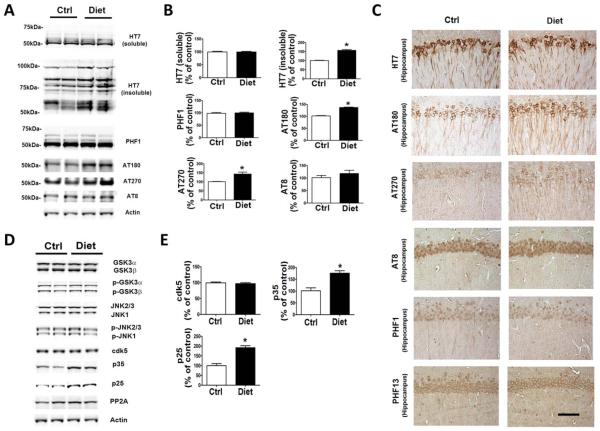
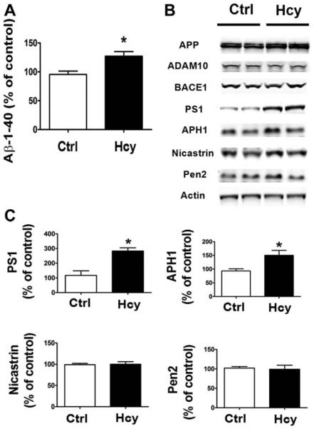
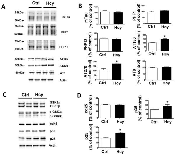
Results: Diet-induced high Hcy resulted in an exacerbation of the entire AD-like phenotype of the 3xTg mice. In particular, we found that compared with controls, mice with high Hcy developed significant memory and learning deficits, and had elevated Aβ levels and deposition, which was mediated by an activation of the γ-secretase pathway. In addition, the same mice had a significant increase in the insoluble fraction of tau and its phosphorylation at specific epitopes, which was mediated by the cdk5 pathway. In vitro studies confirmed these observations and provided evidence that the effects of Hcy on Aβ and tau are independent from each other.

Interpretation: Taken together, our findings demonstrate that a dietary condition that leads to an elevation of Hcy levels results in an exacerbation of all 3 major pathological features of the AD phenotype: memory deficits, and Aβ and tau neuropathology. They support the concept that this dietary lifestyle can act as a risk factor and actively contribute to the development of the disease.


Figures

FIGURE 1
FIGURE 1
Diet-induced high homocysteine brain levels worsen behavioral deficits. (A) Number of total arm entries for 3xTg mice receiving supplemented diet (Diet) or vehicle (Ctrl). (B) Percentage of alternations between 3xTg mice receiving the diet or vehicle treatment. (C) Contextual fear memory response in 3xTg mice treated with the special diet and controls. (D) Cued fear memory response in the same 3xTg mice. (E) Number of entries to the target platform zone for 3xTg mice treated with the supplemented diet or control vehicle. (F) Time in the target platform zone for the same 2 groups of 3xTg mice. (G) Time in the opposite zone for the same 2 groups of 3xTg mice. (H) Average swimming speed for the same 2 groups of 3xTg mice. Values represent mean 6 standard error of the mean (n = 6 control, n = 7 diet); *p < 0.05.
FIGURE 2
FIGURE 2
Diet-induced high homocysteine brain levels affect β-amyloid (Aβ) peptide levels and deposition. (A, B) Radioimmunoprecipitation assay–soluble (RIPA) and formic acid extractable (FA) Aβ1–40 and Aβ1–42 levels in brain cortex of 3xTg mice receiving supplemented diet (Diet, n = 7) or vehicle (Ctrl, n = 6) were measured by sandwich enzyme-linked immunosorbent assay. (C) Representative images of brains from 3xTg mice receiving the diet or placebo immunostained with 4G8 antibody. Scale bar = 500μM. (D) Quantification of the area occupied by Aβ immunoreactivity in brains of diet-treated 3xTg mice and mice receiving vehicle. (E) Representative Western blots of the antibodies APP, ADAM10, sAPPαa, BACE1, sAPPβ, CTFs, PS1, APH1, nicastrin, Pen2, IDE, neprilysin, and actin in brain cortex homogenates from 3xTg mice receiving the diet or vehicle. (F) Densitometric analyses of the immunoreactivities to antibodies shown in the previous panel. Values represent mean 6 standard error of the mean (n = 6 control, n = 7 diet); *p < 0.05. [Color figure can be viewed in the online issue, which is available at www.annalsofneurology.org.]
FIGURE 3
FIGURE 3
Diet-induced high homocysteine brain levels affect tau phosphorylation and metabolism. (A) Representative Western blots of soluble and insoluble total tau (HT7), phosphorylated tau at residues S396/S404 (PHF1), T231/S235 (AT180), T181 (AT270), and S202/T205 (AT8), and actin in brain cortex homogenates from 3xTg mice receiving the supplemented diet (Diet, n = 7) or vehicle (Ctrl, n = 6). (B) Densitometric analyses of the immunoreactivities to antibodies shown in the previous panel. (C) Representative images of brain sections from diet-treated 3xTg mice or vehicle-treated control mice immunostained with HT7, AT180, AT270, AT8, PHF1, and PHF13 antibodies. Scale bar = 100μM. (D) Representative Western blots of GSK3α, GSK3β, pGSK3α, pGSK3β, JNK2/3, JNK1, p-JNK2/3, p-JNK1, cdk5, p35, p25, PP2A, and actin in brain cortex homogenates from 3xTg mice treated with supplemented diet or vehicle. (E) Densitometric analyses of the immunoreactivities to antibodies shown in the previous panel. Values represent mean 6 standard error of the mean (n = 6 control, n = 7 diet); *p < 0.05. [Color figure can be viewed in the online issue, which is available at www.annalsofneurology.org.]
FIGURE 4
FIGURE 4
Diet-induced high homocysteine brain level affects synaptic integrity and neuroinflammation. (A) Representative Western blot analyses of synaptophysin (SYP), postsynaptic density protein 95 (PSD95), microtubule-associated protein 2 (MAP2), and actin in brain cortex homogenates of diet-treated 3xTg mice (Diet, n = 6) or control mice (Ctrl, n = 7). (B) Densitometric analyses of the immunoreactivities to antibodies shown in the previous panel. (C) Representative images of brain sections from 3xTg mice receiving the supplemented diet or vehicle immunostained with SYP, PDS95, and MAP2 antibodies. Scale bar = 100μM. (D) Representative Western blots of glial fibrillary acidic protein (GFAP) and CD45 in brain cortex homogenates from diet-treated 3xTg mice and the vehicle-treated control group. (E) Densitometric analyses of the immunoreactivities to antibodies shown in the previous panel. Values represent mean ± standard error of the mean (n = 6 control, n = 7 diet); *p < 0.05. [Color figure can be viewed in the online issue, which is available at www.annalsofneurology.org.]
FIGURE 5
FIGURE 5
In vitro effect of homocysteine (Hcy)-treated N2A cells on β-amyloid (Aβ) and amyloid precursor protein (APP) metabolism. (A) Levels of Aβ1–40 in conditioned media from N2A cells incubated with 50μM Hcy for 24 hours. (B) Representative Western blots of antibodies APP, ADAM10, BACE1, PS1, APH1, nicastrin, Pen2, and actin in lysates from cells incubated with Hcy or vehicle (Ctrl) for 24 hours. (C) Densitometric analyses of the immunoreactivities to antibodies shown in the previous panel. *p < 0.05.
FIGURE 6
FIGURE 6
In vitro effect of homocysteine (Hcy)-treated N2A cells on tau metabolism. (A) Representative Western blots of total tau (mTau), phosphorylated tau at residues S396/S404 (PHF1), S396 (PHF13), T231/S235 (AT180), T181 (AT270), and S202/ T205 (AT8), and actin in lysates from cells incubated with Hcy or vehicle (Ctrl) for 24 hours. (B) Densitometric analyses of the immunoreactivities to antibodies shown in the previous panel. (C) Representative Western blots of antibodies GSK3α, GSK3β, p-GSK3α, p-GSK3β, cdk5, p35, p25, and actin in lysates from cells incubated with Hcy or vehicle (Ctrl). (D) Densitometric analyses of the immunoreactivities to antibodies shown in the previous panel. *p < 0.05.
FIGURE 7
FIGURE 7
Homocysteine (Hcy) effect on tau phosphorylation is β-amyloid (Aβ) independent. (A) Levels of Aβ1–40 in conditioned media from N2A cells incubated with 50μM Hcy alone or in the presence of L685,458 (1μM) for 24 hrs. (B) Representative Western blots of total tau (mTau), phosphorylated tau at residues T231/S235 (AT180), at T181 (AT270), and S202/T205 (AT8), and actin in lysates from cells incubated with Hcy or vehicle (Ctrl), or with Hcy in the presence of L685,458. (C) Densitometric analyses of the immunoreactivities to antibodies shown in the previous panel. *p < 0.05.

References

    1. Gandy S. Lifelong management of amyloid beta metabolism to prevent Alzheimer’s disease. N Engl J Med. 2012;367:864–866. - PMC - PubMed
    1. Morris MS. Homocysteine and Alzheimer’s disease. Lancet Neurol. 2003;2:425–428. - PubMed
    1. Seshadri S, Beiser A, Selhub J, et al. Plasma homocysteine as a risk factor for dementia and Alzheimer’s disease. N Engl J Med. 2002;346:476–483. - PubMed
    1. Selhub J. Homocysteine metabolism. Annu Rev Nutr. 1999;19:217–246. - PubMed
    1. Van Dam F, Van Gool WA. Hyperhomocysteinemia and Alzheimer’s disease: a systematic review. Arch Gerontol Geriatr. 2009;48:425–430. - PubMed

Publication types

MeSH terms