. 2014 Jun;3(3):500-13.
doi: 10.1002/cam4.229. Epub 2014 Mar 19.

Tumor cell-expressed SerpinB2 is present on microparticles and inhibits metastasis

Affiliations

Tumor cell-expressed SerpinB2 is present on microparticles and inhibits metastasis

Wayne A Schroder et al. Cancer Med. 2014 Jun.

Abstract

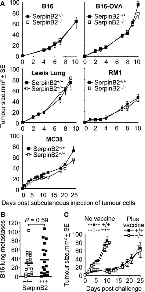
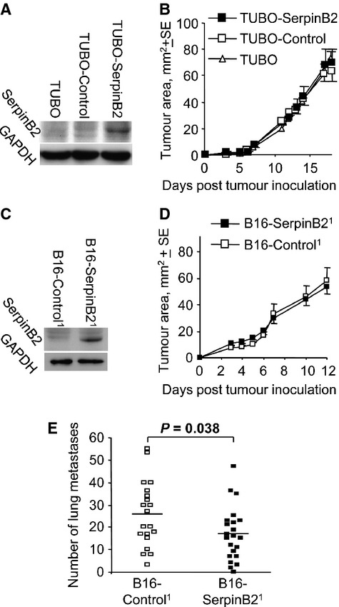
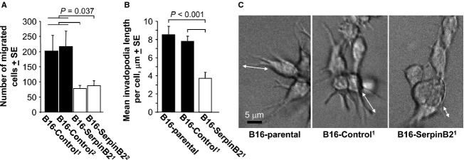
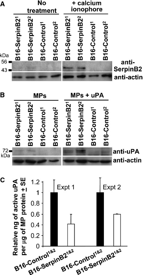
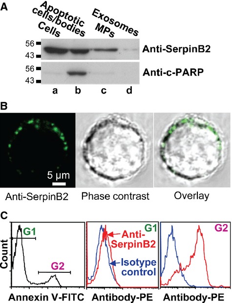
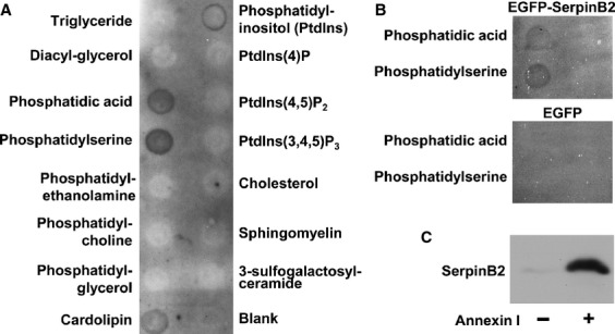
Expression of SerpinB2 (plasminogen activator inhibitor type 2/PAI-2) by certain cancers is associated with a favorable prognosis. Although tumor-associated host tissues can express SerpinB2, no significant differences in the growth of a panel of different tumors in SerpinB2(-/-) and SerpinB2(+/+) mice were observed. SerpinB2 expression by cancer cells (via lentiviral transduction) also had no significant effect on the growth of panel of mouse and human tumor lines in vivo or in vitro. SerpinB2 expression by cancer cells did, however, significantly reduce the number of metastases in a B16 metastasis model. SerpinB2-expressing B16 cells also showed reduced migration and increased length of invadopodia-like structures, supporting the classical view that that tumor-derived SerpinB2 is inhibiting extracellular urokinase. Importantly, although SerpinB2 is usually poorly secreted, we found that SerpinB2 effectively reaches the extracellular milieu on the surface of 0.5-1 μm microparticles (MPs), where it was able to inhibit urokinase. We also provide evidence that annexins mediate the binding of SerpinB2 to phosphatidylserine, a lipid characteristically exposed on the surface of MPs. The presence of SerpinB2 on the surface of MPs provides a physiological mechanism whereby cancer cell SerpinB2 can reach the extracellular milieu and access urokinase plasminogen activator (uPA). This may then lead to inhibition of metastasis and a favorable prognosis.

Keywords: Annexin; SerpinB2; metastasis; microparticle; phosphatidylserine.


Figures

Figure 1
Figure 1
Tumor growth in SerpinB2−/− and SerpinB2+/+ mice. (A) Groups (n = 5–8 per group) of age-matched SerpinB2+/+ and SerpinB2−/− mice were injected s.c. with different tumors and their growth monitored over time. B16, melanoma; B16-OVA, B16 stably expressing ovalbumin, Lewis lung carcinoma; RM1, prostate cancer; MC38, colon carcinoma. (B) SerpinB2−/− and SerpinB2+/+ mice (as above, n = 17 per group) were injected i.v. with B16 cells and after 18 days lung metastases were counted. Statistics by Mann–Whitney U test. (C) SerpinB2−/− and SerpinB2+/+ mice (n = 7–8 per group) were not vaccinated (no vaccine) or were vaccinated with SIINFEKL formulated in CFA (plus vaccine). After 14 days mice were challenged with B16-OVA and tumor growth monitored over time.
Figure 2
Figure 2
SerpinB2 expression in B16 and Tubo cells and their growth in vivo. (A) Parental Tubo cells (Tubo) or Tubo cells transduced with a lentiviral vector encoding EGFP (Tubo-Control) or SerpinB2 (Tubo-SerpinB2) were analyzed by immunoblotting for murine SerpinB2 expression using anti-murine SerpinB2 antibody. (B) The same cells described in A were inoculated s.c. into Balb/c mice and tumor growth was monitored over time (n = 12 mice per group). (C) B16 cells were transduced with a lentiviral vector encoding ZsGreen or SerpinB2-ZsGreen and were then FACs sorted to produce B16-Control and B16-SerpinB2 cell populations, respectively, that were both >99% ZsGreenhi. The cell lines were then analyzed by immunoblotting as in A. (D) B16-Control and B16-SerpinB2 (described in C) were inoculated s.c. into C57BL/6 mice and growth monitored over time. (E) SerpinB2 expression by B16 cells reduces metastases. B16-Control and B16-SerpinB2 cells (described in the figure2) were inoculated i.v. into C57BL/6 mice (n = 20/22 per group) and on day 18 the number of lung metastases was determined. Statistics by t-test.
Figure 3
Figure 3
Migration and invasion of SerpinB2-expressing B16 cells. (A) B16-Control and B16-SerpinB2 cell lines (superscript 1 and 2 indicate lines generated on two separate occasions) were seeded into transwells (104 cells/well) and the number of cells that migrated toward 40 μg/mL fibronection determined after 2 days. The data were presented as a mean of three repeat assays, with each bar showing the mean ± SE of the three assays. The B16-Control1 and B16-Control2 data were combined for statistical analysis (n = 6) and were significantly different from both B16-SerpinB21 and B16-SerpinB22 (Statistics by Kolmogorov–Smirnov tests). (B) Parental B16, B16-Control and B16-SerpinB2 cells were seeded into matrigel and after 2 days the cells were examined by live imaging and length of invadopodia-like structures measured using still images (examples shown in C; 22–47 cells were examined per cell line; statistics by t-test). (C) Representative images of cells providing the data for B (arrows show examples of length measurements).
Figure 4
Figure 4
SerpinB2 microparticles. (A) B16-Control and B16-SerpinB2 cell lines were treated without (no treatment) or with calcium ionophore (+calcium ionophore) for 30 min, supernatants were harvested, MPs isolated by differential centrifugation and analyzed by immunoblotting using anti-murine SerpinB2 and anti-actin antibodies. (B) The same MPs described in A were incubated without (MPs) or with recombinant murine uPA (MPs + uPA), solubilized in SDS and analyzed by immunoblotting using anti-murine uPA and anti-actin antibodies. (C) Active uPA levels in MPs from B16-SerpinB2 and B16-Control cells as measured by capture ELISA. Data were presented as relative to the mean uPA levels in MPs from the two B16-SerpinB2 lines. Each bar represents the mean relative uPA level in MPs from the two SerpinB2-expressing or two control lines (P = 0.021 for B16-SerpinB2 vs. B16-Control when the two experiments are combined, i.e., n = 4 per group).
Figure 5
Figure 5
(A) U937 cells were treated with PMA (25 ng/mL for 2 days) and calcium ionophore (10 μmol/L for 30 min). Whole cell lysate (lane a), apoptotic cells/apoptotic bodies (obtained from the culture supernatant by centrifugation at 2000g for 10 min) (lane b), the MP fraction (obtained from the latter supernatant by further centrifugation at 20000g for 30 min) (lane c), and the exosome fraction (obtained from the latter supernatant by further centrifugation at 100,000g for 1 h) (lane d) were analyzed by immunoblotting (10 μg of protein loaded per lane). The antibodies used were an anti-human SerpinB2 monoclonal antibody and anticleaved PARP antibody (c-PARP) (a marker of apoptosis). (B) U937 cells were treated with PMA and calcium ionophore as above and were then live stained at 4°C and viewed by confocal microscopy. Propidium iodide was included with the FITC-labeled secondary antibody to identify and exclude permeabilized cells. Top row; staining with anti-human SerpinB2 monoclonal antibody showing representative fluorescence image (left), phase-contrast image (middle), and overly (right). Bottom row; costaining with the anti-human SerpinB2 and anti-CD11b antibodies. (C) U937 cells treated with PMA and calcium ionophore as above, were stained with annexin V-FITC (to detect PS) and anti-human SerpinB2 monoclonal antibody (with PE-labeled secondary). FACS analysis showed an annexin Vlow population gated as G1, and an annexin Vhi population gated as G2 (left panel). The G1 population showed only marginal staining with anti-SerpinB2 antibody (middle panel). The G2 population was clearly stained with the anti-SerpinB2 antibody (right panel). The PShi population was thus also SerpinB2hi.
Figure 6
Figure 6
SerpinB2 association with PS via annexin. (A) CaSki cell lysates were incubated with membranes arrayed with the indicated lipids, the membranes were washed and probed with anti-human polyclonal SerpinB2 antibody. (B) HEK293 cells were transiently transfected with DNA plasmids encoding EGFP-SerpinB2 or EGFP, and cell lysates incubated with the lipid membranes as for A. SerpinB2 was detected using anti-EGFP antibody. (C) PS beads were incubated with purified recombinant human SerpinB2 in the absence (−) or presence (+) of purified recombinant human annexin I. The beads were washed, and bead-bound proteins solubilized in SDS and analyzed by immunoblotting using anti-human SerpinB2 antibody.

References

    1. Schroder WA, Major L, Suhrbier A. The role of SerpinB2 in immunity. Crit. Rev. Immunol. 2011;31:15–30. - PubMed
    1. Dougherty KM, Pearson JM, Yang AY, Westrick RJ, Baker MS, Ginsburg D. The plasminogen activator inhibitor-2 gene is not required for normal murine development or survival. Proc. Natl. Acad. Sci. USA. 1999;96:686–691. - PMC - PubMed
    1. Ye RD, Wun TC, Sadler JE. Mammalian protein secretion without signal peptide removal. Biosynthesis of plasminogen activator inhibitor-2 in U-937 cells. J. Biol. Chem. 1988;263:4869–4875. - PubMed
    1. Ritchie H, Booth NA. Secretion of plasminogen activator inhibitor 2 by human peripheral blood monocytes occurs via an endoplasmic reticulum-golgi-independent pathway. Exp. Cell Res. 1998;242:439–450. - PubMed
    1. Manders P, Tjan-Heijnen VC, Span PN, Grebenchtchikov N, Geurts-Moespot A, van Tienoven DT, et al. Complex of urokinase-type plasminogen activator with its type 1 inhibitor predicts poor outcome in 576 patients with lymph node-negative breast carcinoma. Cancer. 2004;101:486–494. - PubMed

Publication types

MeSH terms

Substances

LinkOut - more resources