. 2014 May 2;289(18):12931-45.
doi: 10.1074/jbc.M113.538124. Epub 2014 Mar 18.

Molecular basis for increased risk for late-onset Alzheimer disease due to the naturally occurring L28P mutation in apolipoprotein E4

Affiliations

Molecular basis for increased risk for late-onset Alzheimer disease due to the naturally occurring L28P mutation in apolipoprotein E4

Letta Argyri et al. J Biol Chem. .

Abstract

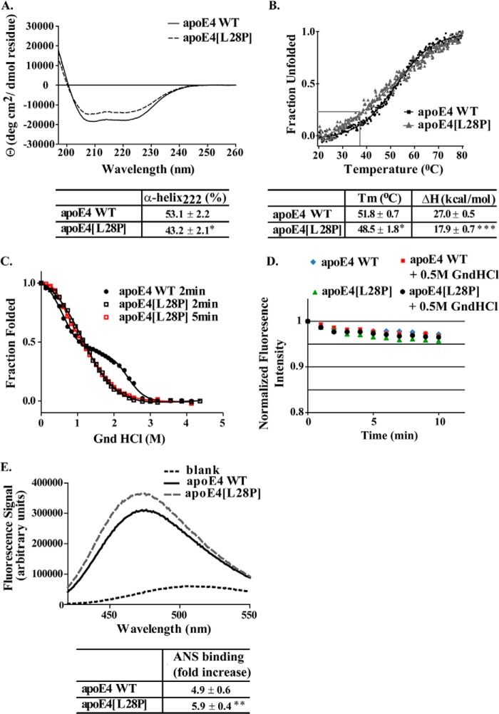
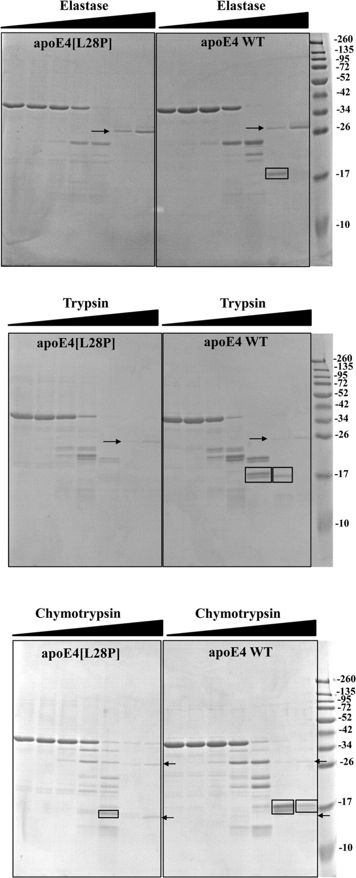
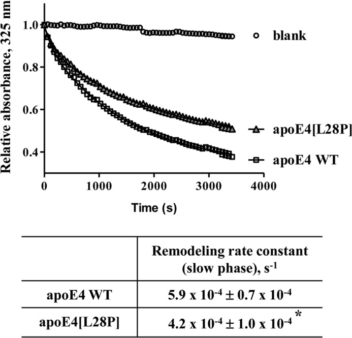
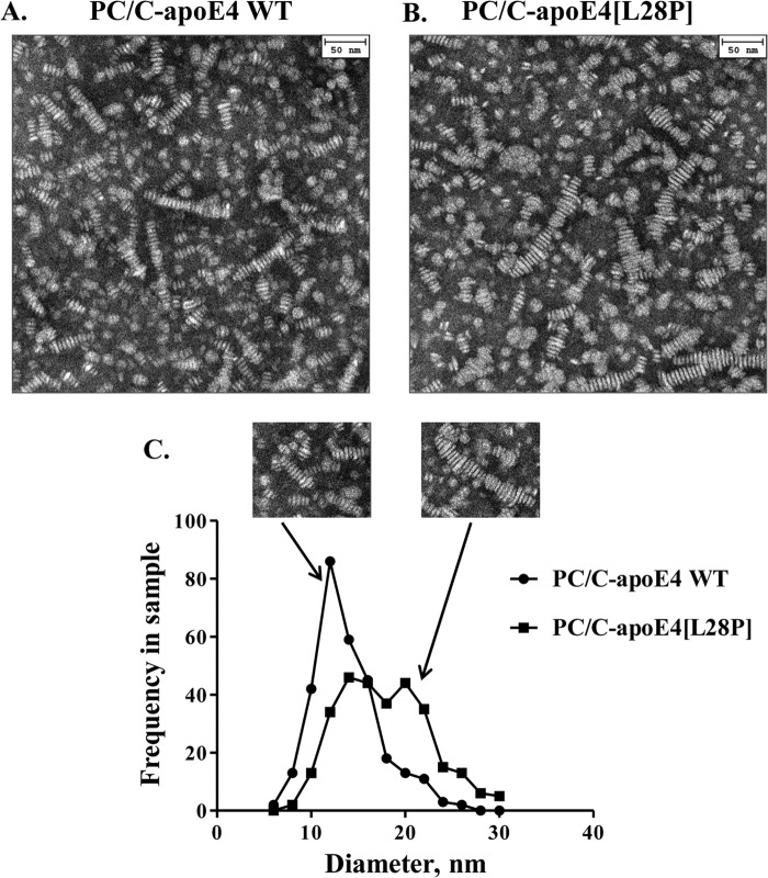
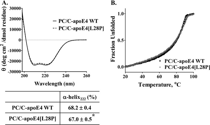
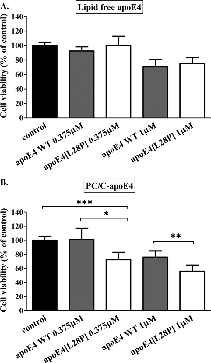
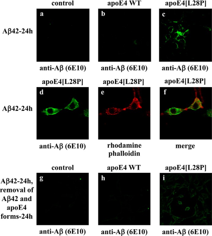
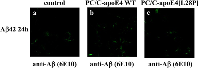
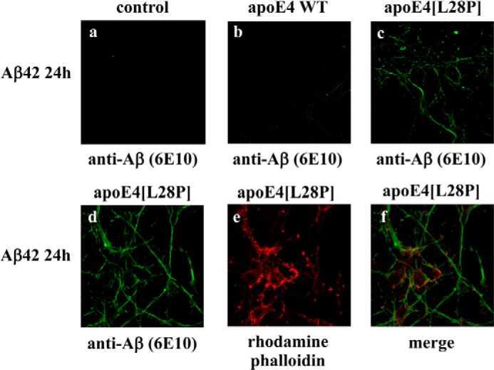
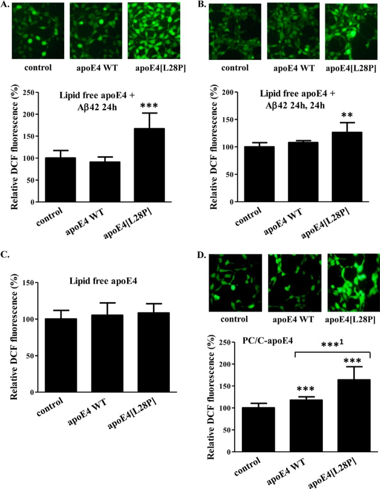
The apolipoprotein (apo) E4 isoform has consistently emerged as a susceptibility factor for late-onset Alzheimer disease (AD), although the exact mechanism is not clear. A rare apoE4 mutant, apoE4[L28P] Pittsburgh, burdens carriers with an added risk for late-onset AD and may be a useful tool for gaining insights into the role of apoE4 in disease pathogenesis. Toward this end, we evaluated the effect of the L28P mutation on the structural and functional properties of apoE4. ApoE4[L28P] was found to have significantly perturbed thermodynamic properties, to have reduced helical content, and to expose a larger portion of the hydrophobic surface to the solvent. Furthermore, this mutant is thermodynamically destabilized and more prone to proteolysis. When interacting with lipids, apoE4[L28P] formed populations of lipoprotein particles with structural defects. The structural perturbations brought about by the mutation were accompanied by aberrant functions associated with the pathogenesis of AD. Specifically, apoE4[L28P] promoted the cellular uptake of extracellular amyloid β peptide 42 (Aβ42) by human neuroblastoma SK-N-SH cells as well as by primary mouse neuronal cells and led to increased formation of intracellular reactive oxygen species that persisted for at least 24 h. Furthermore, lipoprotein particles containing apoE4[L28P] induced intracellular reactive oxygen species formation and reduced SK-N-SH cell viability. Overall, our findings suggest that the L28P mutation leads to significant structural and conformational perturbations in apoE4 and can induce functional defects associated with neuronal Aβ42 accumulation and oxidative stress. We propose that these structural and functional changes underlie the observed added risk for AD development in carriers of apoE4[L28P].

Keywords: Alzheimer Disease; Amyloid Peptide β; ApoE; Apolipoproteins; Biophysics; Lipoprotein; Mutation; Reactive Oxygen Species (ROS); Thermodynamic Stability; Toxicity.


Figures

FIGURE 1.
FIGURE 1.
Schematic of the crystal structure of the N-terminal domain of human apoE4, indicating the location of the L28P mutation and SDS-PAGE and MALDI-TOF analyses of purified WT apoE4 and mutant apoE4[L28P]. A and B, the position of the mutated leucine residue on the N-terminal helical segment of apoE4 was mapped on the crystal structure of apoE4 with PDB code 1GS9. C, SDS-PAGE analysis of refolded WT and mutant apoE4, which were produced and purified as described under “Experimental Procedures.” D and E, MALDI-TOF spectra of refolded WT and mutant apoE4. a.u., arbitrary units.
FIGURE 2.
FIGURE 2.
Physicochemical properties of WT apoE4 and apoE4[L28P]. A, far UV CD spectra of WT and mutant apoE4. Spectra are averages of three separate experiments. The percentage of helical content was calculated on the basis of the molar ellipticity at 222 nm, as described under “Experimental Procedure.” B, thermal denaturation profiles of WT and mutant apoE4. The y axis has been normalized to correspond to the fraction of the protein in the unfolded state. Experimental data were fit to a simple two-state Boltzman transition (solid line). Gray lines indicate the portion of the apoE4[L28P] mutant that is in an unfolded state at physiological temperatures (37 °C). Apparent Tm and ΔH values were calculated as described under “Experimental Procedures.” C, chemical denaturation profiles of WT and mutant apoE4. The titration of WT apoE4 with GndHCl was performed using a 2-min incubation between each measurement, whereas that of apoE4[L28P] was performed using a 2- or 5-min incubation. The y axis has been normalized to correspond to the fraction of the protein remaining in the folded state. Experimental data were fitted to a three-state denaturation model as described under “Experimental Procedures” (solid line). D, time course of the fluorescence signal change of the WT or mutant apoE4 in the absence or presence of GndHCl at a final concentration of 0.5 m. E, ANS fluorescence spectra in the presence or absence of WT and mutant apoE4. Spectra are the average of three separate measurements. The fold increase is the increase in ANS fluorescence in the presence of the protein relative to free ANS in the same buffer. *, p < 0.05 versus WT; **, p < 0.01 versus WT; ***, p < 0.005 versus WT.
FIGURE 3.
FIGURE 3.
Protease digestion sensitivity of WT apoE4 and apoE4[L28P]. WT and mutant apoE4 were incubated for 1 h at room temperature with increasing amounts of elastase (top panel), trypsin (center panel), and chymotrypsin (bottom panel) as described under “Experimental Procedures.” Reactions were stopped by the addition of PMSF and analyzed on SDS-PAGE. Arrows indicate the bands that correspond to the protease (only visible for the highest concentrations used). Rectangles highlight different apoE4 fragments that accumulate in WT apoE4 but are degraded in the mutant apoE4 or apoE4 fragments that are observed for chymotrypsin proteolysis of mutant apoE4 but not of WT apoE4.
FIGURE 4.
FIGURE 4.
Time course of remodeling of multilamellar DMPC vesicles by WT apoE4 and apoE4[L28P]. Absorbance at 325 nm was followed for 1 h after addition of WT or mutant apoE4 to DMPC vesicles, as described under “Experimental Procedures.” Experimental data (□ or ▵) were fit to a two-phase exponential decay model (solid lines). *, p < 0.05 versus WT.
FIGURE 5.
FIGURE 5.
Characteristic electron microscopy pictures and size distribution of reconstituted discoidal lipoprotein particles consisting of WT apoE4 or apoE4[L28P]. A and B, reconstituted discoidal lipoprotein particles consisting of WT or mutant apoE4, phosphatidylcholine, and cholesterol (PC/C-apoE4) were prepared and analyzed by electron microscopy as described under “Experimental Procedures.” C, the calculation of the disc diameter distribution of PC/C-apoE4 particles was performed as described under “Experimental Procedures.” The mutant protein resulted in particles that had, on average, larger diameters (p < 0.0001 versus WT). The insets show lipoprotein particle structures for WT apoE4 or apoE4[L28P].
FIGURE 6.
FIGURE 6.
Biophysical characterization of lipoprotein particles containing WT apoE4 or apoE4[L28P]. Reconstituted discoidal lipoprotein particles consisting of WT or mutant apoE4, phosphatidylcholine, and cholesterol (PC/C-apoE4) were prepared as described under “Experimental Procedures.” A, far UV CD spectra of the protein component (WT or mutant apoE4) of PC/C-apoE4 particles. Spectra are averages of three separate experiments. The percentage of helical content was calculated on the basis of the molar ellipticity at 222 nm, as described under “Experimental Procedures.” *, p < 0.05 versus WT. B, thermal denaturation profiles of WT and mutant apoE4 in lipoprotein particles. The y axis has been normalized to correspond to the fraction of the protein in the unfolded state.
FIGURE 7.
FIGURE 7.
Effect of lipid free or lipoprotein particle-associated WT apoE4 and apoE4[L28P] on SK-N-SH cell viability. Shown is the survival of SK-N-SH cells incubated with lipid-free (A) or lipoprotein particle-associated (PC/C) (B) WT apoE4 or apoE4[L28P] (0.375 or 1 μm) for 24 h, as determined by a MTT assay. Cell viability is expressed as percent relative to the viability of control untreated cells (incubation without apoE4 forms) set to100%. Data are the means ± S.D. of three experiments performed in triplicate. *, p < 0.05 versus WT; **, p < 0.01 versus WT; ***, p = 0.0003 versus control.
FIGURE 8.
FIGURE 8.
Fluorescence confocal laser-scanning microscopy of SK-N-SH cells incubated in the presence of Aβ42 and WT apoE4 or apoE4[L28P]. SK-N-SH cells were incubated with 25 ng/ml Aβ42 in the absence (control) or presence of 375 nm lipid-free WT apoE4 or apoE4[L28P] for 24 h, as indicated (a–f). SK-N-SH cells were incubated with 25 ng/ml Aβ42 in the absence (control) or presence of 375 nm lipid-free WT apoE4 or apoE4[L28P] for 24 h and then washed and incubated in fresh medium without Aβ42 and apoE4 forms for another 24 h, as indicated (g–i). Aβ immunostaining of cells was detected with the antibody 6E10, followed by an FITC-conjugated secondary antibody (a–d, g–i, green). F-actin was stained with rhodamine phalloidin (e, red). The merger of images d and e is shown in f.
FIGURE 9.
FIGURE 9.
Fluorescence confocal laser-scanning microscopy of SK-N-SH cells incubated in the presence of Aβ42 and lipoprotein particle-associated WT apoE4 or apoE4[L28P]. SK-N-SH cells were incubated with 25 ng/ml Aβ42 in the absence (control) or presence of 375 nm lipoprotein particle-associated (PC/C) WT apoE4 or apoE4[L28P] for 24 h, as indicated (a–c). Aβ immunostaining of cells was detected with the antibody 6E10, followed by an FITC-conjugated secondary antibody.
FIGURE 10.
FIGURE 10.
Fluorescence confocal laser-scanning microscopy of primary mouse cortical neurons incubated in the presence of Aβ42 and WT apoE4 or apoE4[L28P]. Primary mouse cortical neurons were incubated with 25 ng/ml Aβ42 in the absence (control) or presence of 375 nm lipid-free WT apoE4 or apoE4[L28P] for 24 h, as indicated. Aβ immunostaining of cells was detected with the antibody 6E10, followed by an FITC-conjugated secondary antibody (a–d, green). F-actin was stained with rhodamine phalloidin (e, red). The merger of images d and e is shown in f.
FIGURE 11.
FIGURE 11.
Effect of lipid free or lipoprotein particle-associated WT apoE4 and apoE4[L28P] in the presence or absence of Aβ42 on ROS formation by SK-N-SH cells. A, SK-N-SH cells were incubated with 25 ng/ml Aβ42 in the absence (control) or presence of 375 nm lipid-free WT apoE4 or apoE4[L28P] for 24 h. B, SK-N-SH cells were incubated with 25 ng/ml Aβ42 in the absence (control) or presence of 375 nm lipid-free WT apoE4 or apoE4[L28P] for 24 h and then washed and incubated in fresh medium without Aβ42 and apoE4 forms for another 24 h. C, SK-N-SH cells were incubated in the absence (control) or presence of 375 nm lipid-free WT apoE4 or apoE4[L28P] for 24 h (without Aβ42). D, SK-N-SH cells were incubated in the absence (control) or presence of 375 nm lipoprotein particle-associated (PC/C) WT apoE4 or apoE4[L28P] for 24 h (without Aβ42). At the end of each incubation period, the cells were incubated with 2′,7′-dichlorofluorescein diacetate for 45 min. The formation of ROS was measured by detection of fluorescent DCF emitted from cells using a fluorescence microscope, as described under “Experimental Procedures.” The DCF fluorescence of cells incubated with lipid-free or lipoprotein particle-associated apoE4 forms in the presence or absence of Aβ42 is shown relative to DCF fluorescence of control cells set as 100%. DCF fluorescence intensity was measured for at least 40 cells from the fluorescent images of each sample, as described under “Experimental Procedures,” and the relative fluorescent intensity was taken as the average of the values of at least five images for each experiment. Values are the means ± S.D. (n = 20) of four experiments. **, p = 0.053 versus control; ***, p < 0.0001 versus control; ***1, p = 0.0001 versus WT.

References

    1. Zannis V. I., Kypreos K. E., Chroni A., Kardassis D., Zanni E. E. (2004) in Molecular Mechanisms of Atherosclerosis (Loscalzo J., ed) pp. 111–174, Taylor & Francis, Abington, UK
    1. Mahley R. W., Weisgraber K. H., Huang Y. (2006) Apolipoprotein E4: a causative factor and therapeutic target in neuropathology, including Alzheimer's disease. Proc. Natl. Acad. Sci. U.S.A. 103, 5644–5651 - PMC - PubMed
    1. Zannis V. I., Cole F. S., Jackson C. L., Kurnit D. M., Karathanasis S. K. (1985) Distribution of apolipoprotein A-I, C-II, C-III, and E mRNA in fetal human tissues: time-dependent induction of apolipoprotein E mRNA by cultures of human monocyte-macrophages. Biochemistry 24, 4450–4455 - PubMed
    1. Corder E. H., Saunders A. M., Strittmatter W. J., Schmechel D. E., Gaskell P. C., Small G. W., Roses A. D., Haines J. L., Pericak-Vance M. A. (1993) Gene dose of apolipoprotein E type 4 allele and the risk of Alzheimer's disease in late onset families. Science 261, 921–923 - PubMed
    1. Myers R. H., Schaefer E. J., Wilson P. W., D'Agostino R., Ordovas J. M., Espino A., Au R., White R. F., Knoefel J. E., Cobb J. L., McNulty K. A., Beiser A., Wolf P. A. (1996) Apolipoprotein E ϵ4 association with dementia in a population-based study: The Framingham study. Neurology 46, 673–677 - PubMed

Publication types

MeSH terms