. 2014 May 2;289(18):12623-32.
doi: 10.1074/jbc.M113.517037. Epub 2014 Mar 18.

Blocking KV1.3 channels inhibits Th2 lymphocyte function and treats a rat model of asthma

Affiliations

Blocking KV1.3 channels inhibits Th2 lymphocyte function and treats a rat model of asthma

Shyny Koshy et al. J Biol Chem. .

Abstract

Allergic asthma is a chronic inflammatory disease of the airways. Of the different lower airway-infiltrating immune cells that participate in asthma, T lymphocytes that produce Th2 cytokines play important roles in pathogenesis. These T cells are mainly fully differentiated CCR7(-) effector memory T (TEM) cells. Targeting TEM cells without affecting CCR7(+) naïve and central memory (TCM) cells has the potential of treating TEM-mediated diseases, such as asthma, without inducing generalized immunosuppression. The voltage-gated KV1.3 potassium channel is a target for preferential inhibition of TEM cells. Here, we investigated the effects of ShK-186, a selective KV1.3 channel blocker, for the treatment of asthma. A significant proportion of T lymphocytes in the lower airways of subjects with asthma expressed high levels of KV1.3 channels. ShK-186 inhibited the allergen-induced activation of peripheral blood T cells from those subjects. Immunization of F344 rats against ovalbumin followed by intranasal challenges with ovalbumin induced airway hyper-reactivity, which was reduced by the administration of ShK-186. ShK-186 also reduced total immune infiltrates in the bronchoalveolar lavage and number of infiltrating lymphocytes, eosinophils, and neutrophils assessed by differential counts. Rats with the ovalbumin-induced model of asthma had elevated levels of the Th2 cytokines IL-4, IL-5, and IL-13 measured by ELISA in their bronchoalveolar lavage fluids. ShK-186 administration reduced levels of IL-4 and IL-5 and induced an increase in the production of IL-10. Finally, ShK-186 inhibited the proliferation of lung-infiltrating ovalbumin-specific T cells. Our results suggest that KV1.3 channels represent effective targets for the treatment of allergic asthma.

Keywords: Airway Inflammation; Allergy; Animal Models; Asthma; Cellular Immune Response; KCNA3; KV1.3; Potassium Channels; T Lymphocyte; Therapy.


Figures
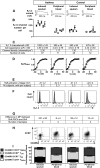
FIGURE 1.
FIGURE 1.
KV1. 3 channel expression in T lymphocytes isolated from the induced sputum and the peripheral blood of patients with asthma and healthy volunteers. A, representative whole-cell KV1.3 currents in T lymphocytes. B, KV1.3 channel number measured by whole-cell patch clamp. Each symbol represents the mean ± S.E. of 8–15 cells from a different donor. C, table detailing the values obtained in panel B. pF, picofarads. D, normalized peak K+ conductance-voltage relations fitted to a Boltzmann function to determine the half-activation voltage of the KV current. E, half-activation voltages determined in D. F, representative KV1.3 channel expression detected by staining of CD3+ cells with ShK-F6CA (black line) in cells from a donor in each group shown in A. Cells incubated with unlabeled ShK are shown in gray. G, difference in mean fluorescence intensity (MFI) for each type of samples shown in panel F. H, representative flow cytometric profile of CD3+ cells stained for CCR7 and CD45RA. I, quantification of the different subsets of T cells based on expression levels of CCR7 and CD45RA in the subjects analyzed. Data are shown as the mean ± S.E. (n = 5 subjects with asthma and 5 control subjects).
FIGURE 2.
FIGURE 2.
ShK-186 inhibits the allergen-induced proliferation of T lymphocytes isolated from the peripheral blood of patients with asthma. Mean ± S.E. of data obtained in triplicate with cells from three different patients with asthma (one per plot) are shown. *, p < 0.05 (Mann-Whitney U test).
FIGURE 3.
FIGURE 3.
ShK-186 inhibits the allergen-induced production of IL-4 by T lymphocytes isolated from the peripheral blood of patients with asthma. A, representative flow cytometric profiles of IL-4 (left), IL-5 (middle), and IL-13 (right) production in CD3+ cells without (top) and with (bottom) 100 nm ShK-186. B, percentages of CD3+IL-4+ (left), CD3+IL-5+ (middle), and CD3+IL-13+ cells; each symbol represents a different donor. **, p < 0.01; ***, p < 0.001 (Mann-Whitney U test).
FIGURE 4.
FIGURE 4.
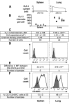
KV1. 3 channel phenotype of T lymphocytes in the lungs and spleens of rats with ovalbumin-induced asthma. A, representative whole-cell KV1.3 currents in T lymphocytes. B, KV1.3 channel number measured by whole-cell patch clamp. Each symbol represents the mean ± S.E. of a minimum of six cells from a different rat. C, table detailing the values obtained in panel B. pF, picofarads. D, representative KV1.3 channel expression detected by staining of CD3+ cells with ShK-F6CA (black lines) in single-cell suspensions. CD3-stained cells incubated with unlabeled ShK are shown in gray. E, difference in mean fluorescence intensity (MFI) for each type of samples shown in panel D. F, representative flow cytometric histograms of CD3+ cells stained for CD45RC. G, quantification of CD3+CD45RC cells. *, p < 0.05; **, p < 0.01 (Mann-Whitney U test).
FIGURE 5.
FIGURE 5.
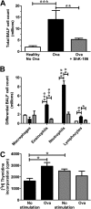
ShK-186 induced a decrease in the number of infiltrating immune cells in the BALF of rats. A, total BALF cell count. n = 8 rats per group. B, differential cell count. Rats were left untreated (white) or were immunized against ovalbumin (Ova) and received intra-tracheal ovalbumin followed by treatment with vehicle (black) or ShK-186 (gray). n = 8 rats per group. C, [3H]thymidine incorporation over a 72-h period by cells in the BALF of rats immunized and challenged against ovalbumin and treated with vehicle (black) and from rats immunized and challenged against ovalbumin and treated with ShK-186 (gray). n = 3 rats per group. *, p < 0.05; **, p < 0.01; ***, p < 0.001 (Mann-Whitney U test).
FIGURE 6.
FIGURE 6.
ShK-186 induces a decrease in the BALF concentration of IL-4 and IL-5 and an increase in the concentration of IL-10. Concentrations of IL-4 (A), IL-5 (B), IL-13 (C), and IL-10 (D) were measured in the BALF of rats by ELISA. n = 6 rats per group. *, p < 0.05; **, p < 0.01; ***, p < 0.001 (Mann-Whitney U test). Ova, ovalbumin.
FIGURE 7.
FIGURE 7.
The subcutaneous administration of ShK-186 (0. 1 mg/kg) significantly decreased RRS in response to increasing doses of acetylcholine. Rats were left untreated (open square) or were immunized against ovalbumin (Ova) and received intra-tracheal ovalbumin followed by treatment with vehicle (black square) or ShK-186 (gray circle). n = 6 rats per group. **, p < 0.01; ***, p < 0.001 (two-way analysis of variance).

References

    1. Bousquet J., Jeffery P. K., Busse W. W., Johnson M., Vignola A. M. (2000) Asthma. From bronchoconstriction to airways inflammation and remodeling. Am. J. Respir. Crit. Care Med. 161, 1720–1745 - PubMed
    1. Barnes P. J. (2010) New therapies for asthma: is there any progress? Trends Pharmacol. Sci. 31, 335–343 - PubMed
    1. Follenweider L. M., Lambertino A. (2013) Epidemiology of asthma in the United States. Nurs. Clin. North Am. 48, 1–10 - PubMed
    1. Baïz N., Annesi-Maesano I. (2012) Is the asthma epidemic still ascending? Clin. Chest Med. 33, 419–429 - PubMed
    1. Holgate S. T. (2008) Pathogenesis of asthma. Clin. Exp. Allergy 38, 872–897 - PubMed

Publication types

MeSH terms