. 2014 May 2;289(18):12922-30.
doi: 10.1074/jbc.M113.536326. Epub 2014 Mar 18.

Cleavage within Reelin repeat 3 regulates the duration and range of the signaling activity of Reelin protein

Affiliations

Cleavage within Reelin repeat 3 regulates the duration and range of the signaling activity of Reelin protein

Mari Koie et al. J Biol Chem. .

Abstract

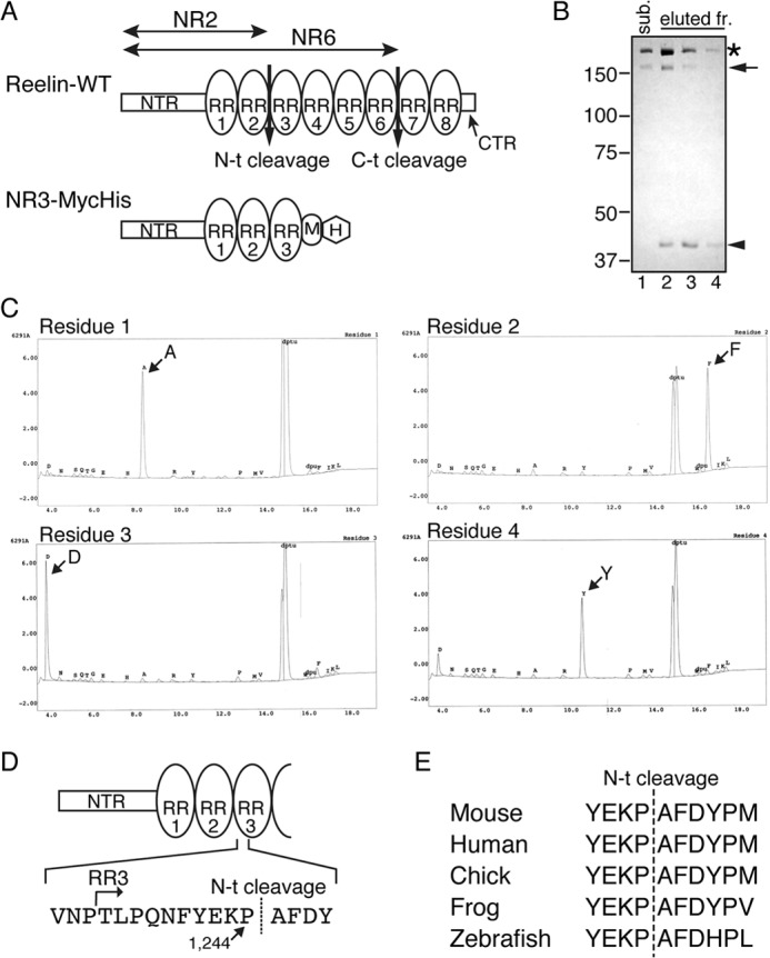
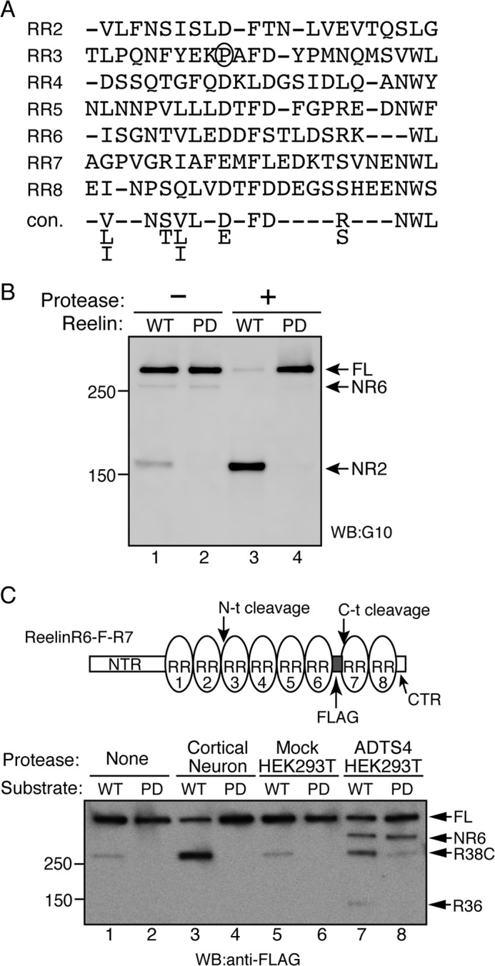
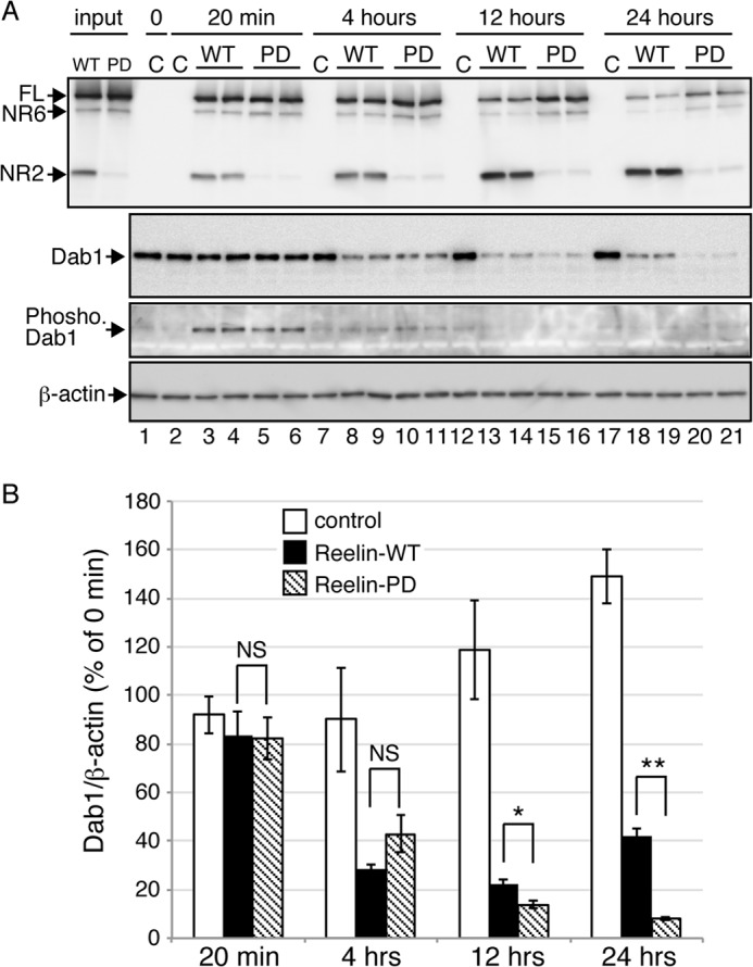
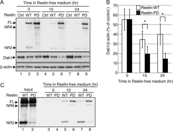
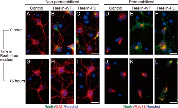
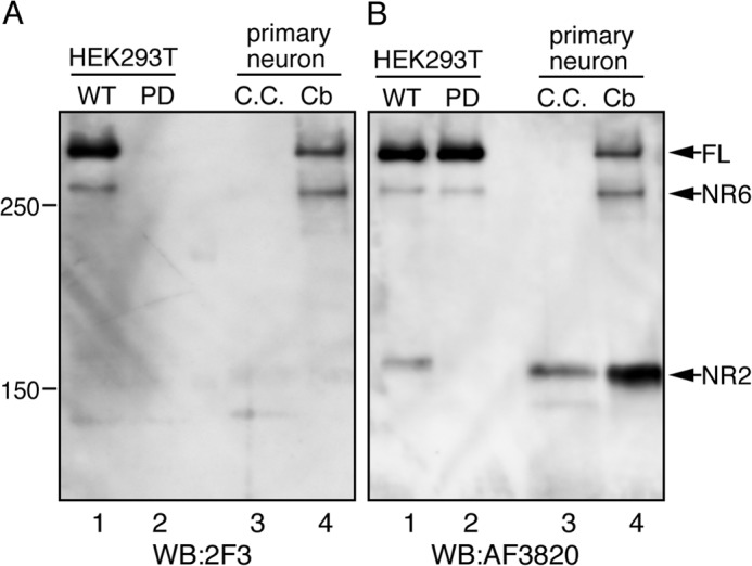
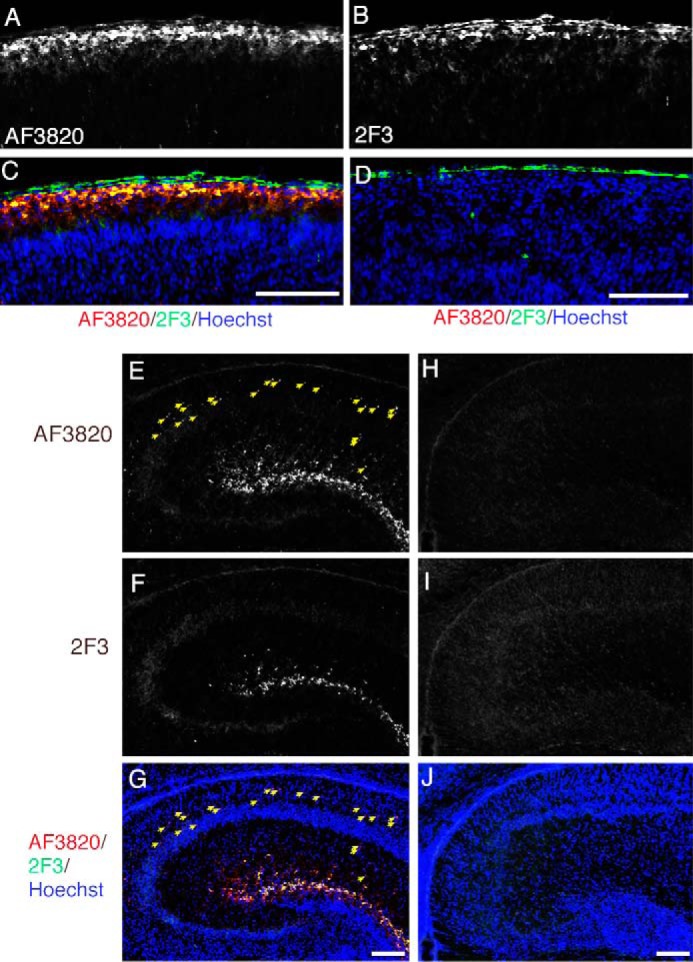
Reelin is a secreted glycoprotein that plays essential roles in the brain. Reelin is specifically cleaved at two distinct sites, called N-t and C-t, with the former being the major one. N-t cleavage can occur both in the extracellular space and in the endosomes, although the physiological importance of endosomal N-t cleavage has not been investigated. In this study, we first determined the exact N-t cleavage site catalyzed by a protease secreted by cerebral cortical neurons. Cleavage occurred between Pro-1244 and Ala-1245 within Reelin repeat 3. A Reelin mutant in which Pro-1244 was replaced with aspartate (Reelin-PD) was resistant to a protease secreted by cultured cerebral cortical neurons, and its biological activity stayed active longer than that of wild-type Reelin. Interestingly, Reelin-PD remained in the intracellular compartments longer than wild-type Reelin and persistently activated downstream signaling. Therefore, N-t cleavage of Reelin is required for halting the signaling machinery in the extracellular space as well as within endosomes of target neurons. We established a monoclonal antibody specific to uncleaved Reelin protein and found that it is localized in the vicinity of Reelin-producing cells, whereas the N-terminal fragment diffuses, or is transported, to distant regions. These data demonstrate that N-t cleavage of Reelin plays critical roles in regulating the duration and range of Reelin functions both in the extracellular milieu and in the intracellular compartments.

Keywords: ADAM ADAMTS; Brain; Dab1; Endosomes; Neurons; Protein Degradation; Reelin; Signal Transduction.


Figures

FIGURE 1.
FIGURE 1.
Determination of the Reelin N-t cleavage site. A, schematic of Reelin and the artificial substrate NR3-MycHis. Reelin-WT consists of the NTR, eight tandem RRs (oval), and the C-terminal region (CTR). NR3 consists of the NTR, RR1, RR2, and RR3, followed by a Myc epitope (M) and His6 tag (H). B, purification of the N-t cleavage product. NR3-MycHis protein was incubated with the N-t site protease fraction obtained from cultured cortical neurons, and the reaction mixture was subjected to nickel-agarose column chromatography. The substrate NR3-MycHis (sub., lane 1) and the eluted fractions (eluted fr.) from the column chromatography (lanes 2–4) were analyzed by SDS-PAGE followed by Coomassie Brilliant Blue staining. The asterisk, arrow, and arrowhead denote full-length NR3-MycHis, and the N-terminal and C-terminal products of N-t cleavage, respectively. The C-terminal product of lanes 2-4 were excised from the gel and subjected to Edman sequencing. Positions of molecular mass markers (kDa) are shown on the left. C, automatic Edman sequencing profiles of the C-terminal product of N-t cleavage. D, schematic of the N-t cleavage site and its surrounding sequence. E, comparison of the primary sequence surrounding the N-t cleavage site among various animal species.
FIGURE 2.
FIGURE 2.
Pro-1244 is necessary for N-t cleavage. A, alignment of the partial primary sequences of the RRs according to Ichihara et al. (22). The consensus sequence (con.) is provided at the bottom of the alignment. Pro-1244 of RR2 is circled. B, Pro-1244 is required for N-t cleavage. Reelin-WT (lanes 1 and 3) or its mutant, Reelin-PD (Pro-1244 substituted with aspartic acid, lanes 2 and 4), were incubated for 24 h with the control buffer (lanes 1 and 2) or partially purified N-t site protease (lanes 3 and 4). The reaction mixtures were analyzed by Western blotting (WB) using anti-Reelin NTR antibody G10. Positions of molecular mass markers (kDa) are shown on the left. FL, full-length. C, N-t cleavage by ADAMTS-4 is inhibited significantly by PD mutation. The schematic of the substrate used in this assay (ReelinR6-F-R7) is shown on the top. ReelinR6-F-R7 (WT, lanes 1, 3, 5, and 7) or its PD mutant (PD, lanes 2, 4, 6, and 8) were incubated with control buffer (None, lanes 1 and 2), the N-t protease partially purified from the culture supernatant of cortical neurons (Cortical Neuron, lanes 3 and 4), the culture supernatant of mock-transfected HEK293T cells (Mock HEK293T, lanes 5 and 6), or that of ADAMTS-4-transfected HEK293T cells (ADTS4 HEK293T, lanes 7 and 8) at 37 °C for 24 h. The reaction mixtures were separated by SDS-PAGE and analyzed by Western blotting with anti-FLAG antibody. Positions of full-length substrate (FL), NR6, R38C, and R36 fragments are indicated by arrows. Positions of molecular mass markers (kDa) are shown at the left.
FIGURE 3.
FIGURE 3.
The biological activity of Reelin-PD is sustained longer than that of Reelin-WT. A, primary cortical neurons from reeler mice were incubated with culture medium from control mock-transfected cells (C), Reelin-WT-expressing cells (WT), or Reelin-PD-expressing cells (PD) for the times indicated at the top. Cells were then lysed in SDS-PAGE sample buffer and analyzed by Western blotting. The two left lanes (input) represent the culture medium used to stimulate the neurons. At each time point, two wells of neurons were used for Reelin treatment. B, quantification of the amount of Dab1 protein. Open, black, and hatched bars indicate neurons treated with control, Reelin-WT, and Reelin-PD, respectively. Band intensities were quantified using ImageJ and normalized to those of β-actin. Data are mean ± S.E. and were analyzed using Mann-Whitney U test (n = 8). *, p < 0.05; **, p < 0.01; NS, not significant.
FIGURE 4.
FIGURE 4.
N-t cleavage is required for intracellular degradation of Reelin and halting of the downstream signaling. A, primary cerebral cortical neurons from reeler mice were incubated with control medium (Ctrl, lanes 1, 4, and 7), medium containing Reelin-WT (WT, lanes 2, 5, and 8), or medium containing Reelin-PD (PD, lanes 3, 6, and 9) for 3 h. The neurons were further incubated with Reelin-free medium for 0 (lanes 1, 2, and 3), 15 (lanes 4, 5, and 6), or 24 h (lanes 7, 8, and 9), lysed with SDS-PAGE sample buffer, and analyzed by Western blotting. Anti-Reelin NTR G10 was used for detection of Reelin. FL, full-length. B, quantification of the amount of Dab1 protein. Open and black bars indicate neurons treated with Reelin-WT and Reelin-PD, respectively. Band intensities were quantified using ImageJ and normalized to β-actin. Data are mean ± S.E. and were analyzed using Mann-Whitney U test (n = 4). *, p < 0.05. C, a small amount of Reelin-WT and Reelin-PD is resecreted from neurons. Cortical neurons from reeler mice were incubated with either Reelin-WT (lanes 3, 5, and 7) or Reelin-PD (lanes 4, 6, and 8) for 3 h. The neurons were further incubated for 0 (lanes 3 and 4), 15 (lanes 5 and 6), or 24 h (lanes 7 and 8), and the culture supernatants were collected and analyzed by Western blotting with anti-Reelin NTR G10. The amount of resecreted Reelin is very low compared with that in the original culture medium (Input, lanes 1 and 2).
FIGURE 5.
FIGURE 5.
Internalized Reelin-PD is retained in the intracellular compartments longer than Reelin-WT. Primary cerebral cortical neurons from reeler mice were incubated with control medium (A, D, G, and J), medium containing Reelin-WT (B, E, H, and K), or medium containing Reelin-PD (C, F, I, and L) for 3 h. They were further incubated with Reelin-free medium for 0 (A–F) or 15 h (G–L). A–C and G–I, cells were fixed and stained with anti-Reelin NTR AF3820 (green) without membrane permeabilization. Biotinylated cholera toxin B subunit (red) and Hoechst33342 nucleus staining (blue) were used to visualize the neuronal membrane and nucleus, respectively. D–F and J–L, cells were fixed, permeabilized, and stained with AF3820 (green) and anti-Rab11 (red). Nuclei were stained with Hoechst33342 (blue). Scale bars = 20 μm.
FIGURE 6.
FIGURE 6.
Establishment of a monoclonal antibody that specifically recognizes Reelin protein not cleaved at the N-t site. The culture supernatant of HEK293T cells expressing Reelin-WT (WT, lane 1) or Reelin-PD (PD, lane 2), mouse cerebral cortical primary neurons (C.C., lane 3), and mouse cerebellar primary granule cells (Cb, lane 4) were analyzed by Western blotting (WB) with 2F3 antibody (A) and then reprobed with AF3820 antibody (B). FL, full-length.
FIGURE 7.
FIGURE 7.
Localization of full-length and cleaved Reelin. Frozen sections from the cerebral cortices of postnatal day 1 (P1) wild-type (A–C) or reeler (D) mice were immunostained with AF3820 (A) and mAb 2F3 (B). C, merged image of A (red), B (green), and Hoechst33342 nucleus staining (blue). D, merged image of signals from AF3820 (red), mAb 2F3 (green), and Hoechst33342 (blue) staining. The pia mater gave a background staining when mAb 2F3 was used (the green signals outside of the brain in C and D). E–J, frozen sections of hippocampi of P1 wild-type (E–G) or reeler (H–J) mice were coimmunostained with AF3820 (E and H) and mAb 2F3 (F and I). The merged images with Hoechst33342 nucleus staining (blue) are shown in G and J. The yellow arrows indicate punctate signals that are positive for AF3820 but not 2F3. Scale bars = 100 μm.

Similar articles

Cited by

References

    1. Tissir F., Goffinet A. M. (2003) Reelin and brain development. Nat. Rev. Neurosci. 4, 496–505 - PubMed
    1. Tiberi L., Vanderhaeghen P., van den Ameele J. (2012) Cortical neurogenesis and morphogens: diversity of cues, sources and functions. Curr. Opin. Cell Biol. 24, 269–276 - PubMed
    1. Park H., Poo M. M. (2013) Neurotrophin regulation of neural circuit development and function. Nat. Rev. Neurosci. 14, 7–23 - PubMed
    1. Baker K. A., Moore S. W., Jarjour A. A., Kennedy T. E. (2006) When a diffusible axon guidance cue stops diffusing: roles for netrins in adhesion and morphogenesis. Curr. Opin. Neurobiol. 16, 529–534 - PubMed
    1. Rogers K. W., Schier A. F. (2011) Morphogen gradients: from generation to interpretation. Annu. Rev. Cell Dev. Biol. 27, 377–407 - PubMed

Publication types

MeSH terms