

Are spinal or paraspinal anatomic markers helpful for vertebral numbering and diagnosing lumbosacral transitional vertebrae?
- PMID: 24644411
- PMCID: PMC3955794
- DOI: 10.3348/kjr.2014.15.2.258
Are spinal or paraspinal anatomic markers helpful for vertebral numbering and diagnosing lumbosacral transitional vertebrae?
Abstract
Objective: To evaluate the value of spinal and paraspinal anatomic markers in both the diagnosis of lumbosacral transitional vertebrae (LSTVs) and identification of vertebral levels on lumbar MRI.
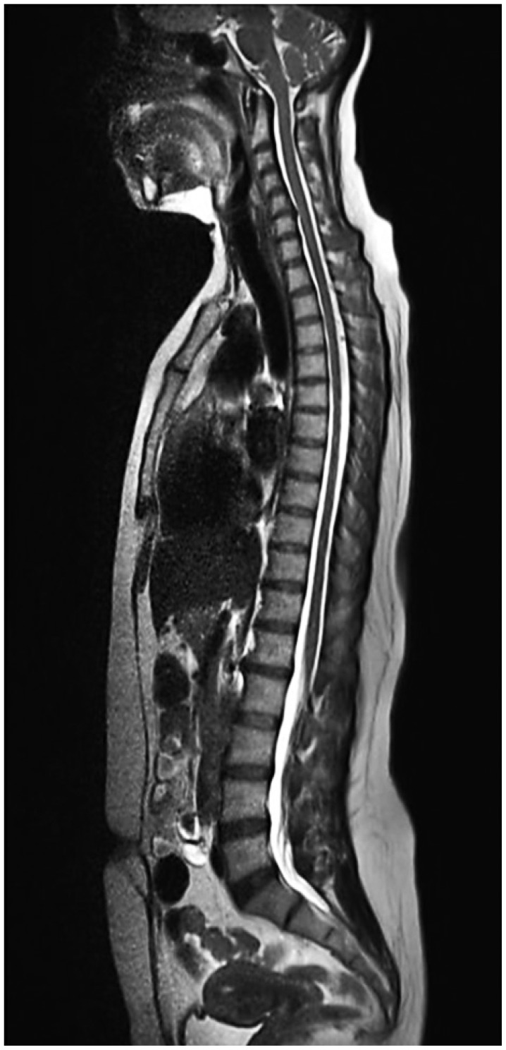
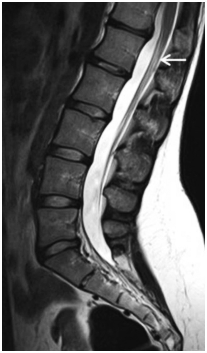
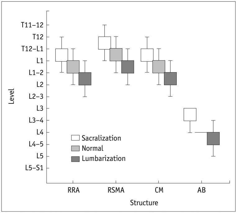
Materials and methods: Lumbar MRI from 1049 adult patients were studied. By comparing with the whole-spine localizer, the diagnostic errors in numbering vertebral segments on lumbar MRI were evaluated. The morphology of S1-2 disc, L5 and S1 body, and lumbar spinous processes (SPs) were evaluated by using sagittal MRI. The positions of right renal artery (RRA), superior mesenteric artery, aortic bifurcation (AB) and conus medullaris (CM) were described.
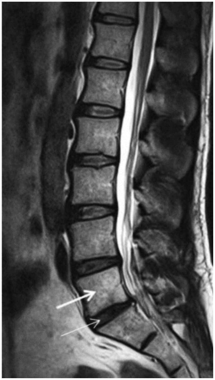
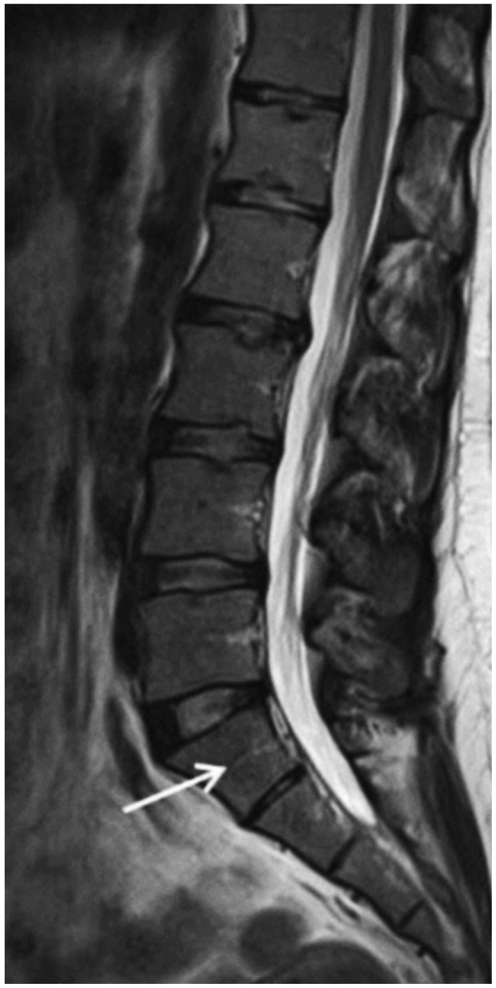
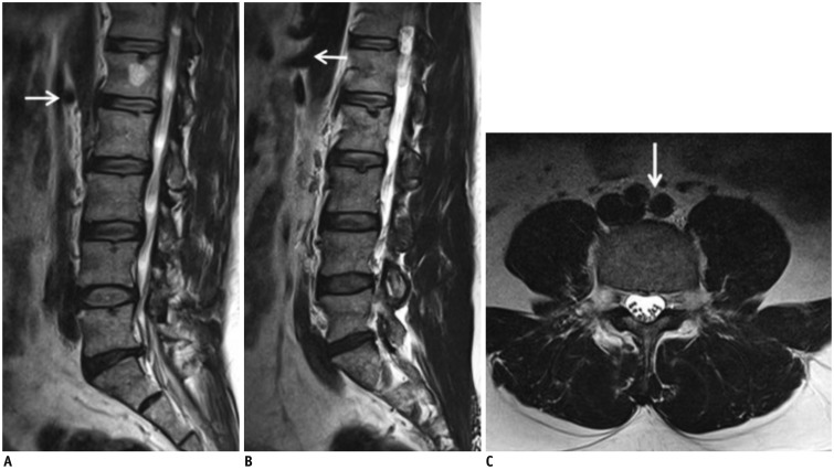
Results: The diagnostic error for evaluation of vertebral segmentation on lumbar MRI alone was 14.1%. In lumbarization, all patients revealed a well-formed S1-2 disc with squared S1 body. A rhombus-shaped L5 body in sacralization and a rectangular-shaped S1 body in lumbarization were found. The L3 had the longest SP. The most common sites of spinal and paraspinal structures were: RRA at L1 body (53.6%) and L1-2 disc (34.1%), superior mesenteric artery at L1 body (55.1%) and T12-L1 disc (31.6%), and AB at L4 body (71.1%). CM had variable locations, changing from the T12-L1 disc to L2 body. They were located at higher sacralization and lower lumbarization.
Conclusion: The spinal morphologic features and locations of the spinal and paraspinal structures on lumbar MRI are not completely reliable for the diagnosis of LSTVs and identification on the vertebral levels.
Keywords: Lumbosacral vertebrae; MRI; Spine; Transitional vertebrae.
Figures
Similar articles
-
A review of lumbosacral transitional vertebrae and associated vertebral numeration.Eur Spine J. 2018 May;27(5):995-1004. doi: 10.1007/s00586-018-5554-8. Epub 2018 Mar 21. Eur Spine J. 2018. PMID: 29564611 Review.
-
Using MRI to evaluate anatomic significance of aortic bifurcation, right renal artery, and conus medullaris when locating lumbar vertebral segments.AJR Am J Roentgenol. 2004 May;182(5):1295-300. doi: 10.2214/ajr.182.5.1821295. AJR Am J Roentgenol. 2004. PMID: 15100135
-
Is any landmark reliable in vertebral enumeration? A study of 3.0-Tesla lumbar MRI comparing skeletal, neural, and vascular markers.Clin Imaging. 2014 Nov-Dec;38(6):792-6. doi: 10.1016/j.clinimag.2014.05.001. Epub 2014 May 12. Clin Imaging. 2014. PMID: 24928822
-
The anatomical relationship of the aortic bifurcation to the lumbar vertebrae: a MRI study.Surg Radiol Anat. 2002 Dec;24(5):308-12. doi: 10.1007/s00276-002-0036-3. Epub 2002 Jul 20. Surg Radiol Anat. 2002. PMID: 12497222
-
Lumbosacral Transitional Segments: An Interventional Spine Specialist's Practical Approach.Phys Med Rehabil Clin N Am. 2018 Feb;29(1):35-48. doi: 10.1016/j.pmr.2017.08.004. Phys Med Rehabil Clin N Am. 2018. PMID: 29173663 Review.
Cited by
-
Lumbar plain radiograph is not reliable to identify lumbosacral transitional vertebra types according to Castellvi classification principle.BMC Musculoskelet Disord. 2020 May 29;21(1):333. doi: 10.1186/s12891-020-03358-3. BMC Musculoskelet Disord. 2020. PMID: 32471475 Free PMC article.
-
Lumbosacral Vertebral Angles can Predict Lumbosacral Transitional Vertebrae on Routine Sagittal MRI.Arch Bone Jt Surg. 2025;13(5):271-280. doi: 10.22038/ABJS.2025.83244.3790. Arch Bone Jt Surg. 2025. PMID: 40630817 Free PMC article.
-
Role of iliac crest tangent in correct numbering of lumbosacral transitional vertebrae.Turk J Med Sci. 2019 Feb 11;49(1):184-189. doi: 10.3906/sag-1807-258. Turk J Med Sci. 2019. PMID: 30764596 Free PMC article.
-
A review of lumbosacral transitional vertebrae and associated vertebral numeration.Eur Spine J. 2018 May;27(5):995-1004. doi: 10.1007/s00586-018-5554-8. Epub 2018 Mar 21. Eur Spine J. 2018. PMID: 29564611 Review.
-
Normative spino-pelvic parameters in patients with the lumbarization of S1 compared to a normal asymptomatic population.Eur Spine J. 2016 Nov;25(11):3694-3698. doi: 10.1007/s00586-016-4794-8. Epub 2016 Sep 26. Eur Spine J. 2016. PMID: 27671281
References
-
- Hughes RJ, Saifuddin A. Numbering of lumbosacral transitional vertebrae on MRI: role of the iliolumbar ligaments. AJR Am J Roentgenol. 2006;187:W59–W65. - PubMed
-
- Hughes RJ, Saifuddin A. Imaging of lumbosacral transitional vertebrae. Clin Radiol. 2004;59:984–991. - PubMed
-
- Lee CH, Park CM, Kim KA, Hong SJ, Seol HY, Kim BH, et al. Identification and prediction of transitional vertebrae on imaging studies: anatomical significance of paraspinal structures. Clin Anat. 2007;20:905–914. - PubMed
-
- Carrino JA, Campbell PD, Jr, Lin DC, Morrison WB, Schweitzer ME, Flanders AE, et al. Effect of spinal segment variants on numbering vertebral levels at lumbar MR imaging. Radiology. 2011;259:196–202. - PubMed
Publication types
MeSH terms
LinkOut - more resources
Full Text Sources
Other Literature Sources
