. 2014 Mar 20;14(1):26.
doi: 10.1186/1475-2867-14-26.

Prostate specific membrane antigen (PSMA) expression in primary gliomas and breast cancer brain metastases

Affiliations

Prostate specific membrane antigen (PSMA) expression in primary gliomas and breast cancer brain metastases

Natsuko Nomura et al. Cancer Cell Int. .

Abstract

Background: Primary and secondary brain cancers are highly treatment resistant, and their marked angiogenesis attracts interest as a potential therapeutic target. Recent observations reveal that the microvascular endothelium of primary high-grade gliomas expresses prostate specific membrane antigen (PSMA). Breast cancers express PSMA and they frequently form secondary brain tumors. Hence we report here our pilot study addressing the feasibility of PSMA targeting in brain and metastatic breast tumors, by examining PSMA levels in all glioma grades (19 patients) and in breast cancer brain metastases (5 patients).

Methods: Tumor specimens were acquired from archival material and normal brain tissues from autopsies. Tissue were stained and probed for PSMA, and the expression levels imaged and quantified using automated hardware and software. PSMA staining intensities of glioma subtypes, breast tumors, and breast tumor brain metastases were compared statistically versus normals.

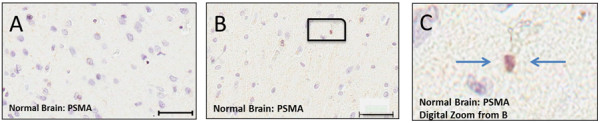
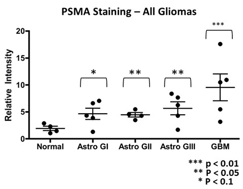
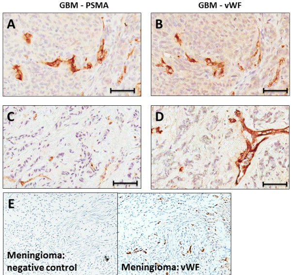
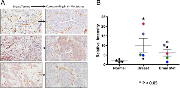
Results: Normal brain microvessels (4 autopsies) did not stain for PSMA, while a small proportion (<5%) of healthy neurons stained, and were surrounded by an intact blood brain barrier. Tumor microvessels of the highly angiogenic grade IV gliomas showed intense PSMA staining which varied between patients and was significantly higher (p < 0.05) than normal brain. Grade I gliomas showed moderate vessel staining, while grade II and III gliomas had no vessel staining, but a few (<2%) of the tumor cells stained. Both primary breast cancer tissues and the associated brain metastases exhibited vascular PSMA staining, although the intensity of staining was generally less for the metastatic lesions.

Conclusions: Our results align with and extend previous data showing PSMA expression in blood vessels of gliomas and breast cancer brain metastases. These results provide a rationale for more comprehensive studies to explore PSMA targeted agents for treating secondary brain tumors with PSMA expressing vasculature. Moreover, given that PSMA participates in angiogenesis, cell signaling, tumor survival, and invasion, characterizing its expression may help guide later investigations of the poorly understood process of low grade glioma progression to glioblastoma.


Figures

Figure 1
Figure 1
PSMA staining in tissues samples representative of normal brain. A – Normal brain showing no PSMA staining. B – Normal brain probed for PSMA showing no blood vessel staining and light staining of cellular elements, magnified digitally in (C) and indicated by arrows.
Figure 2
Figure 2
PSMA staining of grades I – IV glioma tissue Sections. A - PSMA staining of gliomas grades (I – IV). Note moderate staining of blood vessels and some cells in grade I, only light staining of tumor cells in grades II and III, and heavy blood vessel staining of GBM (grade IV). The bottom two staining control slides show adjacent GBM sections, one stained with the primary antibody for PSMA and the other, the negative control, which was fully stained including the secondary antibody, but with the primary antibody omitted. B – PSMA staining for astrocytoma grade I and grade IV (GBM). Each section represents a different patient. Arrows indicate cell staining in grade I tumor tissue Section. C – PSMA staining of grade II and III glioma. There is some staining of cells only in grade I and although there is more staining in grade III it is also restricted only to cells, with no blood vessel staining. Size bars in all panels indicate approximately 50 μm, except in negative control sections where they indicate 100 μm.
Figure 3
Figure 3
Summary of PSMA Data. Graph showing data points, one per patient tumor, and the mean (middle bars) and standard error (outer bars) of PSMA relative staining intensity (normalized to image mean background intensity) for astrocytomas of grades I – IV. Unpaired t–test both parametric and nonparametric forms, p values are indicated.
Figure 4
Figure 4
Correspondence between PSMA and VWF staining in GBM blood vessels. A and B - Adjacent GBM (glioma grade IV) sections stained for PSMA (A) and VWF (B). Note that these sections exhibit similar patterns of highlighted blood vessels. The two bottom panels, (C) (PSMA) and (D) (VWF) exhibit dissimilar staining patterns and tumor cells are stained for PSMA. (E) This panel displays two adjacent tissue sections taken for staining control purposes from a meningioma. In the left section the full staining procedure was used except that the primary antibody for VFW was omitted (negative control) and VFW staining is absent, while in the adjacent, right section the primary antibody was included and VFW staining is present. Bars in A - D indicate approximately 50 μm.
Figure 5
Figure 5
PSMA Staining in Breast Cancer and Brain Metastases. A – PSMA staining for breast tumors and breast tumor brain metastases pairs. Each pair of sections is from a different breast tumor case (n = 3). Size bars in all panels indicate approximately 50 μm. B – Graph of PSMA staining intensity for primary breast tumors and for breast tumor brain metastases, showing individual patient values and group mean (bars). The color coding indicates those primary breast tumor and brain metastases that came from the same patient (n = 4). Although there was considerable variability, each group included individual PSMA staining data points that were higher than the largest values for normal brain staining in terms of PSMA, these data were significant (p < 0.05), and for 3 of 4 patients staining intensity was less for breast cancer brain metastases than for the primary breast tumors.

References

    1. Wen PY, Kesari S. Malignant gliomas in adults. N Engl J Med. 2008;359:492–507. - PubMed
    1. Jones C, Perryman L, Hargrave D. Paediatric and adult malignant glioma: close relatives or distant cousins? Nat Rev Clin Oncol. 2012;9:400–413. - PubMed
    1. Linnert M, Iversen HK, Gehl J. Multiple brain metastases - current management and perspectives for treatment with electrochemotherapy. Radiol Oncol. 2012;46:271–278. - PMC - PubMed
    1. Gavrilovic IT, Posner JB. Brain metastases: epidemiology and pathophysiology. J Neurooncol. 2005;75:5–14. - PubMed
    1. Eichler AF, Loeffler JS. Multidisciplinary management of brain metastases. Oncologist. 2007;12:884–898. - PubMed