Review
. 2014 Apr;87(1036):20130684.
doi: 10.1259/bjr.20130684.

Multimodality imaging of primary CNS lymphoma in immunocompetent patients

Review

Multimodality imaging of primary CNS lymphoma in immunocompetent patients

S Partovi et al. Br J Radiol. 2014 Apr.

Abstract

Primary central nervous system lymphoma (PCNSL) belongs to the group of extranodal non-Hodgkin's lymphoma, and the management of the disease is radically different from other central nervous system neoplasms. Owing to its varied appearance on imaging, diagnosis of PCNSL can be challenging. The purpose of this pictorial review is to depict the brain findings of PCNSL during initial diagnosis in immunocompetent individuals. Multimodal imaging integrating advanced sequences can facilitate differentiation of PCNSL from other CNS neoplasms.


Figures
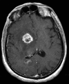
Figure 1.
Figure 1.
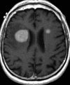
A 56-year-old male with confirmed diffuse large B-cell lymphoma of the central nervous system. Axial T1 post-contrast image shows an enhancing right basal ganglia mass. Also, a periventricular lesion just adjacent to the right trigone at the edge of the corpus callosum can be appreciated.
Figure 2.
Figure 2.
A 90-year-old male with colon cancer presenting with word finding difficulties. Biopsy showed malignant T-cell primary central nervous system lymphoma. (a) Axial T2 shows an expansive mass in the left thalamus. (b) T1 post-contrast image shows no significant enhancement in the lesion.
Figure 3.
Figure 3.
An 80-year-old male with diffuse large B-cell lymphoma. Axial T1 post-contrast image shows enhancing masses in the frontal lobes.
Figure 4.
Figure 4.
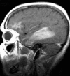
A 55-year-old female with progressive cognitive changes. Biopsy revealed diffuse large B-cell lymphoma. Sagittal T1 post-contrast scan showing multifocal enhancement of the right frontal and temporal lobe white matter.
Figure 5.
Figure 5.
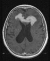
A 65-year-old female with diffuse large B-cell lymphoma. Axial T1 post-contrast image shows an enhancing mass in the anterior corpus callosum.
Figure 6.
Figure 6.
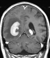
A 72-year-old male with memory loss and confusion due to diffuse large B-cell lymphoma. Coronal T1 post-contrast image shows an enhancing mass around the right occipital horn.
Figure 7.
Figure 7.
A 58-year-old male with headaches and vertigo. Biopsy showed diffuse large B-cell lymphoma. Sagittal T1 post-contrast image shows an enhancing mass within or along the fourth ventricle causing obstructive hydrocephalus.
Figure 8.
Figure 8.
A 60-year-old female with mild headaches and clumsiness resulting from primary central nervous system lymphoma. Sagittal T1 post-contrast image demonstrating a mass along the superficial left parietal cortex, which abuts the overlying dura and mimics an extra-axial mass. During surgery, parts of the mass were found to be stuck to the pia surface.
Figure 9.
Figure 9.
A 74-year-old male with seizures and high-grade diffuse large B-cell lymphoma with systemic manifestation. Coronal T1 post-contrast image shows diffuse folial enhancement sugar-coating appearance of leptomeningeal disease.
Figure 10.
Figure 10.
An 82-year-old female with decreased vision and swelling of left eye. Biopsy showed malignant marginal zone type orbital lymphoma. (a) Axial T1 non-contrast image showing an expansive mass in the left orbit and infiltrating the orbital apex, with proptosis. (b) Coronal T1 post-contrast image shows that the mass enhances.
Figure 11.
Figure 11.
A 76-year-old male with confusion. Cerebrospinal fluid sampling showed cells highly suspicious for lymphoma of the central nervous system. Coronal T1 post-contrast image shows infiltration and expansive enhancement of the bilateral pre-chiasmatic optic nerves.
Figure 12.
Figure 12.
A 47-year-old female with seizures who has diffuse large B-cell lymphoma. (a) Sagittal T1 pre-contrast and (b) sagittal T1 post-contrast scans show an enhancing haemorrhagic mass with pre-contrast T1 hyperintense signal haemorrhage within the lesion. (c) Axial T2 sequence shows low intensity of the haemorrhage and high signal of surrounding oedema.
Figure 13.
Figure 13.
(a) Fractional anisotropy and (b) coloured fractional anisotropy map of the same patient show lack of anisotropy of the tumour.
Figure 14.
Figure 14.
(a) Plasma volume and (b) K-trans or permeability maps from dynamic contrast-enhanced perfusion imaging demonstrate increased perfusion and permeability of the tumour.
Figure 15.
Figure 15.
(a) Non-contrast CT of a patient with primary central nervous system lymphoma demonstrates an iso- to slightly hyperdense mass in the medial left temporal lobe extending to the corpus callosum with surrounding oedema. (b) Revealing avid enhancement. (c) The tumour is hypointense in the T2 weighted MRI image owing to high cellularity and (d) with homogeneous intense enhancement on the contrast-enhanced T1 weighted MR image. (e) The tumour is hyperintense in the diffusion-weighted image and (g) the exponential map with (f) decreased signal on the apparent diffusion map.
Figure 16.
Figure 16.
(a) Axial contrast T1 weighted image and (b) T2 weighted image 3 weeks after the start of systemic treatment demonstrate marked decrease in the enhancing mass with resolution of oedema and mass effect. Note that the residual abnormality no longer demonstrates low signal intensity (compare with Figure 15c).
Figure 17.
Figure 17.
18Fluorodeoxyglucose (FDG)–positron emission tomography image reveals marked FDG avidity owing to high metabolic activity of the tumour.

References

    1. Watanabe M, Tanaka R, Takeda N, Wakabayashi K, Takahashi H. Correlation of computed tomography with the histopathology of primary malignant lymphoma of the brain. Neuroradiology 1992; 34: 36–42. - PubMed
    1. Shenkier TN, Blay JY, O'Neill BP, Poortmans P, Thiel E, Jahnke K, et al. . Primary CNS lymphoma of T-cell origin: a descriptive analysis from the international primary CNS lymphoma collaborative group. J Clin Oncol 2005; 23: 2233–9. doi: 10.1200/JCO.2005.07.109 - DOI - PubMed
    1. Olson JE, Janney CA, Rao RD, Cerhan JR, Kurtin PJ, Schiff D, et al. . The continuing increase in the incidence of primary central nervous system non-Hodgkin lymphoma: a surveillance, epidemiology, and end results analysis. Cancer 2002; 95: 1504–10. doi: 10.1002/cncr.10851 - DOI - PubMed
    1. Bessell EM, Hoang-Xuan K, Ferreri AJ, Reni M. Primary central nervous system lymphoma: biological aspects and controversies in management. Eur J Cancer 2007; 43: 1141–52. doi: 10.1016/j.ejca.2006.12.011 - DOI - PubMed
    1. Bataille B, Delwail V, Menet E, Vandermarcq P, Ingrand P, Wager M, et al. . Primary intracerebral malignant lymphoma: report of 248 cases. J Neurosurg 2000; 92: 261–6. doi: 10.3171/jns.2000.92.2.0261 - DOI - PubMed