Review
. 2014 Apr;15(4):217-32.
doi: 10.1038/nrn3707.

Growth and folding of the mammalian cerebral cortex: from molecules to malformations

Affiliations
Review

Growth and folding of the mammalian cerebral cortex: from molecules to malformations

Tao Sun et al. Nat Rev Neurosci. 2014 Apr.

Abstract

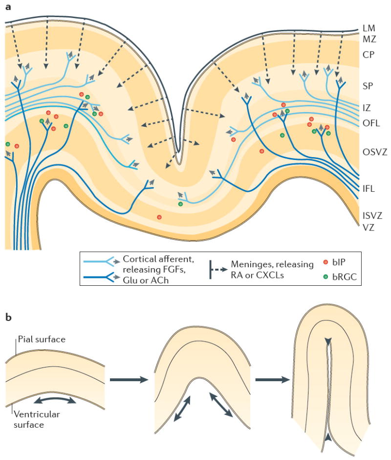
The size and extent of folding of the mammalian cerebral cortex are important factors that influence a species' cognitive abilities and sensorimotor skills. Studies in various animal models and in humans have provided insight into the mechanisms that regulate cortical growth and folding. Both protein-coding genes and microRNAs control cortical size, and recent progress in characterizing basal progenitor cells and the genes that regulate their proliferation has contributed to our understanding of cortical folding. Neurological disorders linked to disruptions in cortical growth and folding have been associated with novel neurogenetic mechanisms and aberrant signalling pathways, and these findings have changed concepts of brain evolution and may lead to new medical treatments for certain disorders.


Conflict of interest statement

Competing interests statement

The authors declare no competing interests.

Figures

Figure 1
Figure 1. Multiple progenitors in the mouse and human developing cerebral cortex
Progenitors are termed according to their apical or basal position in relation to the apical (ventricular) surface of the developing cortex. Apical radial glial cells (aRGCs) and apical intermediate progenitors (aIPs) reside in the ventricular zone (VZ). Basal IPs (bIPs) are mostly positioned in the subventricular zone (SVZ) in the mouse cortex and in the inner SVZ (ISVZ) in the human cortex. Basal RGCs (bRGCs) are identified in the SVZ in the mouse cortex and the outer SVZ (OSVZ) in the human cortex. Migrating neurons mostly appear in the intermediate zone (IZ), whereas mature neurons form an inside-out six-layered structure in the cortical plate (CP). Cortical surface expansion in the human cortex results in folded structures called sulci and gyri. The ISVZ and OSVZ are histologically separated by an inner fibre layer. It is unclear how the cytoarchitectonically distinct OSVZ and the characteristics of bRGCs in the OSVZ are related to cortical size and folding.
Figure 2
Figure 2. Molecular mechanisms of cortical growth
Various mechanisms regulate neural progenitor (NP) expansion and cortical size. a | The expansion of radial glial cells (RGCs) in the ventricular zone (VZ) is dependent on interkinetic nuclear migration (INM) within these cells, and various genes have been identified that regulate this process. In INM, the nuclei of VZ progenitors (RGCs) are positioned at basal (abventricular) locations in the VZ during the S phase of the cell cycle and move towards the apical surface (the ventricle side) during the G2 phase. Subsequently, they undergo mitosis (the M phase) at the apical (ventricular) surface and return towards basal positions in the G1 phase. b | NP proliferation is controlled by genes that regulate the cell cycle length (Pax6 and filamin A (Flna)) and cell cycle exit (Rac1, cyclin D2 (Ccnd2), numb homologue (Numb) and numb-like (Numbl)). c | Genes that are associated with primary cilia have direct or indirect roles in the cell cycle control of NPs. A mutation in Tg737 causes dysfunctional ciliogenesis. Trafficking of molecules such as sonic hedgehog (SHH) and WNT by intraflagellar transport (IFT) relies on proper ciliogenesis. Knockout of ADP-ribosylation factor-like 13B (Arl13b), which encodes a protein that is abundant in cilia, causes reversal of the apical–basal polarity of RGCs in the mouse cortex. TCTEX-type 1 (Tctex1) and insulin-like growth factor 1 receptor (Igf1r) encode proteins that are directly involved in both ciliary disassembly and cell cycle re-entry. d | NP apoptosis and survival are regulated by multiple genes and are crucial for cortical size control. Whereas overexpression of Notch1, Pax6 or ephrin A5 (Efna5) or knockout (KO) of breast cancer 1 (Brca1), Fanconi anaemia complementation group A (Fanca) or Fancg promotes apoptosis of NPs, the addition of lysophosphatidic acid (LPA; a mitogen) or knockout of ephrin receptor type A receptor 7 (Epha7), caspase 3 (Casp3) or Casp9 increases the survival of NPs. e | MicroRNAs (miRNAs) are essential for regulating cortical size. Ablation of Dicer, an enzyme that processes miRNA precursors, in mice using Emx1-Cre or Nex-Cre lines results in smaller cortices, (left and middle panels). Mice in which the specific miRNA cluster miR-17-92 is knocked out also exhibit a small cortex (right panel). Cep120, centrosomal protein 120; Dock7, dedicator of cytokinesis 7; Hook3; hook microtubule-tethering protein 3; P10, postnatal day 10; Sun, SUN-domain-containing. The middle panel of part e is reproduced from REF. . The right-hand panel of part e is reproduced, with permission, from REF. © (2013) Elsevier.
Figure 3
Figure 3. Symmetrical–asymmetrical cell division, centrosome associated proteins and neural progenitors
a | According to spindle orientation and cleavage plane, neural progenitor (NP) divisions can be classified as being vertical (that is, symmetrical), oblique or horizontal (that is, asymmetrical). Various gene mutations can affect the orientations of cleavage planes, symmetrical versus asymmetrical division of NPs and, in turn, cortical size. Loss of lissencephaly 1 (Lis1) or protein phosphatase 4 catalytic subunit (Pp4c) in NPs disrupts vertical division, and loss of nuclear distribution E homologue 1 (Nde1) promotes horizontal division. Inactivation of Lgn or inscuteable (Insc) promotes or decreases oblique division, respectively. Loss of Tcof1, Mals3, Pax6, Staufen homologue 2 (Stau2) or Gβγ disrupts asymmetrical division in NPs. b | All identified autosomal recessive primary microcephaly (MCPH)-linked genes encode centrosome-associated proteins, and the mutations in these genes affect proper divisions of NPs and cause microcephaly. ASPM, abnormal spindle-like microcephaly-associated; CDK5RAP2, cyclin-dependent kinase 5 regulatory subunit-associated protein 2; WDR62, WD-repeat-containing protein 62.
Figure 4
Figure 4. Brain mass, cortical thickness and gyrification
a | The mouse brain is small (0.65 g) and is characterized by a smooth neocortex (gyrification index (GI) = 1.03). The thickness of the mouse cortex is indicated by the colour scale. b | By contrast, the human brain is considerably larger (1,230 g), and its cortex is gyrified (GI=2.56). Its cortical thickness also varies but is, on average, thicker than that of mice. c | The GI generally increases with brain mass across mammalian species for which data are available (n = 103). Manatees (Trichechus manatus (T.m.)) and humans (Homo sapiens (H.s.)) have relatively low GI scores for their brain mass compared with other mammalian species, such as the Atlantic bottlenose dolphin (Tursiops truncates (T.t.)). Other mammalian species not mentioned here are denoted by the blue data points. d | Cortical thickness varies little with brain mass across species for which data are available (n = 42), except in manatees and humans, which have unusually thick cerebral cortices on average. Part a is reproduced, with permission, from REF. © (2008) Elsevier. Part b is modified, with permission, from REF. © (2005) Oxford Journals. Data for panels c and d from REFS 4,154,157.
Figure 5
Figure 5. Cortical afferent axons and the meninges in gyrogenesis
a | Afferent axons (dark blue and light blue, representing different fibre pathways) arrive in the cortex from many sources (including the thalamus, nucleus basalis, brainstem and contralateral cortex) while neurogenesis is still ongoing. Some axons may release neurotransmitters, such as glutamate (Glu) and acetylcholine (ACh) as well as peptides such as fibroblast growth factors (FGFs); these factors may influence gyrogenesis by regulating basal radial glial cell (bRGC) and basal intermediate progenitor (bIP) proliferation, and by contributing to neuropile growth. Indeed, the crowns of gyri receive more innervation than do sulcal depths. The leptomeninges (LM) (pia–arachnoid mater) produce additional diffusible factors, such as retinoic acid (RA) and chemokines (CXCLs), that can affect progenitor proliferation, cell migration and layer formation. b | Ventricular surface growth (arrows) and buckling can lead to obliteration of the enclosed ventricular recess (arrowheads), for example, during formation of the parahippocampal gyrus and some occipital gyri. CP, cortical plate; IFL, inner fibrous layer; ISVZ, inner subventricular zone; IZ, intermediate zone; MZ, marginal zone; OFL, outer fibrous layer; OSVZ, outer subventricular zone; SP, subplate; VZ, ventricular zone.

References

    1. Kaas JH. The evolution of brains from early mammals to humans. Wiley Interdiscip Rev Cogn Sci. 2013;4:33–45. - PMC - PubMed
    1. Geschwind DH, Rakic P. Cortical evolution: judge the brain by its cover. Neuron. 2013;80:633–647. - PMC - PubMed
    1. Striedter G. Principles of Brain Evolution. Sinauer; 2005.
    1. Zilles K, Palomero-Gallagher N, Amunts K. Development of cortical folding during evolution and ontogeny. Trends Neurosci. 2013;36:275–284. - PubMed
    1. Kelava I, Lewitus E, Huttner WB. The secondary loss of gyrencephaly as an example of evolutionary phenotypical reversal. Front Neuroanat. 2013;7:16. - PMC - PubMed

Publication types