. 2014 Mar 19;9(3):e91616.
doi: 10.1371/journal.pone.0091616. eCollection 2014.

Singlet molecular oxygen generation by light-activated DHN-melanin of the fungal pathogen Mycosphaerella fijiensis in black Sigatoka disease of bananas

Affiliations

Singlet molecular oxygen generation by light-activated DHN-melanin of the fungal pathogen Mycosphaerella fijiensis in black Sigatoka disease of bananas

Miguel J Beltrán-García et al. PLoS One. .

Abstract

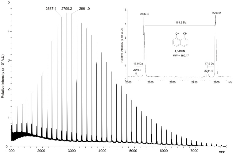
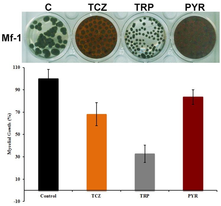
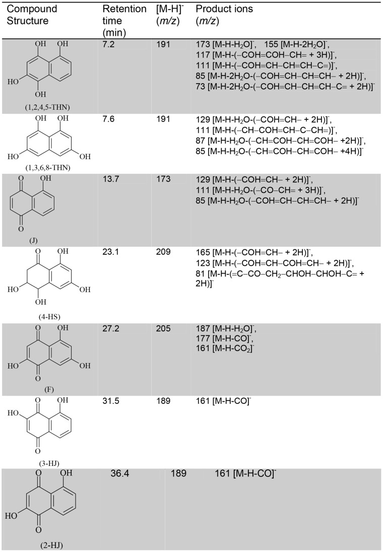
In pathogenic fungi, melanin contributes to virulence, allowing tissue invasion and inactivation of the plant defence system, but has never been implicated as a factor for host cell death, or as a light-activated phytotoxin. Our research shows that melanin synthesized by the fungal banana pathogen Mycosphaerella fijiensis acts as a virulence factor through the photogeneration of singlet molecular oxygen O2 (1Δg). Using analytical tools, including elemental analysis, ultraviolet/infrared absorption spectrophometry and MALDI-TOF mass spectrometry analysis, we characterized both pigment content in mycelia and secreted to the culture media as 1,8-dihydroxynaphthalene (DHN)-melanin type compound. This is sole melanin-type in M. fijiensis. Isolated melanins irradiated with a Nd:YAG laser at 532 nm produced monomol light emission at 1270 nm, confirming generation of O2 (1Δg), a highly reactive oxygen specie (ROS) that causes cellular death by reacting with all cellular macromolecules. Intermediary polyketides accumulated in culture media by using tricyclazole and pyroquilon (two inhibitors of DHN-melanin synthesis) were identified by ESI-HPLC-MS/MS. Additionally, irradiation at 532 nm of that mixture of compounds and whole melanized mycelium also generated O2 (1Δg). A pigmented-strain generated more O2 (1Δg) than a strain with low melanin content. Banana leaves of cultivar Cavendish, naturally infected with different stages of black Sigatoka disease, were collected from field. Direct staining of the naturally infected leaf tissues showed the presence of melanin that was positively correlated to the disease stage. We also found hydrogen peroxide (H2O2) but we cannot distinguish the source. Our results suggest that O2 (1Δg) photogenerated by DHN-melanin may be involved in the destructive effects of Mycosphaerella fijiensis on banana leaf tissues. Further studies are needed to fully evaluate contributions of melanin-mediated ROS to microbial pathogenesis.


Conflict of interest statement

Competing Interests: The authors have declared that no competing interest exist.

Figures

Figure 1
Figure 1. Linear-MALDI-TOF mass spectrum of 1,8-DHN-melanin extracted from M. fijiensis.
Linear matrix-assisted laser desorption/ionization mass spectrum of DHN-melanin extracted from M. fijiensis (Mf-1 strain) mycelium with NaOH 6M and precipitated with 6 M HCl during 3 hours at boiling temperature and then purified by washing in different solvents and freeze dried. Inset: Linear MALDI-TOF mass spectrum at 2600–2900 m/z of 1,8-DHN-melanin extracted from M. fijiensis (Mf-1 strain) showing well-defined spacings of 161.8 Da between peaks.
Figure 2
Figure 2. Influence of inhibitors of DHN-melanin synthesis and tropolone affects partially mycelial growth of M. fijiensis.
Seven days-old cultures on PDB amended with 50 μg/ml of tricyclazole (TCZ), tropolone (TRP) and pyroquilon (PYR). Mycelial growth are expressed in percentage and compared to control (C). The culture media showing reddish brown color due to accumulation intermediaries product of melanin inhibition. The experiment was conducted in the Mf-1 strain. Results are reported as the mean ± SD of 3 independent experiments.
Figure 3
Figure 3. ESI-HPLC-MS/MS analysis of pentaketide metabolites accumulated in tricyclazole and pyroquilon amended culture of M. fijiensis.
Compound names: 1,2,4,5 tetra-hydroxynaphthalene (1,2,4,5-THN), 1,3,6,8 tetra- hydroxynaphthalene (1,3,6,8 THN), Juglone (J), 4-Hydroxyscytalone (4HS), Flaviolin (F), 3-Hydroxyjuglone (3HJ), 2-Hydroxyjuglone (2HJ). The elemental compositions HPLC-ESI-MS/MS was determined in negative ion mode. [M-H]- (m/z) is indicative of precursor ion. Product ions is the mass spectral fragmentation of the [M-H]- produced after collision. The same compounds was found in cultures amended with Tricyclazole (TCZ) and Pyroquilon (PYR). This fungicides compound inhibits the reduction of 1,3,6,8-THN and 1,3,8-THN to scytalone and vermelone, respectively. Its strongest inhibitory effect is on the reduction of 1,3,8-THN. This results in the accumulation of F, 2-HJ, and their related shunt products. 1,2,4,5-tetrahydroxynaphthalene (1,2,4,5-THN) is an unstable metabolite identified by the method used in this work.
Figure 4
Figure 4. Ultrastructural morphologies of fungal melanin pigments isolated from mycelium and secreted to culture media.
A–B are SEM images of synthetic melanin obtained by tyrosine oxidation with H2O2 (Sigma M8136); C–D SEM images of Melanin extracted from mycelium, this pigment displays spherical granules tightly aggregated with an average size diameter of 100–300 nm; E–F Melanin extracted from culture media display amorphous material without definable structure composed by tightly packed of various sizes. M. fijiensis pigments were obtained using NaOH extraction, acid precipitation and solvent purification methods. The scale bars are showed in each image.
Figure 5
Figure 5. Generation of 1O2 (1Δg) by DHN-melanin, polyketides intermediaries and living whole mycelium of M. fijiensis.
A representative monomol light emission spectra of 1O2 at 1270 nm obtained after pulsed laser excitation at 532 nm. (A) Melanin pigments adjusted to 0.6 Abs in 0.1 M NaOD. Red line corresponds to secreted melanin and black line is from mycelium melanin. The phosphorescence intensity of secreted melanin was higher than mycelium melanin. (B) phosphorescence emission of synthetic melanin (M8136), (C) Reddish-brown pigment composed by polyketides and naphtoquinones mixture, was dissolved in D2O at Abs 0.6. Black line shows the phosphorescence of culture amended with tricyclazole and red line shows the phosphorescence of pyroquilon inhibitor. (D) Active whole mycelium grown in potato-dextrose culture medium. 250 mg of mycelium was used for each experiments. A black and red line corresponds to mycelium from green-black Mf-1 strain cultured in liquid and agar media respectively. Blue and Green lines are emission spectra of albino102 strain cultured in liquid and agar media. (E) Singlet oxygen production of 1,2 DHN used as standard dissolved in acetonitrile; (F) Emission spectra of dimethylnaphthalene endoperoxide.
Figure 6
Figure 6. Fungal melanin content is related with necrotic damage in banana leaf naturally infected.
Fouré stages of black Sigatoka disease in banana leaves naturally infected. Ten months old Giant dwarf (Cavendish) plants were collected from commercial orchard in Jalisco, Mexico. Sigatoka disease symptoms (upper panel). (A) Cigar leaf and (B) Stage 1. (C) Stage 4, (D) S5 stage, (E) Advanced stage 5 and (F) advanced necrotized stage. Scale bar = 10 μm. Relative area covered by melanin (lower panel). The values were normalized to capote leaf (used as control) that shown fungal infection. Results were obtained from at least five leaves analyzed for each stage. One representative analysis is shown. S4–S5 and S5*, showing lesions characteristic of the two stages. In the Cu-Sulfide-Silver stain, copper is bound to melanin followed by formation of copper sulfide at melanin sites, which are amplified into strong black stain using a silver enhacement step. Detection of H2O2 was performed by an endogenous peroxidase-dependent in situ histochemical staining procedure using DAB . DAB polymerizes locally as soon as it comes into contact with H2O2 giving a reddish-brown polymer. In the aniline-blue staining, aniline blue reacts with beta-glucans contents in fungal hyphae.

References

    1. Liu JJ, Fisher DE (2010) Lighting a path to pigmentation: mechanism of MITF induction by UV. Pigment Cell Melanoma Res 23: 741–774. - PubMed
    1. Kunwar A, Adhikary B, Jayakumar S, Barik A, Chattopadhayay S, et al. (2012) melanin, a promising radioprotector: mechanism of actions in a mice model. Toxicol Appl Pharmacol 264: 202–211. - PubMed
    1. Turick CE, Beliaev AS, Zakrajsek BA, Reardon CL, Lowy DA, et al. (2009) The role of 4-hydroxyphenylpyruvate dioxygenase in enhancement of solid-phase electron transfer by Shewanella oneidensis MR-1. FEMS Microbiol Ecol 68: 223–225. - PubMed
    1. Turick CE, Tisa LS, Caccavo F Jr (2002) Melanin production and use as a soluble electron shuttle for Fe (III) oxide reduction and as a terminal electron acceptor by Shewanella algae BrY. Appl Environ Microbiol 68: 2436–2444. - PMC - PubMed
    1. Korytowski W, Pilas B, Sarna T, Kalyanaraman B (1987) Photoinduced generation of hydrogen peroxide and hydroxyl radicals in melanins. Photochem Photobiol 45: 185–190. - PubMed

Publication types

MeSH terms