

Toxicological assessment of inhaled nanoparticles: role of in vivo, ex vivo, in vitro, and in silico studies
- PMID: 24646916
- PMCID: PMC3975425
- DOI: 10.3390/ijms15034795
Toxicological assessment of inhaled nanoparticles: role of in vivo, ex vivo, in vitro, and in silico studies
Abstract
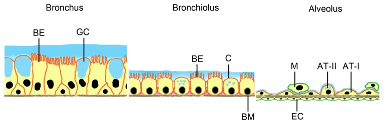
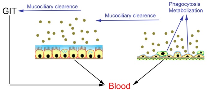
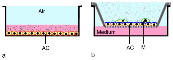
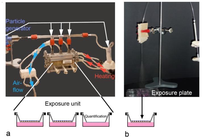
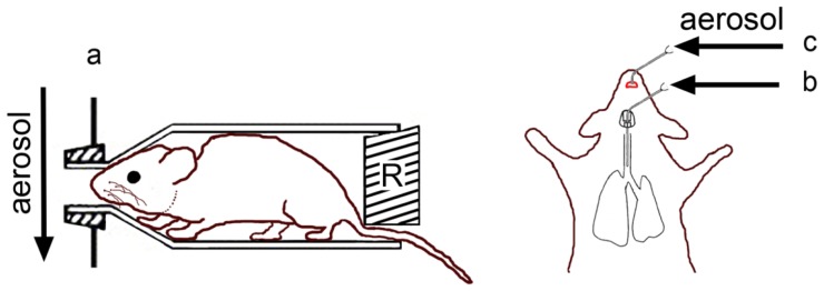
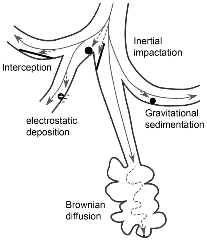
The alveolar epithelium of the lung is by far the most permeable epithelial barrier of the human body. The risk for adverse effects by inhaled nanoparticles (NPs) depends on their hazard (negative action on cells and organism) and on exposure (concentration in the inhaled air and pattern of deposition in the lung). With the development of advanced in vitro models, not only in vivo, but also cellular studies can be used for toxicological testing. Advanced in vitro studies use combinations of cells cultured in the air-liquid interface. These cultures are useful for particle uptake and mechanistic studies. Whole-body, nose-only, and lung-only exposures of animals could help to determine retention of NPs in the body. Both approaches also have their limitations; cellular studies cannot mimic the entire organism and data obtained by inhalation exposure of rodents have limitations due to differences in the respiratory system from that of humans. Simulation programs for lung deposition in humans could help to determine the relevance of the biological findings. Combination of biological data generated in different biological models and in silico modeling appears suitable for a realistic estimation of potential risks by inhalation exposure to NPs.
Figures
Similar articles
-
An in vitro testing strategy towards mimicking the inhalation of high aspect ratio nanoparticles.Part Fibre Toxicol. 2014 Sep 23;11:40. doi: 10.1186/s12989-014-0040-x. Part Fibre Toxicol. 2014. PMID: 25245637 Free PMC article.
-
Toxicological perspectives of inhaled therapeutics and nanoparticles.Expert Opin Drug Metab Toxicol. 2014 Jul;10(7):933-47. doi: 10.1517/17425255.2014.916276. Epub 2014 May 8. Expert Opin Drug Metab Toxicol. 2014. PMID: 24810077 Review.
-
Nanoscale and fine zinc oxide particles: can in vitro assays accurately forecast lung hazards following inhalation exposures?Environ Sci Technol. 2009 Oct 15;43(20):7939-45. doi: 10.1021/es901453p. Environ Sci Technol. 2009. PMID: 19921917
-
Informing selection of nanomaterial concentrations for ToxCast in vitro testing based on occupational exposure potential.Environ Health Perspect. 2011 Nov;119(11):1539-46. doi: 10.1289/ehp.1103750. Epub 2011 Jul 25. Environ Health Perspect. 2011. PMID: 21788197 Free PMC article.
-
Air-Liquid Interface: Relevant In Vitro Models for Investigating Air Pollutant-Induced Pulmonary Toxicity.Toxicol Sci. 2018 Jul 1;164(1):21-30. doi: 10.1093/toxsci/kfy053. Toxicol Sci. 2018. PMID: 29534242 Review.
Cited by
-
Adverse immunological responses against non-viral nanoparticle (NP) delivery systems in the lung.J Immunotoxicol. 2021 Dec;18(1):61-73. doi: 10.1080/1547691X.2021.1902432. J Immunotoxicol. 2021. PMID: 33956565 Free PMC article. Review.
-
Nanoparticles and Airway Epithelial Cells: Exploring the Impacts and Methodologies in Toxicity Assessment.Int J Mol Sci. 2024 Jul 18;25(14):7885. doi: 10.3390/ijms25147885. Int J Mol Sci. 2024. PMID: 39063127 Free PMC article. Review.
-
Many ways, one microorganism: Several approaches to study Malassezia in interactions with model hosts.PLoS Pathog. 2022 Sep 8;18(9):e1010784. doi: 10.1371/journal.ppat.1010784. eCollection 2022 Sep. PLoS Pathog. 2022. PMID: 36074792 Free PMC article. Review.
-
Integrated Transcriptomics, Metabolomics, and Lipidomics Profiling in Rat Lung, Blood, and Serum for Assessment of Laser Printer-Emitted Nanoparticle Inhalation Exposure-Induced Disease Risks.Int J Mol Sci. 2019 Dec 16;20(24):6348. doi: 10.3390/ijms20246348. Int J Mol Sci. 2019. PMID: 31888290 Free PMC article.
-
Attenuation of Combined Nickel(II) Oxide and Manganese(II, III) Oxide Nanoparticles' Adverse Effects with a Complex of Bioprotectors.Int J Mol Sci. 2015 Sep 17;16(9):22555-83. doi: 10.3390/ijms160922555. Int J Mol Sci. 2015. PMID: 26393577 Free PMC article.
References
-
- Royal Academy of Engineering Nanoscience and Nanotechnologies: Opportunities and Uncertainties. Royal Academy of Engineering; London, UK: 2004.
-
- Nawrot T.S., Alfaro-Moreno E., Nemery B. Update in occupational and environmental respiratory disease 2007. Am. J. Respir. Crit. Care Med. 2008;177:696–700. - PubMed
Publication types
MeSH terms
LinkOut - more resources
Full Text Sources
Other Literature Sources
Medical
