. 2014 Mar 20;507(7492):329-34.
doi: 10.1038/nature13147. Epub 2014 Mar 12.

Unexpected link between an antibiotic, pannexin channels and apoptosis

Affiliations

Unexpected link between an antibiotic, pannexin channels and apoptosis

Ivan K H Poon et al. Nature. .

Abstract

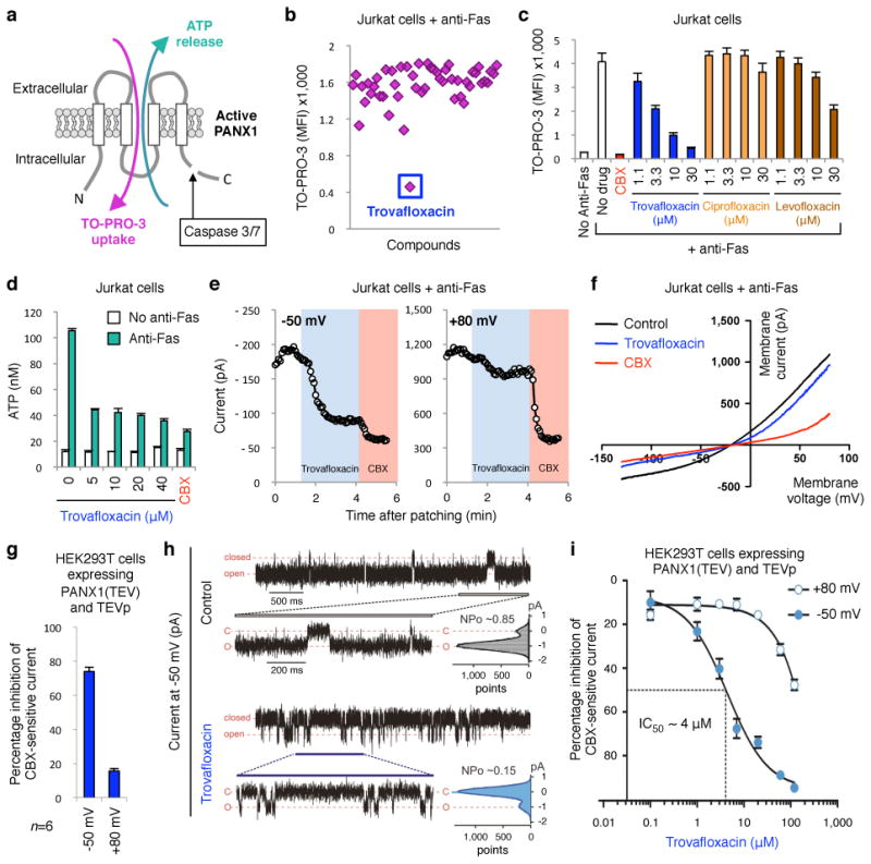
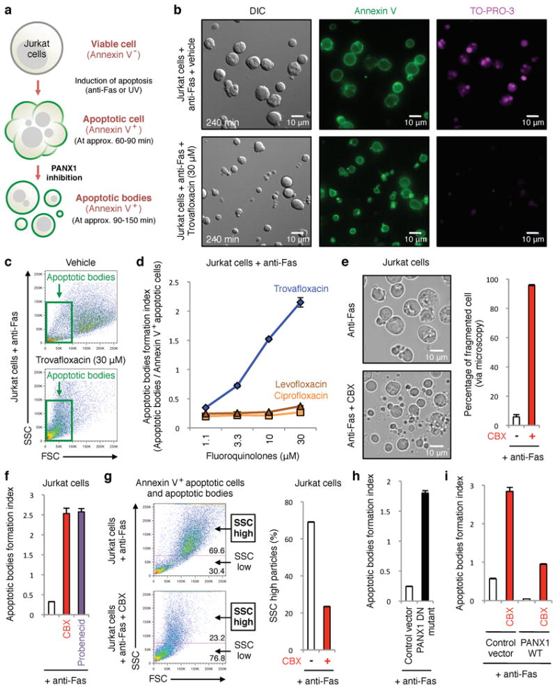
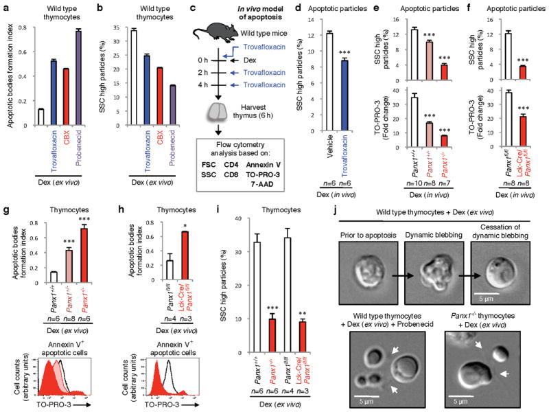
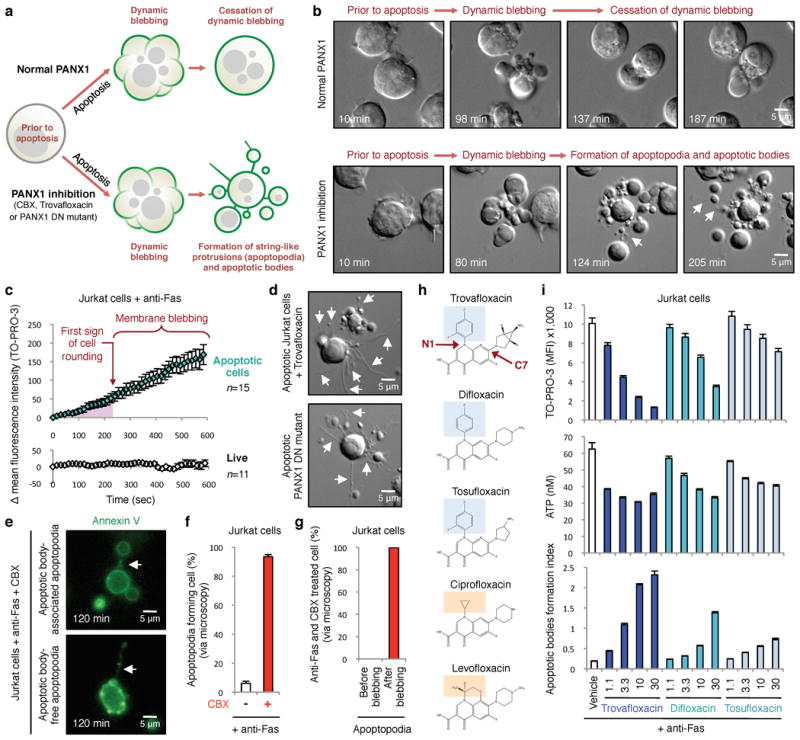
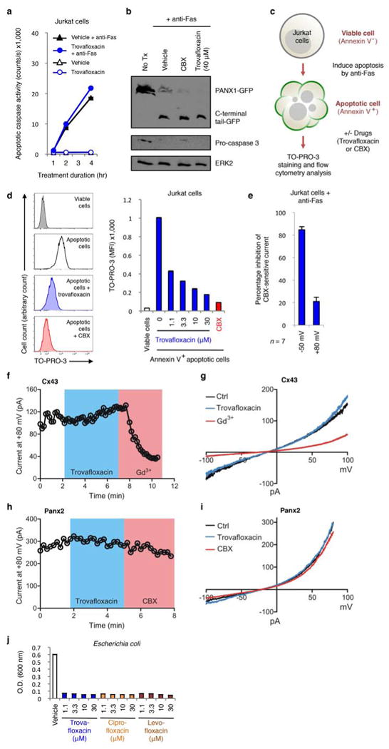
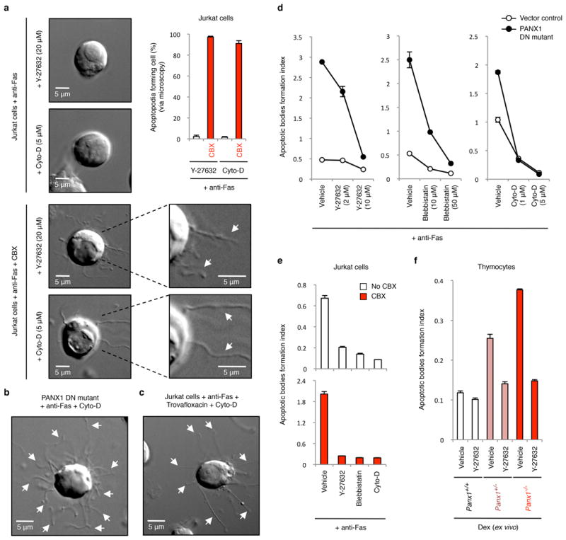
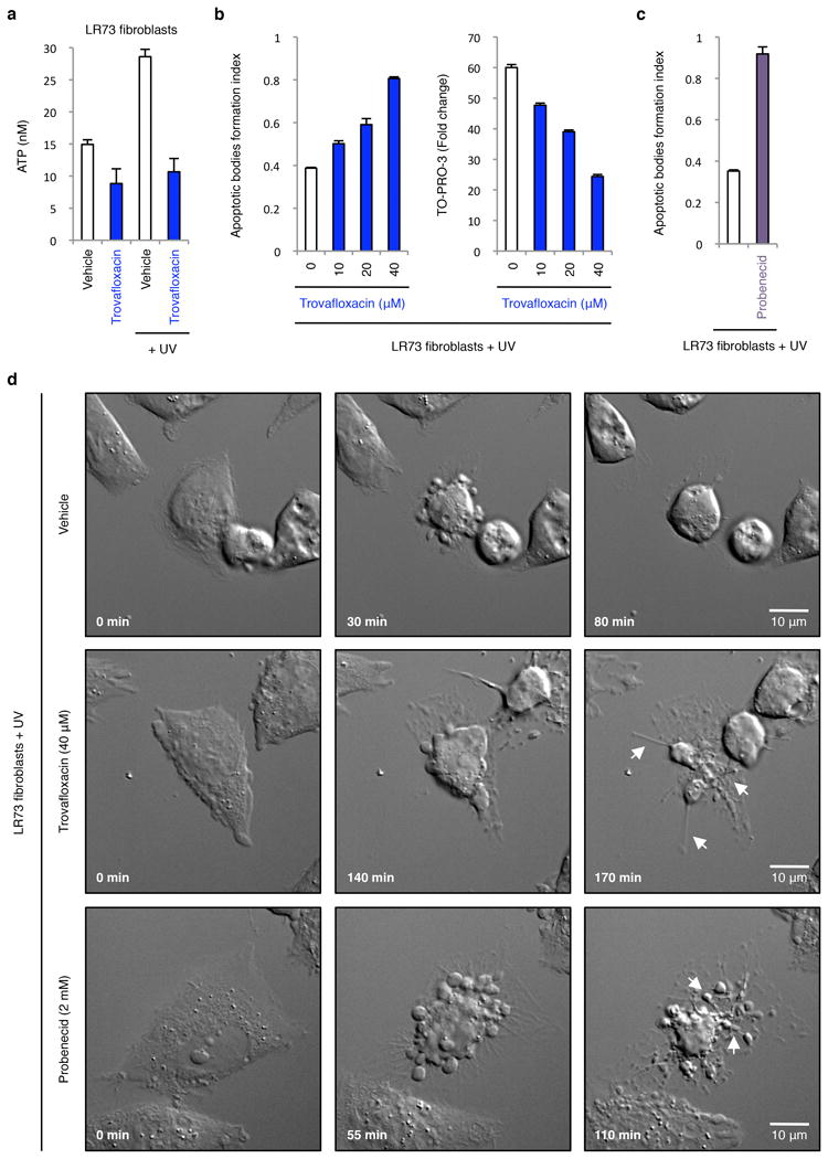
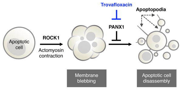
Plasma membrane pannexin 1 channels (PANX1) release nucleotide find-me signals from apoptotic cells to attract phagocytes. Here we show that the quinolone antibiotic trovafloxacin is a novel PANX1 inhibitor, by using a small-molecule screen. Although quinolones are widely used to treat bacterial infections, some quinolones have unexplained side effects, including deaths among children. PANX1 is a direct target of trovafloxacin at drug concentrations seen in human plasma, and its inhibition led to dysregulated fragmentation of apoptotic cells. Genetic loss of PANX1 phenocopied trovafloxacin effects, revealing a non-redundant role for pannexin channels in regulating cellular disassembly during apoptosis. Increase in drug-resistant bacteria worldwide and the dearth of new antibiotics is a major human health challenge. Comparing different quinolone antibiotics suggests that certain structural features may contribute to PANX1 blockade. These data identify a novel linkage between an antibiotic, pannexin channels and cellular integrity, and suggest that re-engineering certain quinolones might help develop newer antibacterials.


Conflict of interest statement

The authors declare no competing financial interests.

Figures

Figure 1
Figure 1. Trovafloxacin inhibits pannexin 1 activity during apoptosis
a, Schematic of PANX1 with ATP release and TO-PRO-3 uptake by apoptotic cells after caspase-mediated channel opening. b, Subset of LOPAC1280TM library screen in modulating TO‐PRO-3 uptake (MFI average of 1200-1800), with inhibition by trovafloxacin indicated. c, Dose-dependent inhibition of TO-PRO-3 uptake by trovafloxacin (or positive control CBX), levofloxacin and ciprofloxacin (n=3). d, Inhibition of ATP release from apoptotic cells by trovafloxacin (n=3). e, Patch-clamp recordings from apoptotic cells at ‐50mV and +80mV with trovafloxacin (blue shading) or CBX (pink); traces are representative of recordings from 7 cells per group. f, Current-voltage relationships (I-V curves) from apoptotic cells over a range of voltages (n=7). g, Trovafloxacin inhibition of CBX-sensitive current in HEK293T cells expressing TEV-protease activated PANX1 channels (n=6). h, Inside-out patch clamp recordings from excised patches containing TEV-cleaved PANX1 channels showing traces of inward currents at ‐50mV. PANX1 channels are mostly ‘open’ without trovafloxacin (open probability, NPo=0.85), while mostly closed with trovafloxacin (NPo=0.15); histograms of channel activity are shown for each trace (n=3). i, Inhibition of TEV-cleaved open PANX1 channels by trovafloxacin, with IC50 of 4μM at -50mV (n≥6 per data point). e,f, Jurkat cells after 2h with anti-Fas used for patch-clamp recordings. Error bars represent s.e.m.
Figure 2
Figure 2. Trovafloxacin-mediated inhibition of PANX1 promotes formation of smaller apoptotic bodies
a, Schematic for apoptosis progression and generation of apoptotic bodies, derived from multi-parameter time-lapse analyses of Jurkat cells. b, Images of vehicle- or trovafloxacin-treated apoptotic cells stained with annexin V or TO-PRO-3. Note the annexin Vpositive, TO-PRO-3negative apoptotic bodies (∼0.9-5μm) after trovafloxacin treatment. c, Trovafloxacin increases particles with reduced size (FSC) and complexity (SSC). d, Generation of apoptotic bodies is enhanced by trovafloxacin, but not levofloxacin and ciprofloxacin (n=3). e, Left, representative 4h time-lapse images monitoring apoptotic cell morphology (454 cells for untreated and 355 cells for CBX-treated, n=3). Right, percentage of cell fragmentation (≥2 fragments). f, Apoptotic bodies formation index (apoptotic bodies/annexin V+ apoptotic cells) is enhanced by PANX1 blockers CBX or probenecid (n=3). g, Left, Size (FSC) and complexity (SSC) of particles from apoptotic cells with or without CBX. Right, percentage of SSChigh particles. h,i, Formation of apoptotic bodies is increased in Jurkat cells expressing PANX1 DN mutant, but decreased in PANX1 WT transfected cells (n=3). The PANX1 WT effect is reversed by CBX. Error bars represent s.e.m. Scale bars represent 10μm.
Figure 3
Figure 3. Pannexin 1 regulates disassembly of apoptotic thymocytes
Increase in formation of apoptotic bodies (a), or decreases SSChigh particles (b) from dexamethasone (dex)-treated thymocytes ex vivo by trovafloxacin, CBX, and probenecid (n=3). c, Schematic for testing trovafloxacin in thymic apoptosis in vivo. d, Reduction in SSChigh apoptotic bodies in thymi of wild-type mice treated with dex+trovafloxacin (n=6). e,f, Complexity and TO-PRO-3 uptake (fold change relative to unstained) in thymi of indicated mice injected intraperitoneally with dex (n=10 for Panx1+/+, n=8 for Panx1+/-, n=7 for Panx1-/-, n=8 for Panx1fl/fl and Lck-Cre/Panx1fl/fl). g,h, Formation of apoptotic bodies (upper) or TO-PRO-3 uptake (lower) in ex vivo in dex-treated thymocytes from Panx1-/- or Lck-Cre/Panx1fl/fl thymocytes. i, Reduction in SSChigh subcellular apoptotic particles in thymocytes from Panx1-/- and Lck-Cre/Panx1fl/fl mice. (g,h,i, n=6 for Panx1+/+, n=8 for Panx1+/-, n=6 for Panx1-/-, n=4 for Panx1fl/fl, n=3 for Lck-Cre/Panx1fl/fl). j, Time-lapse images monitoring apoptotic cell morphology of thymocytes. Arrows, apoptopodia. Error bars represent s.e.m. Scale bars=5μm. Data in a, b and j are representative of at least two independent experiments. See Extended Data Fig. 5 and 6 for gating strategy. * P < 0.05, ** P < 0.01, *** P < 0.001, unpaired Student’s two-tailed t-test.
Figure 4
Figure 4. Formation of string-like apoptopodia after membrane blebbing correlates with formation of apoptotic bodies
a, Schematic of apoptotic Jurkat cells with normal or impaired PANX1, based on time-lapse microscopy. b, Time-lapse images monitoring progression of apoptotic cell morphology. c, PANX1-mediated TO-PRO-3 entry occurs prior to membrane blebbing as determined by quantitating TO-PRO-3 uptake by apoptotic cells (n=15) in time-lapse imaging (normalized to first sign of cell rounding). TO-PRO-3 uptake was undetectable in live cells (n=11). d, Trovafloxacin-treated apoptotic cells or expressing PANX1-DN mutant show formation of apoptopodia. e, Apoptopodia have membranes with exposed phosphatidylserine and apoptotic blebs at the end of the protrusion. f, Percentage of apoptotic cells with apoptopodia during 4h of time-lapse imaging. g, Apoptopodia are barely detectable before onset of membrane blebbing in cells with impaired PANX1 function, but detected after initiation of blebbing. (f,g, 429 and 302 cells for untreated and CBX-treated, respectively, n=3). h, Chemical structures of quinolones used, with the N1 fluorinated ring (blue) indicated in quinolones that inhibit PANX1 (trovafloxacin, difloxacin, and tosufloxacin), compared to quinolones with no apparent PANX1 inhibition (ciprofloxacin and levofloxacin) orange. i, TO-PRO-3 uptake, ATP release, and formation of apoptotic bodies from apoptotic Jurkat cells treated with trovafloxacin, difloxacin or tosufloxacin (n=3). Error bars represent s.e.m. Scale bars represent 5μm. Arrows, apoptopodia. Data are representative of at least two independent experiments.
Extended Data Figure 1
Extended Data Figure 1. Trovafloxacin does not block caspase activation or inhibit connexin 43 (Cx43) or pannexin 2 (Panx2) membrane currents
a, Caspase 3/7 activation in Jurkat cells undergoing apoptosis is not altered by treatment with trovafloxacin (40 μM) (n=3). b, Proteolytic cleavage of PANX1-GFP during apoptosis is not inhibited by trovafloxacin (40 μM) or CBX (500 μM) treatment. c, Schematic diagram for the acute treatment of apoptotic cells with trovafloxacin or CBX. d, Acute trovafloxacin treatment inhibits TO-PRO-3 uptake by apoptotic Jurkat cells. Left, histograms showing TO-PRO-3 uptake by viable cells, apoptotic cells, or apoptotic cells treated with trovafloxacin or CBX (500 μM) post induction of apoptosis and analyzed by flow cytometry. Right, uptake of TO-PRO-3 presented as (median fluorescence intensity, MFI) of viable cells or apoptotic cells (n=3). e, Inhibition of CBX-sensitive current in apoptotic cells treated with trovafloxacin (20 μM), as measured by whole-cell patch-clamp recording (n=7). f, Patch-clamp recordings from HEK293T cells expressing the Cx43 and receiving indicated treatments. Whole-cell current at +80 mV is shown under conditions when bath solution was perfused with trovafloxacin (20 μM, blue shading) or gadolinium (Gd3+) (100 μM, pink shading). Exemplar traces are representative of 14 cells per group. g, Current-voltage relationships of Cx43 current in HEK293T cells treated with or without trovafloxacin (20 μM) or Gd3+ (100 μM), with the current measured over a range of voltages. h, Patch-clamp recordings from HEK293T cells expressing the mouse Panx2 and receiving indicated treatments. Whole-cell current at +80 mV is shown under conditions when bath solution was perfused with trovafloxacin (20 μM, blue shading) or carbenoxolone (CBX) (50 μM, pink shading). Exemplar traces are representative of 4 cells per group. i, Current-voltage relationships of Panx2 current in HEK293T cells treat with or without trovafloxacin (20 μM) or CBX (50 μM), with the current measured over a range of voltages. j, Trovafloxacin, ciprofloxacin and levofloxacin inhibit bacterial growth. Escherichia coli growth (as measured by O.D. at 600 nm) in the presence of indicated concentrations of quinolones (n=3). Error bars represent s.e.m.
Extended Data Figure 2
Extended Data Figure 2. Electronic gating strategy for the separation of different cellular and subcellular population of Jurkat cells undergoing apoptosis in vitro
a, Flow cytometric analysis showing each type of particles gated (see b below) has a distinctive level of cellular complexity (side scatter, SSC), cell size (forward scatter, FSC) as well as TO-PRO-3 (indicative of caspase-mediated activation of pannexin 1 channels), 7-AAD (indicative of membrane integrity) and annexin V (indicative of phosphatidylserine exposure) staining. b, Flow cytometry gating strategy used to distinguish viable cells, annexin V- apoptotic cells, annexin V+ apoptotic cells, annexin V- particles, and apoptotic bodies. c, ImageStream analysis of particles gated using the same strategy as described in b. Representative images for each type of particles are shown. Jurkat cells were induced to undergo apoptosis by anti-Fas treatment (2 h) in all indicated experiments.
Extended Data Figure 3
Extended Data Figure 3. Inhibition of pannexin 1 promotes the formation of apoptotic bodies via a mechanism independent of extracellular ATP
a, CBX and probenecid enhance the generation of apoptotic bodies from cells undergoing UV-induced apoptosis (n=3). b, Formation of apoptotic bodies after treatment with the indicated concentrations of CBX (n=3). The corresponding TO-PRO-3 uptake by annexin V+ apoptotic cells at each CBX concentration is shown above the respective bars. c,d, Addition of exogenous ATP during apoptosis induction does not inhibit formation of apoptotic bodies in CBX-treated cells (n=3) (c) or cells stably expressing the dominant-negative PANX1 mutant (PANX1 DN mutant) (n=3) (d). e, Removal of extracellular ATP by apyrase does not promote formation of apoptotic bodies (n=3). f, P2Y receptor antagonist suramin does not promote formation of apoptotic bodies (n=3). Jurkat cells were induced to undergo apoptosis by anti-Fas treatment in all indicated experiments. Error bars represent s.e.m.
Extended Data Figure 4
Extended Data Figure 4. Pannexin 1 activity does not affect DNA fragmentation during apoptosis
a,b, TO-PRO-3 dye uptake (n=3) (a) and DNA fragmentation (b) were assessed in Jurkat cells stably expressing the control vector, the dominant-negative PANX1 mutant (PANX1 DN mutant) or wild-type PANX1 (PANX1 WT). DNA fragmentation from cells induced to undergo apoptosis and treated with or without 500 μM CBX is also shown in b. c, Time-lapse images monitoring TO-PRO-3 dye uptake during progression of apoptosis in Jurkat cells with normal PANX1 function show that TO-PRO-3 uptake occurs prior to initiation of membrane blebbing. Jurkat cells were induced to undergo apoptosis by anti-Fas treatment (2 h). Error bars in a represent s.e.m.
Extended Data Figure 5
Extended Data Figure 5. Electronic gating strategy for the separation of different cellular and subcellular populations of primary thymocytes undergoing apoptosis ex vivo
a, Flow cytometry analysis showing each type of particles gated according to b has a distinctive level of SSC, FSC as well as TO-PRO-3 and annexin V staining. b, Flow cytometry analysis showing electronic gating strategy used to distinguish viable cells, annexin V- apoptotic cells, annexin V+ apoptotic cells, annexin V- particles, and apoptotic bodies. c, ImageStream analysis of particles gated using the same strategy as described in b. Representative images for each type of particles are shown. Primary mouse thymocytes were induced to undergo apoptosis by dexamethasone (dex) treatment in all indicated experiments.
Extended Data Figure 6
Extended Data Figure 6. Electronic gating strategy for analysing the complexity of subcellular apoptotic particles generated ex vivo and in vivo
a, Flow cytometry analysis showing electronic gating strategy used to distinguish annexin Vhigh, 7-AADlow subcellular particles generated from primary mouse thymocytes induced to undergo apoptosis via dexamethasone treatment. Subcellular apoptotic particles with high complexity (SSC high) or low complexity (SSC low) are gated as shown. b, Flow cytometry analysis showing electronic gating strategy used to distinguish different subsets of apoptotic cell-derived particles generated in the thymus of mice injected intraperitoneally with dexamethasone (6 h). Annexin Vhigh, 7-AADlow, CD4/CD8intermediate particles were initially selected and subsequently gated based on forward scatter (FSC, indicative of cell size). Apoptotic particles of interest (as indicated) are therefore defined as Annexin Vhigh, 7-AADlow, CD4/CD8intermediate and FSClow/intermediate.
Extended Data Figure 7
Extended Data Figure 7. Generation of conditional and global pannexin 1 deficient mice
a, Strategy for deletion of neomycin cassette and exon 3 of Panx1. b, Identification of mice with floxed Panx1 loci, assessed by PCR. c, mRNA levels of Panx1 in CD4+ thymocytes relative to Gapdh. n = 3 mice per group. d, Immunoblotting of lysates from thymocytes with the indicated genotypes. e, Identification of mice with wild-type, heterozygous and homozygous Panx1 targeted loci, assessed by PCR. f, mRNA levels of Panx1 in thymocytes with the indicated genotypes relative to Gapdh. n = 3 mice per group. Error bars in c and f represent s.e.m.
Extended Data Figure 8
Extended Data Figure 8. Formation of apoptotic bodies but not string-like apoptopodia structures is dependent on actomyosin contraction
a, Time-lapse images monitoring apoptotic cell morphology of cells treated with or without CBX (500 μM) and in the presence of actomyosin contraction inhibitors. Top right, percentage of apoptotic cells forming string-like apoptopodia structures (387, 414, 459 and 372 apoptotic cells were analysed for Y-27632, Y-27632+CBX, Cyto-D and Cyto-D+CBX-treated cells, respectively, from three independent experiments). b,c, Time-lapse images monitoring apoptotic cell morphology of cells stably expressing the dominant-negative PANX1 mutant (PANX DN mutant) (b) or treated with 40 μM trovafloxacin (c) in the presence of Cyto-D (5 μM). d, Inhibitors of blebbing, Y-27632, blebbistatin, or cytochalasin D (Cyto-D) reduce the formation of apoptotic bodies in Jurkat cells expressing PANX1 DN mutant (n=3). e, Generation of apoptotic bodies by dying cells treated with Y-27632 (10 μM), blebbistatin (50 μM) and Cyto-D (5 μM). Cells were induced to undergo apoptosis in the presence or absence of CBX (500 μM) (n=3). f, The enhanced formation of apoptotic bodies in apoptotic thymocytes from mice with PANX1 deficiency is also blunted by the ROCK inhibitor Y-27632 (10 μM) that blocks membrane blebbing (n=3). Error bars represent s.e.m. Scale bars represent 5 μm. Arrows, apoptopodia.
Extended Data Figure 9
Extended Data Figure 9. Inhibition of pannexin 1 during UV-induced apoptosis in LR73 fibroblasts promotes the formation of membrane protrusions and apoptotic bodies
a, ATP levels in supernatants of LR73 fibroblasts treated with 40 μM trovafloxacin with or without apoptosis induction (n=3). b, Formation of apoptotic bodies (left) and TO-PRO-3 uptake (right) by LR73 fibroblasts treated with the indicated concentrations of trovafloxacin (n=3). c, Generation of apoptotic bodies by LR73 fibroblasts treated with 2 mM probenecid (n=3). d, Time-lapse images monitoring apoptotic cell morphology of LR73 fibroblasts treated with or without trovafloxacin (40 μM) or probenecid (2 mM). LR73 fibroblasts were induced to undergo apoptosis by UV treatment in all indicated experiments. Error bars in a-c represent s.e.m. Arrows, apoptopodia. Scale bars represent 10 μm.
Extended Data Figure 10
Extended Data Figure 10. Schematic diagram depicting where pannexin 1 likely acts in limiting the fragmentation of apoptotic cells
Blocking PANX1 function (for example via trovafloxacin) leads to formation of apoptopodia, and subsequently the release of apoptotic bodies.

Comment in

References

    1. Penuela S, Gehi R, Laird DW. The biochemistry and function of pannexin channels. Biochim Biophys Acta. 2013;1828:15–22. - PubMed
    1. Sosinsky GE, et al. Pannexin channels are not gap junction hemichannels. Channels (Austin) 2011;5:193–197. - PMC - PubMed
    1. Chen Y, et al. Purinergic signaling: a fundamental mechanism in neutrophil activation. Sci Signal. 2010;3:ra45. - PMC - PubMed
    1. Seminario-Vidal L, et al. Rho signaling regulates pannexin 1-mediated ATP release from airway epithelia. The Journal of biological chemistry. 2011;286:26277–26286. - PMC - PubMed
    1. Seror C, et al. Extracellular ATP acts on P2Y2 purinergic receptors to facilitate HIV-1 infection. J Exp Med. 2011;208:1823–1834. - PMC - PubMed

Publication types

MeSH terms