. 2014 Mar 19:5:3446.
doi: 10.1038/ncomms4446.

Superoxide dismutase 1 acts as a nuclear transcription factor to regulate oxidative stress resistance

Affiliations

Superoxide dismutase 1 acts as a nuclear transcription factor to regulate oxidative stress resistance

Chi Kwan Tsang et al. Nat Commun. .

Abstract

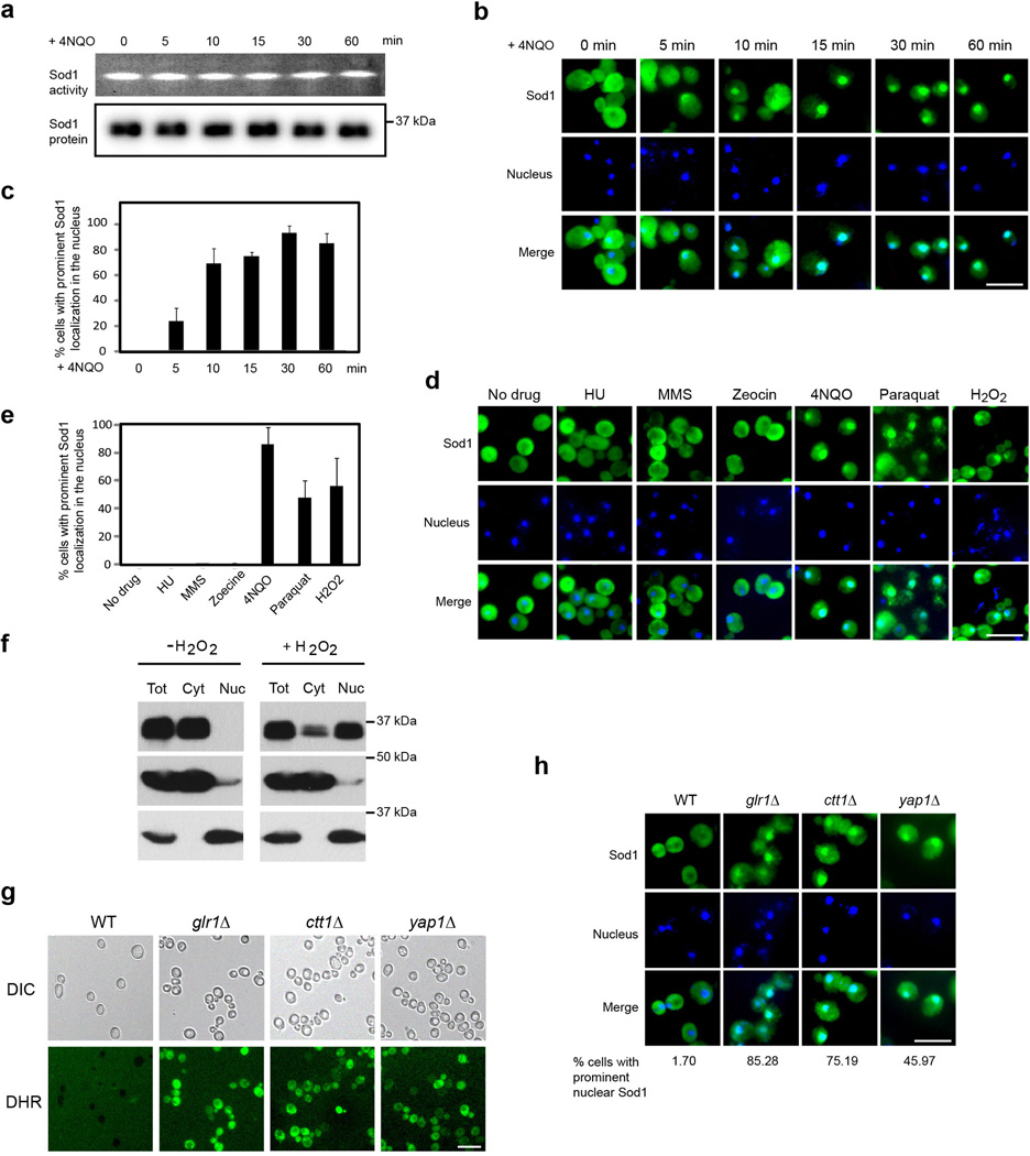
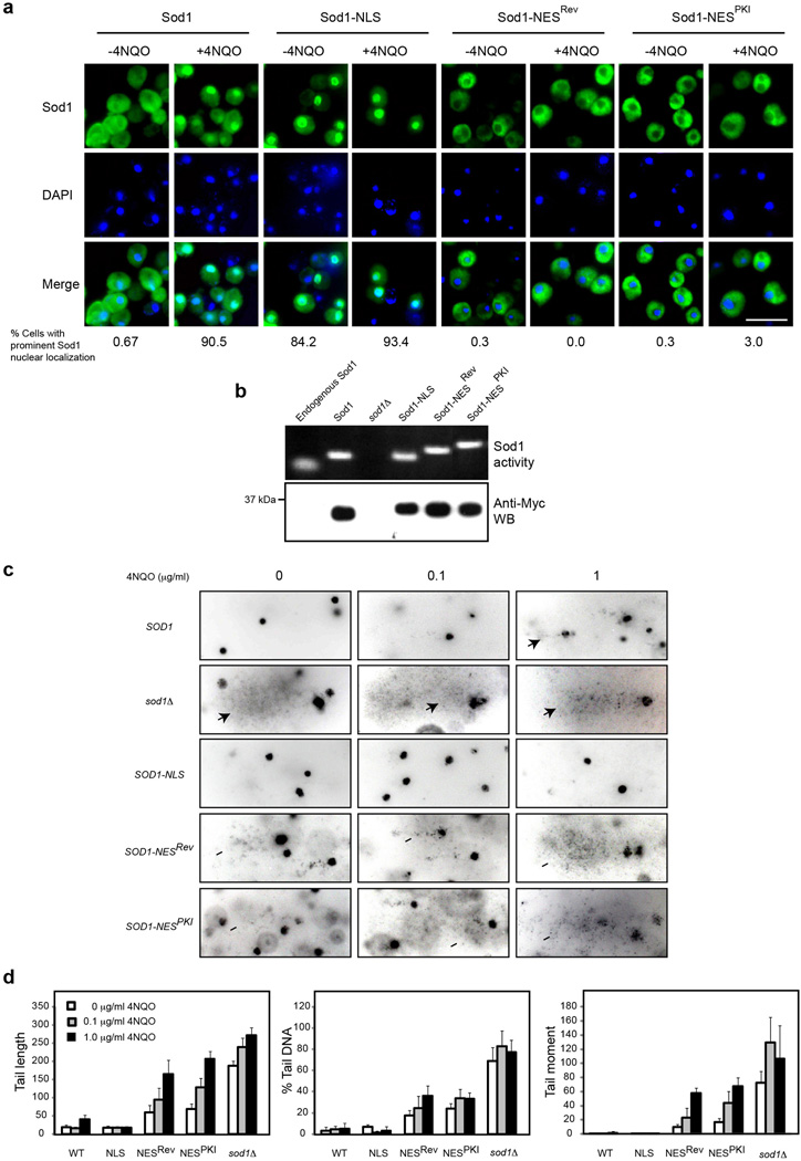
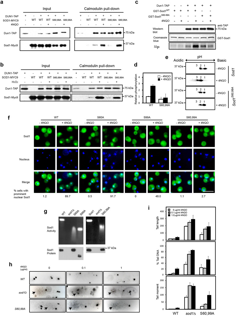
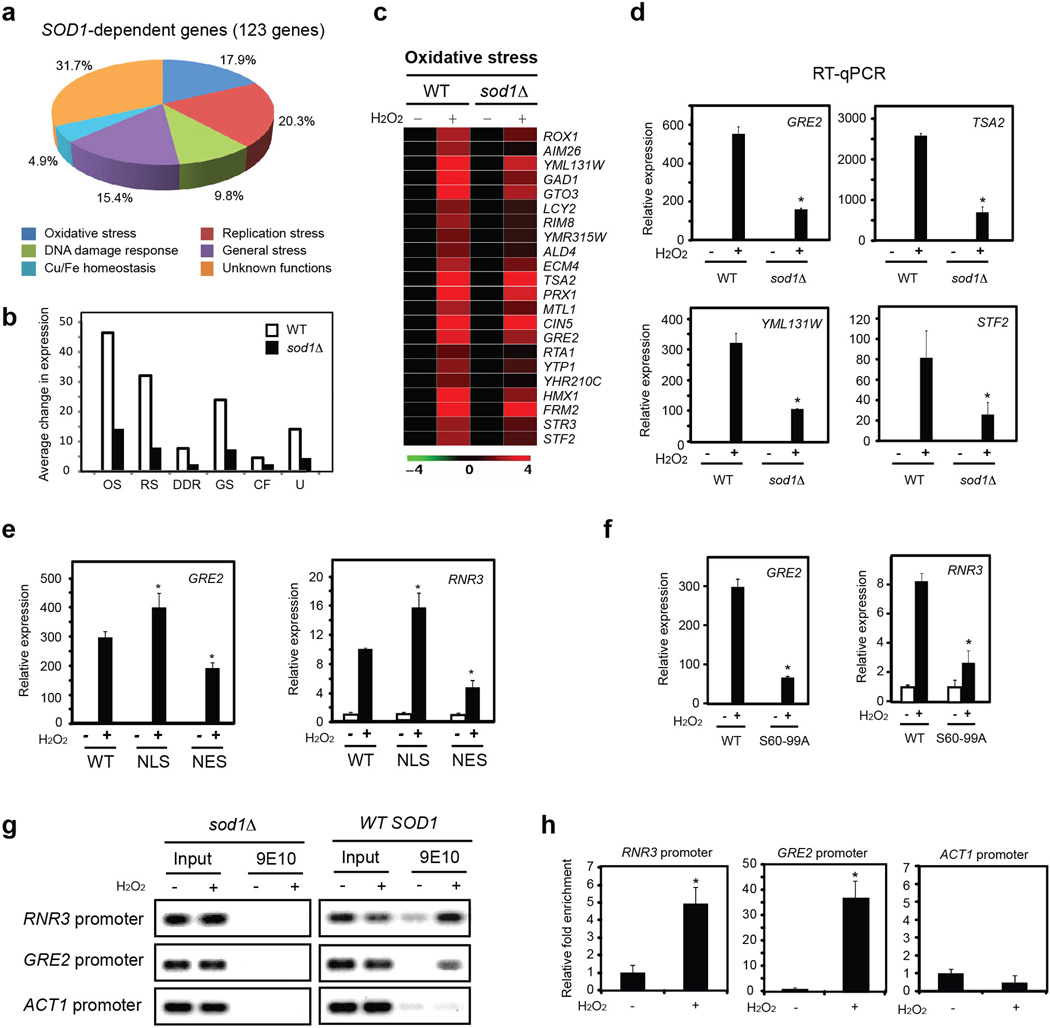
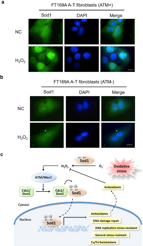
Superoxide dismutase 1 (Sod1) has been known for nearly half a century for catalysis of superoxide to hydrogen peroxide. Here we report a new Sod1 function in oxidative signalling: in response to elevated endogenous and exogenous reactive oxygen species (ROS), Sod1 rapidly relocates into the nucleus, which is important for maintaining genomic stability. Interestingly, H2O2 is sufficient to promote Sod1 nuclear localization, indicating that it is responding to general ROS rather than Sod1 substrate superoxide. ROS signalling is mediated by Mec1/ATM and its effector Dun1/Cds1 kinase, through Dun1 interaction with Sod1 and regulation of Sod1 by phosphorylation at S60, 99. In the nucleus, Sod1 binds to promoters and regulates the expression of oxidative resistance and repair genes. Altogether, our study unravels an unorthodox function of Sod1 as a transcription factor and elucidates the regulatory mechanism for its localization.


Figures

Figure 1
Figure 1. ROS induces rapid Sod1 nuclear localization
(a)ROS does not affect Sod1 protein level or enzymatic activity. Yeast cells (SZy1051) were treated with 5 µg ml−1 4NQO for different times and analyzed for Sod1-Myc9 protein level and enzymatic activity. (b) ROS induces rapid nuclear localization. Yeast cells (SZy1051) were treated with 5 µg ml−1 4NQO for different times and analyzed for Sod1-Myc9 localization by IF. The nucleus was stained by DAPI. Scale bar, 10 µm. (c) Shown is the percentage of yeast cells with prominent Sod1 nuclear localization. Error bars indicate ± standard deviation (SD) of triplicates and at least 100 cells were counted per replicate. (d) ROS but not DNA damage per se causes Sod1 relocalization. Yeast cells (SZy1051) were treated with ROS-generating agents 4NQO, Paraquat or H2O2, or non-oxidative DNA damaging agents HU, MMS and Zeocin and analyzed for Sod1-Myc9 localization. Scale bar, 10 µm. (e) Percentage of yeast cells with prominent Sod1 nuclear localization in the Fig. 1d experiment. Error bars indicate ± SD of triplicates and at least 100 cells were counted per replicate. (f) Sod1 is enriched in the nucleus in response to oxidative stress as determined by subcellular fractionation. Yeast cells were treated without or with 0.4 mM H2O2 for 30 min. Yeast cytosol and nuclei were separated by centrifugation and analyzed by Western blot. Pgk1 and Nop1 were used as cytosolic and nuclear marker, respectively. Tot, total cell extracts; Cyt, cytosol; Nuc, nuclei. (g) Mutation of GLR1, CTT1 and YAP1 causes elevated ROS level. Wild type (WT, SZy2492), glr1Δ (SZy2502), ctt1Δ (SZy2503) and yap1Δ (SZy2504) cells under normal culture conditions were stained with dihydrorhodamine (DHR). Scale bar, 10 µm. (h) Increased endogenous ROS is correlated with Sod1 nuclear localization. Sod1-Myc9 localization was analyzed by IF in WT (SZy2492), glr1Δ (SZy2502), ctt1Δ (SZy2503) and yap1Δ (SZy2504) cells. Scale bar, 10 µm.
Figure 2
Figure 2. Nuclear Sod1 is crucial to protect against genomic DNA damage by ROS
(a) Targeted Sod1 localization in the nucleus or cytoplasm. Yeast cells expressing Sod1-Myc9 (SZy1051), Sod1-NLS-Myc9 (SZy2489), Sod1-NESRev-Myc9 (SZy2499), Sod1-NESPKI-Myc9 (SZy2491) were treated without or with 5 µg ml−1 4NQO for 30 min, and analyzed for Sod1 localization (n > 100). Scale bar, 10 µm. (b) Differential subcellular localization does not affect Sod1 protein level or enzymatic activity. The protein level and superoxide dismutase activity of nuclear and cytoplasmic Sod1 were assayed. (c) Nuclear, but not cytoplasmic Sod1 plays a critical role against oxidative DNA damage. Different yeast cells were treated without or with low concentrations of 4NQO for 20 min and assayed for genomic DNA damage by Comet assay. Arrowheads indicate Comet tails. (d) Quantification of the Comet assay results by three different parameters: tail length, % tail DNA and tail moment. Error bars indicate ± SD of triplicates and at least 50 cells were counted per replicate.
Figure 3
Figure 3. ROS-induced Sod1 nuclear localization is dependent on Mec1 and Dun1
(a) Mec1 is required for ROS-induced Sod1 nuclear localization. Exponential WT (SZy2492) and mec1-1 (SZy2494) cells at the permissive temperature (23°C) were maintained at the permissive temperature or switched to the restrictive temperature (37°C) for 3 hrs before treated with 5 µg ml−1 4NQO for 30 min. Sod1-Myc9 localization was then analyzed by IF (n > 100). Scale bar, 10 µm. (b) ROS stimulates Dun1 interaction with Sod1. Yeast cells expressing Dun1-TAP and/or Sod1-Myc9 (SZy2495, SZy2496, SZy2497) were treated without or with 5 µg ml−1 4NQO for 30 min. Dun1-TAP was purified by Calmodulin beads. Dun1-TAP and its association with Sod1-Myc9 were analyzed. (c) Dun1 is required for 4NQO-induced Sod1 nuclear localization. Exponentially growing WT (SZy2492) and dun1Δ (SZy2493) cells were treated with or without 5 µg ml−1 4NQO for 30 min, and analyzed for Sod1-Myc9 localization by IF (n > 100). Scale bar, 10 µm. (d) Dun1 is required for H2O2-induced Sod1 nuclear localization. Exponentially growing WT (SZy2492) and dun1Δ (SZy2493) cells were treated with or without 0.4 mM H2O2 for 30 min, and analyzed for Sod1-Myc9 localization by IF (n > 100). Scale bar, 10 µm. (e) [SN1]Sod1 is a phosphoprotein and its phosphorylation is stimulated by oxidative stress. Yeast cells (SZy1051) were treated with or without 5 µg ml−1 4NQO for 30 min. Sod1-Myc9 phosphorylation was analyzed by two-dimensional (2D) gel electrophoresis. Different electrophoretic forms are marked by numbers and their status was confirmed by mixing 4NQO-untreated and -treated samples (third panel). (f) Mec1 is required for oxidative stress-induced Sod1 phosphorylation. WT (SZy2492) and mec1-1 (SZy2494) yeast cells were cultured at the permissive temperature (23°C) or restrictive temperature (37°C) for 3 hours before treated with 5 µg ml−1 4NQO for 30 min. Sod1-Myc9 was analyzed by 2D gel electrophoresis. (g) Dun1 is required for oxidative stress-induced Sod1 phosphorylation. WT (SZy2492) and dun1Δ (SZy2493) yeast cells were treated with 5 µg ml−1 4NQO for 30 min. Sod1-Myc9 was analyzed by 2D gel electrophoresis.
Figure 4
Figure 4. ROS stimulates Sod1 phosphorylation at S60 and S99 by Dun1 to promote Sod1 nuclear localization
(a) 4NQO promotes Dun1 interaction with Sod1 and Sod1S60,99A. Yeast cells expressing Dun1-TAP and/or Sod1-Myc9 (SZy2495, SZy2496, SZy2497, SZy2498) were treated without or with 5 µg ml−1 4NQO for 30 min. Dun1-TAP interaction with Sod1 proteins were assayed by TAP-pull down and Western blot. (b) [SN2]H2O2 promotes Dun1 interaction with Sod1 and Sod1S60,99A. Yeast cells expressing Dun1-TAP and/or Sod1-Myc9 (SZy2495, SZy2496, SZy2497, SZy2498) were treated without or with 0.4 mM H2O2 for 30 min. Dun1-TAP interaction with Sod1 proteins were assayed. (c) Dun1 phosphorylates Sod1 at S60 and S99 in response to ROS. Yeast cells expressing Dun1-TAP was treated without or with 5 µg ml−1 4NQO for 30 min. Dun1-TAP was affinity-purified and incubated with bacterial recombinant GST-Sod1 or GST- Sod1S60,99A in the presence of γ-[32P]-ATP. Phosphorylation of GST-Sod1 proteins was detected by autoradiography. (d) Quantification of in vitro GST-Sod1 phosphorylation by Dun1-TAP. Error bars indicate ± SD of triplicates. (e) The S60, 99A mutations blunt Sod1 phosphorylation in vivo. Exponentially growing yeast cells expressing Sod1-Myc9 or Sod1S60,99A–Myc9 were treated with or without 5 µg ml−1 4NQO for 30 min. Sod1 phosphorylation was analyzed by 2D gel electrophoresis. (f) Phosphorylation at S60 and S99 regulates Sod1 nuclear localization. Yeast cells expressing Sod1-Myc9 (SZy1051), Sod1S60A–Myc9 (SZy2499), Sod1S99A–Myc9 (SZy2500) or Sod1S60,99A–Myc9 (SZy2501) were treated without or with 5 µg ml−1 4NQO for 30 min. Sod1 localization was analyzed by IF. Scale bar, 10 µm. (g) The S60, 99A mutations do not affect Sod1 protein level and enzymatic activity. Yeast cells were cultured under normal conditions. Superoxide dismutase activity (upper panel) and protein expression (Lower panel) were assayed. (h) Sod1S60,99A cells exhibit elevated genomic DNA damage under normal and oxidative stress conditions. Yeast cells expressing Sod1-Myc9 (SZy1051) or Sod1S60,99A–Myc9 (SZy2501) were analyzed for genomic DNA damage by the Comet assay in the absence or presence of 4NQO. (i) Quantification of the Comet assay results by three different parameters: tail length, % tail DNA and tail moment. Error bars indicate ± SD of triplicates and at least 50 cells were counted per replicate.
Figure 5
Figure 5. Nuclear Sod1 regulates expression of oxidative stress responsive genes
(a) Sod1 is required for the induction of oxidative response (OR) genes. WT or sod1Δ cells were treated without or with 0.4 mM H2O2 for 20 min and analyzed for global gene expression profile. 123 Sod1-dependent genes were identified and most of the known genes belong to five related functional categories. (b) Shown is the relative induction level of OR genes by H2O2 in each category in WT and sod1Δ cells. Data represents average fold change of induction in each category. (c) Shown is the heat map of genes in the oxidative stress response category. (d) Validation of representative genes (GRE2, Genes de Respuesta a Estres 2; TSA2: Thiol-Specific Antioxidant 2; YML131; STF2: STabilizing Factor 2) in the oxidative stress response category by RT-qPCR. Error bars indicate ± SD from triplicates of two independent experiments. * p< 0.05. (e) Nuclear Sod1 is critical for the induction of OR genes. Yeast cells expressing different forms of Sod1 were treated with 0.4 mM H2O2 for 20 min. Representative genes were validated by RT-qPCR. Error bars indicate ± SD from triplicates of two independent experiments. * p< 0.05. (f) The induction of OR genes by ROS was attenuated in Sod1S60,99A cells. Yeast cells expressing Sod1 or Sod1S60,99A were treated with 0.4 mM H2O2 for 20 min. Expression of GRE2 and RNR3 were determined by RT-qPCR. Error bars indicate ± SD from triplicates of two independent experiments. * p < 0.05. (g) ROS treatment increases the association of Sod1 with promoter of oxidative responsive genes. WT (SZy1051) and sod1Δ (SZy1050) cells were treated with 0.4 mM H2O2for 20 min. The binding of Sod1 to representative promoters were analyzed by chromatin immunoprecipitation (ChIP). (h) Quantification of the Fig. 5g experiment. Error bars indicate ± SD from triplicates of two independent experiments. * p< 0.05, Student’s t-test.
Figure 6
Figure 6. ROS rapidly induces Sod1 nuclear localization in human FT169A fibroblasts in an ATM-dependent manner
(a) H2O2 burst induces rapid nuclear localization of human Sod1. Human FT169A A-T fibroblasts carrying an ATM-expressing plasmid (ATM+) or a control plasmid (ATM-) were treated with 0.25 mM H2O2 for 15 min. Sod1 localization was determined by IF with a human Sod1-specific antibody (green). Nuclear DNA was stained by DAPI (blue). Scale bar, 10 µm. (b) The same as Fig. 6a except Human FT169A A-T fibroblasts carrying a control plasmid (ATM-) were used. (c) A working model for Sod1 to act as a nuclear transcription factor to regulate oxidative stress resistance.

References

    1. Apel K, Hirt H. REACTIVE OXYGEN SPECIES: Metabolism, Oxidative Stress, and Signal Transduction. Annual Review of Plant Biology. 2004;55:373–399. - PubMed
    1. Dickinson BC, Chang CJ. Chemistry and biology of reactive oxygen species in signaling or stress responses. Nat Chem Biol. 2011;7:504–511. - PMC - PubMed
    1. COOKE MS, EVANS MD, DIZDAROGLU M, LUNEC J. Oxidative DNA damage: mechanisms, mutation, and disease. The FASEB Journal. 2003;17:1195–1214. - PubMed
    1. Miao L, St.Clair DK. Regulation of superoxide dismutase genes: Implications in disease. Free Radical Biology and Medicine. 2009;47:344–356. - PMC - PubMed
    1. Sturtz LA, Diekert K, Jensen LT, Lill R, Culotta VC. A Fraction of Yeast Cu,Zn-Superoxide Dismutase and Its Metallochaperone, CCS, Localize to the Intermembrane Space of Mitochondria: A PHYSIOLOGICAL ROLE FOR SOD1 IN GUARDING AGAINST MITOCHONDRIAL OXIDATIVE DAMAGE. Journal of Biological Chemistry. 2001;276:38084–38089. - PubMed

Publication types

MeSH terms

Associated data