. 2014 Mar 19;9(3):e92259.
doi: 10.1371/journal.pone.0092259. eCollection 2014.

ZASP interacts with the mechanosensing protein Ankrd2 and p53 in the signalling network of striated muscle

Affiliations

ZASP interacts with the mechanosensing protein Ankrd2 and p53 in the signalling network of striated muscle

Valentina C Martinelli et al. PLoS One. .

Retraction in

Abstract

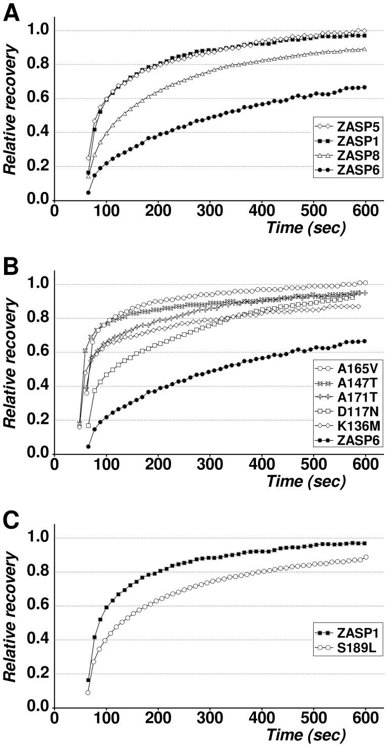
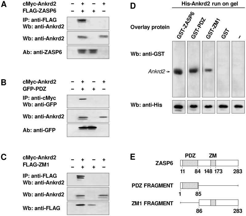
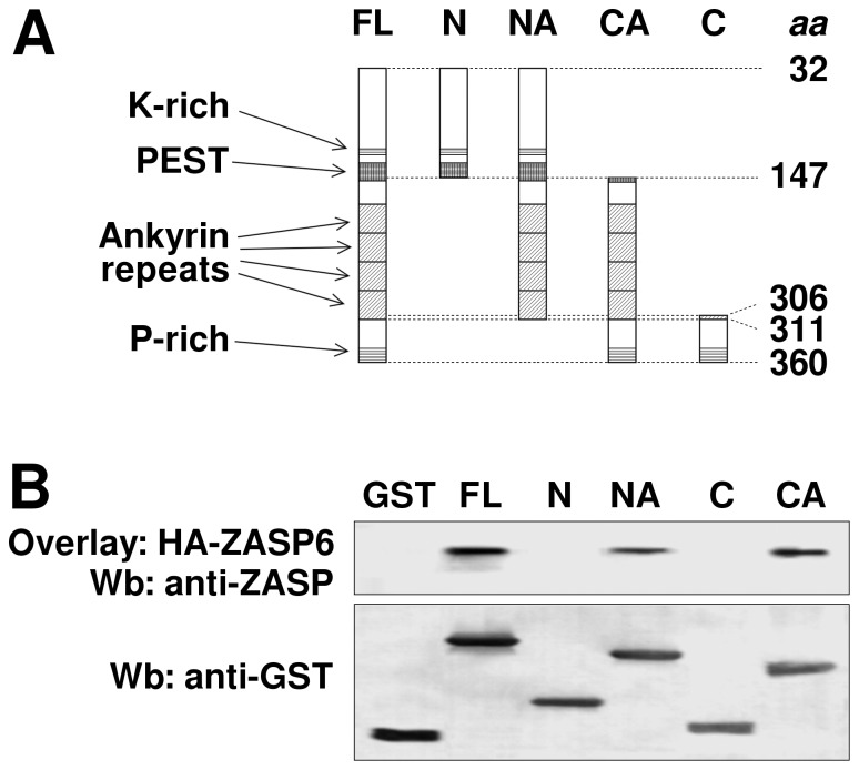
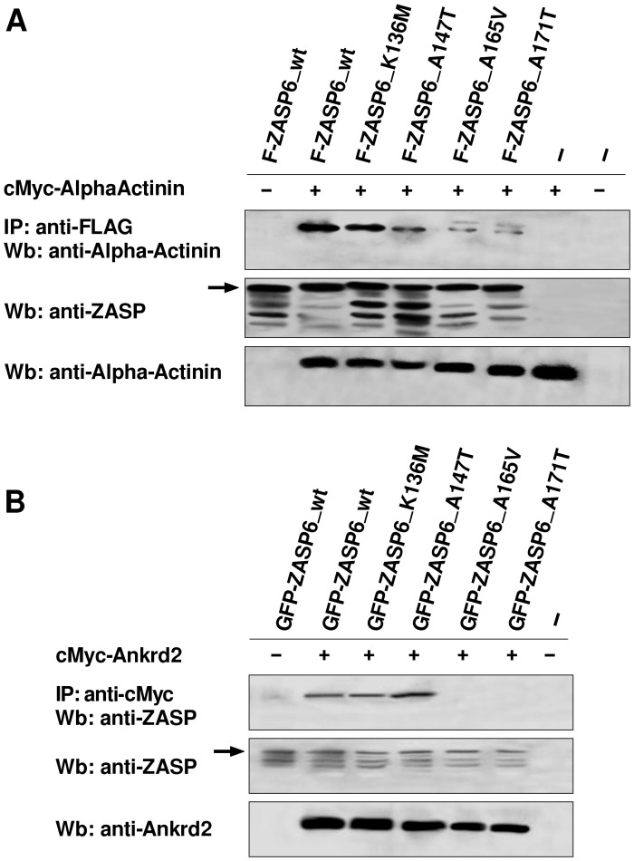
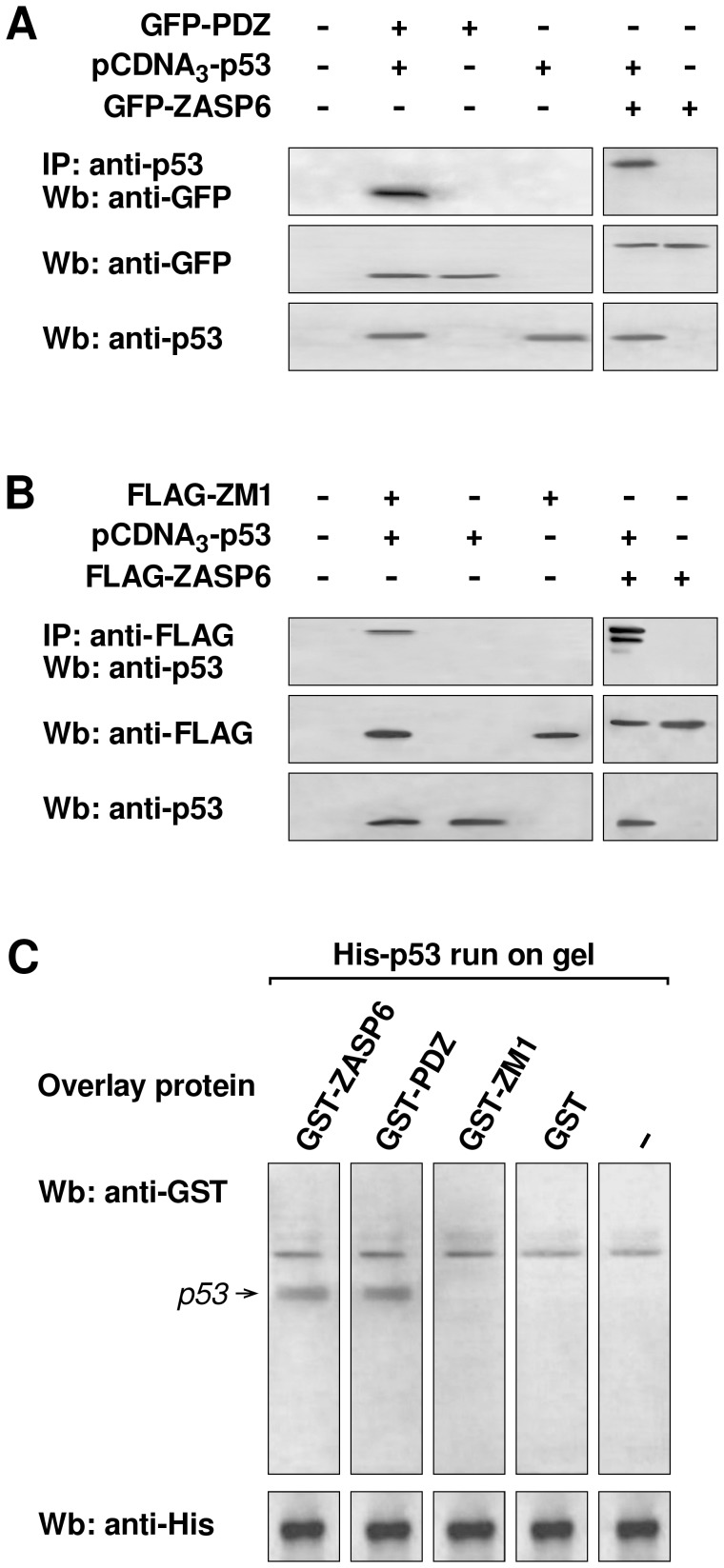
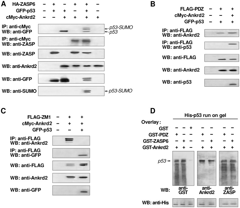
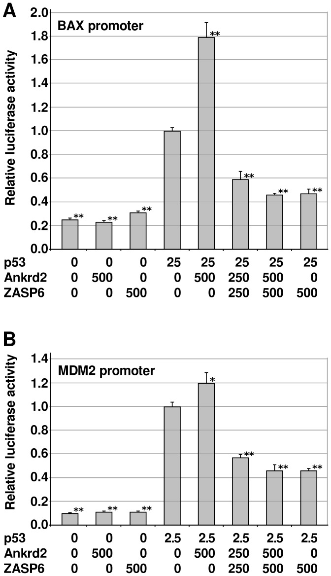
ZASP is a cytoskeletal PDZ-LIM protein predominantly expressed in striated muscle. It forms multiprotein complexes and plays a pivotal role in the structural integrity of sarcomeres. Mutations in the ZASP protein are associated with myofibrillar myopathy, left ventricular non-compaction and dilated cardiomyopathy. The ablation of its murine homologue Cypher results in neonatal lethality. ZASP has several alternatively spliced isoforms, in this paper we clarify the nomenclature of its human isoforms as well as their dynamics and expression pattern in striated muscle. Interaction is demonstrated between ZASP and two new binding partners both of which have roles in signalling, regulation of gene expression and muscle differentiation; the mechanosensing protein Ankrd2 and the tumour suppressor protein p53. These proteins and ZASP form a triple complex that appears to facilitate poly-SUMOylation of p53. We also show the importance of two of its functional domains, the ZM-motif and the PDZ domain. The PDZ domain can bind directly to both Ankrd2 and p53 indicating that there is no competition between it and p53 for the same binding site on Ankrd2. However there is competition for this binding site between p53 and a region of the ZASP protein lacking the PDZ domain, but containing the ZM-motif. ZASP is negative regulator of p53 in transactivation experiments with the p53-responsive promoters, MDM2 and BAX. Mutations in the ZASP ZM-motif induce modification in protein turnover. In fact, two mutants, A165V and A171T, were not able to bind Ankrd2 and bound only poorly to alpha-actinin2. This is important since the A165V mutation is responsible for zaspopathy, a well characterized autosomal dominant distal myopathy. Although the mechanism by which this mutant causes disease is still unknown, this is the first indication of how a ZASP disease associated mutant protein differs from that of the wild type ZASP protein.


Conflict of interest statement

Competing Interests: The authors have declared that no competing interests exist.

Figures

Figure 1
Figure 1. FRAP analysis of human isoforms of ZASP and their mutants.
The recovery times were measured every 10–12 sec, within 10 min after bleaching (50 points). The relative FRAP recovery was plotted as a function of time. A) FRAP analysis of ZASP isoforms, 1 (black squares), 5 (white diamonds), 6 (black circles) and 8 (white triangles). B) FRAP analysis of human ZASP6 (black circles) and its mutants ZASP6_K136M (white diamonds), ZASP6_D117N (white squares), ZASP6_A171T (white cross), ZASP-6_A147T (white saltire cross), ZASP6_A165V (white circles). C) FRAP analysis comparing the ZASP1 wt (black square) and ZASP1 mutant S189L (white circles).
Figure 2
Figure 2. PDZ and ZM1 regions of ZASP6 directly bind Ankrd2.
A) Co-IP of ZASP6 and Ankrd2 using lysates of COS-7 cells transfected with the indicated combination of pCMV vectors expressing cMyc-Ankrd2 and FLAG-ZASP6. Immunoprecipitation was performed with anti-FLAG antibody. The presence of Ankrd2 in the immune complex was confirmed by probing the membrane with mouse polyclonal anti-Ankrd2 antibody (upper panel). As controls, the cell lysates were tested with polyclonal antibodies to anti-Ankrd2 (middle panel) and anti-ZASP6 (bottom panel). B) Co-IP of PDZ-ZASP6 and Ankrd2 using lysates of COS-7 cells transfected with cMyc-Ankrd2 and pEGFP-PDZ as indicated. The cell lysates were immunoprecipitated with an anti-cMyc monoclonal antibody and probed with a polyclonal goat anti-GFP antibody conjugated to HRP (upper panel). Comparable expression levels of Ankrd2 and PDZ-ZASP6, weredemonstrated by probing the membrane with a mouse anti-Ankrd2 polyclonal antibody (middle panel) and rabbit anti-GFP polyclonal antibody (lower panel). C) Co-IP of ZM1-ZASP6 and Ankrd2 using lysates of COS-7 cells transfected with cMyc-Ankrd2 and FLAG-ZM1 as indicated. Ankrd2 was immunoprecipitated with anti-FLAG EZview resin and detected with an anti-Ankrd2 mouse polyclonal antibody (upper panel). As controls, cell lysates were tested with an anti-Ankrd2 mouse polyclonal antibody (middle panel) and with an anti-FLAG rabbit polyclonal antibody (bottom panel). All secondary antibodies were conjugated with HRP. D) GST-overlay assay with purified recombinant proteins expressed in E. coli. His-Ankrd2 (4 μg) was separated by SDS-PAGE, blotted and membrane strips were incubated with 4 μg of either GST-ZASP6, GST-PDZ, GST-ZM1 or GST alone, washed and then probed with anti-GST goat polyclonal antibody. As a control, an anti-histidine antibody was used to probe membrane strips identical to those used for the overlay to show that the His-Ankrd2 protein was equally loaded (lower panel). E) A schematic diagram of ZASP6 showing the positions of the PDZ domain (aa 11–84) and ZM-motif (aa 148–173). In this study the expression vectors with constructs for PDZ and ZM1 (ZASP6 without the PDZ domain) expressed respectively 85 and 197 amino acid fragments of ZASP6.
Figure 3
Figure 3. ZASP6 interacts with the ankyrin repeat region of Ankrd2.
A) is a schematic diagram showing the different regions of Ankrd2 used in this experiment. The Ankrd2 numbering refers to the canonical Ankrd2-202 sequence (ENSEMBL); FL refers to the full-length of Ankrd2-001 (Kojic et al., 2004). There are four ankyrin repeat motifs in the human Ankrd2 protein between aa 148–308, a PEST sequence that targets proteins for proteolytic cleavage, as well as regions rich in amino acids lysine (K) and proline (P). B) GST-overlay assay demonstrating that NA and CA regions of Ankrd2 contain the binding site/sites for ZASP6. GST-FL-Ankrd2, GST-N-Ankrd2, GST-NA-Ankrd2, GST-CA- Ankrd2, GST-C-Ankrd2 and GST alone were separated by SDS-PAGE and blotted. The membrane was blocked and then overlaid with lysate from COS-7 cells transfected with pcDNA3-HA-ZASP6. The membrane was probed with an anti-ZASP mouse polyclonal antibody. The Western blot in the lower panel shows that equal amounts of the GST proteins were used in this experiment.
Figure 4
Figure 4. Mutations in ZASP6 affect its interaction with alpha-actinin2 and Ankrd2.
Wt and mutants of the ZASP6 isoform fused with FLAG (A) or GFP (B) tag were co-expressed with cMyc-alpha-actinin2 (A) or cMyc-Ankrd2 (B) in COS-7 cells as indicated. Protein extracts were immunoprecipitated with polyclonal antibodies to FLAG (A) or cMyc (B) tag. Precipitated proteins were analyzed by SDS-PAGE followed by Western blotting using rabbit anti-alpha actinin2 (A) or mouse anti-ZASP antibody (B). As controls, the lysates were probed with polyclonal antibodies to ZASP (A and B, middle panels), alpha-actinin2 (A, bottom panel) and mouse anti-Ankrd2 (B, bottom panel). The arrows in A and B, middle panels, show the major ZASP6 band.
Figure 5
Figure 5. Binding between the PDZ domain of ZASP6 and p53 is direct, while interaction of p53 with ZM1 requires mediation of another protein/proteins.
H1299 cells lacking endogenous p53,were transfected with pcDNA3-p53, GFP-ZASP6, GFP-PDZ and FLAG-ZM1, as indicated. Transfected cell lysates were incubated with (A) anti-p53 monoclonal antibody or (B) with anti-FLAG antibody immobilized on resin. Precipitated immune complexes were separated by SDS-PAGE. Immunoblotting was performed with anti-GFP polyclonal antibody (A, top panel) or anti-p53 (B, top panel). As controls, cell lysates were probed with rabbit polyclonal anti-GFP antibody (A, middle panel), monoclonal antibody to FLAG (B, middle panel) and anti-p53 monoclonal antibody (A and B, bottom panels). C) GST-overlay assay with purified His-p53 (4 μg), separated by SDS PAGE, transferred to Immobilon P membrane and overlaid with GST-ZASP6, GST-PDZ, GST-ZM1 or GST alone. GST proteins bound to p53 was detected by anti-GST polyclonal antibody. In the bottom panel, an identical membrane was probed with an anti-Histidine antibody to demonstrate that the same amount of His-p53 protein was loaded.
Figure 6
Figure 6. ZASP6, Ankrd2 and p53 are able to form a triple complex.
(A) H1299 cells (p53−/−) were transfected with pcDNA3-HA-ZASP6, pcDNA3-p53 and pCMV3B-Ankrd2 as indicated. Transfected cell lysates were used for immunoprecipitation with anti-cMyc antibody. The top two panels represent the immuno-complexes separated by SDS-PAGE and blotted either with anti-GFP conjugated with HRP or anti-ZASP antibodies. Third, fourth and fifth panels represent Western blots of the extracts of transfected cells probed with anti-ZASP, anti-Ankrd2 and anti-GFP antibodies. The membrane where p53 was detected with anti-GFP antibody was stripped and reprobed with a mixture of anti SUMO-1 and anti-SUMO-2/3 antibodies (bottom panel). B) Co-IPs performed using lysates from H1299 cells transfected with pCMV2B-PDZ, pCMV3B-Ankrd2 and pEGFP-p53 as indicated. After immunoprecipitation with an anti-FLAG monoclonal antibody, SDS-PAGE and blotting, the membranes were probed with anti-Ankrd2 (first panel) or anti-p53 (second panel) antibodies. As controls, cell lysates were probed with anti-FLAG, anti-Ankrd2 and anti-p53 antibodies (respectively, third, fourth and fifth panels). C) In vivo interaction between ZM1, Ankrd2 and p53 was tested by co-immunoprecipitation of Ankrd2 and p53 with immunoprecipitated FLAG-ZM1 from lysates of the transfected H1299 cells using anti-FLAG antibody. Immuno-complexes were separated by SDS-PAGE, blotted and then probed with anti-Ankrd2 (first panel) and anti-GFP antibody conjugated with HPR (second panel). Expression levels of tested proteins were evaluated by Western blot, with anti-FLAG, anti-Ankrd2 and anti-GFP antibodies (respectively, third, fourth and fifth panel). D) GST overlay assay was performed by separating 4 μg of His-p53 by SDS-PAGE and blotting onto membranes. Strips were incubated with the same amounts (4 μg) of GST-PDZ, GST-Ankrd2, GST-ZASP6 and GST alone as indicated and probed in parallel with anti-GST (left panel), anti-Ankrd2 (middle panel) and anti-ZASP (right panel). The lower panel shows a Western blot of the cell lysates probed with an anti-Histidine monoclonal antibody to ensure that equal amounts of His-p53 recombinant protein was used.
Figure 7
Figure 7. The effect of co-expression of ZASP6 and Ankrd2 on p53 transactivation of BAX and MDM2 promoters.
Expression vectors for p53, ZASP6 and Ankrd2 were transiently transfected into SaOs2 cells along with a vector containing the Renilla luciferase reporter gene and (A) the BAX promoter-LUC or (B) the MDM2 promoter-LUC as indicated in the figure. The cells were lysed 16 h after transfection and luciferase activity was measured. Firefly luciferase activity was normalized to that of Renilla luciferase and presented as fold change relative to the activity when p53 alone was expressed. The plotted results were representative of three independent experiments performed in triplicate. Data were presented as means ± standard error of the mean. The Students t-test was done to determine the statistical significance, asterisks above the columns correspond to * p<0.05, **p<0.005.

Similar articles

Cited by

References

    1. Pyle WG, Solaro RJ (2004) At the crossroads of myocardial signaling: the role of Z-discs in intracellular signaling and cardiac function. Circ Res 94: 296–305. Review. - PubMed
    1. Clark KA, McElhinny AS, Beckerle MC, Gregorio CC (2002) Striated muscle cytoarchitecture: An intricate web of form and function. Annu Rev Cell Dev Biol 18: 637–706. - PubMed
    1. Sanger JM, Sanger JW (2008) The dynamic Z-band of striated muscle cells. Sci Signal 1: pe37. - PubMed
    1. Knöll R, Hoshijima M, Hoffman HM, Person V, Lorenzen-Schmidt I, et al. (2002) The cardiac mechanical stretch sensor machinery involves a Z disc complex that is defective in a subset of human dilated cardiomyopathy. Cell 111: 943–955. - PubMed
    1. Frank D, Kuhn C, Katus HA, Frey N (2006) The sarcomeric Z-disc: a nodal point in signaling disease. J Mol Med 84: 446–468. - PubMed

Publication types

MeSH terms