. 2014 May 15;20(10):2695-702.
doi: 10.1158/1078-0432.CCR-13-1941. Epub 2014 Mar 19.

Anti-VEGF antibodies mitigate the development of radiation necrosis in mouse brain

Affiliations

Anti-VEGF antibodies mitigate the development of radiation necrosis in mouse brain

Xiaoyu Jiang et al. Clin Cancer Res. .

Abstract

Purpose: To quantify the effectiveness of anti-VEGF antibodies (bevacizumab and B20-4.1.1) as mitigators of radiation-induced, central nervous system (brain) necrosis in a mouse model.

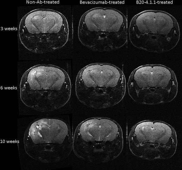
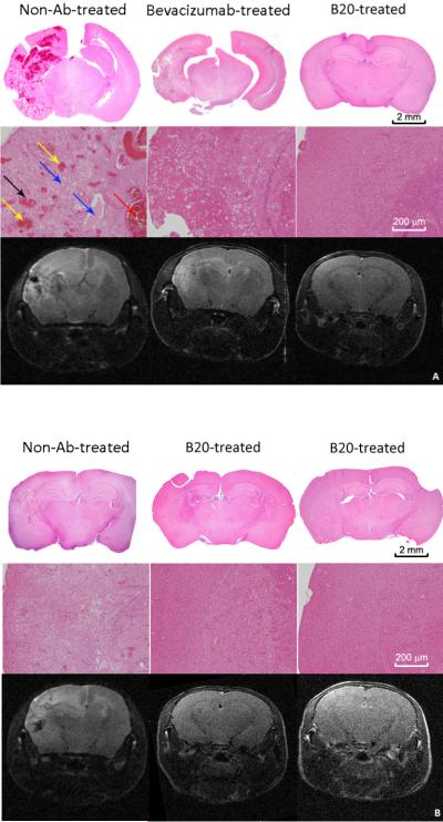
Experimental design: Cohorts of mice were irradiated with single-fraction 50- or 60-Gy doses of radiation targeted to the left hemisphere (brain) using the Leksell Perfexion Gamma Knife. The onset and progression of radiation necrosis were monitored longitudinally by in vivo, small-animal MRI, beginning 4 weeks after irradiation. MRI-derived necrotic volumes for antibody (Ab)-treated and untreated mice were compared. MRI results were supported by correlative histology.

Results: Hematoxylin and eosin-stained sections of brains from irradiated, non-Ab-treated mice confirmed profound tissue damage, including regions of fibrinoid vascular necrosis, vascular telangiectasia, hemorrhage, loss of neurons, and edema. Treatment with the murine anti-VEGF antibody B20-4.1.1 mitigated radiation-induced changes in an extraordinary, highly statistically significant manner. The development of radiation necrosis in mice under treatment with bevacizumab (a humanized anti-VEGF antibody) was intermediate between that for B20-4.1.1-treated and non-Ab-treated animals. MRI findings were validated by histologic assessment, which confirmed that anti-VEGF antibody treatment dramatically reduced late-onset necrosis in irradiated brain.

Conclusions: The single-hemispheric irradiation mouse model, with longitudinal MRI monitoring, provides a powerful platform for studying the onset and progression of radiation necrosis and for developing and testing new therapies. The observation that anti-VEGF antibodies are effective mitigants of necrosis in our mouse model will enable a wide variety of studies aimed at dose optimization and timing and mechanism of action with direct relevance to ongoing clinical trials of bevacizumab as a treatment for radiation necrosis.


Figures

Figure 1
Figure 1
MRI can detect radiation necrosis in irradiated brain. Representative, transaxial, T2-weighted spin-echo images acquired longitudinally from non-Ab-treated, bevacizumab-treated, and B20-4.1.1-treated mice at 3 (top), 6 (middle) and 10 weeks (bottom) following a single 60 Gy at (50% isodose) of radiation. Slices are chosen to display the same anatomic region of the brain at all three time points.
Figure 2
Figure 2
Histogram analysis enables quantitative MRI measurement of necrotic volume. (A) Image-pixel intensity histograms for the same non-Ab-treated mouse at 6 and 10 weeks following a single 60 Gy of radiation (red, blue) and average intensity histogram for 10 non-irradiated non-Ab-treated mice (black). The intensity cutoff used to define hyperintense pixels, corresponding to necrotic tissue, is indicated by the labeled arrow at a normalized intensity of 1.4. (B) Image-pixel intensity histograms for the same B20-4.1.1-treated (red) and bevacizumab-treated (blue) mice at 6 (dash line) and 10 (solid line) weeks following a single 60-Gy dose of radiation.
Figure 3
Figure 3
MRI-derived necrotic volumes in mice irradiated hemispherically with a single 50-Gy dose of GK radiation. MRI-defined volumes, mean ± SD (n = 10), of radiation necrosis vs. time post-irradiation for non-Ab-treated and B20-4.1.1-treated mice; all the mice received a single 50-Gy of radiation (50% isodose). At 13 weeks post-irradiation, the difference in MRI-derived necrotic volumes for the B20-4.1.1-treated cohort and the non-Ab-treated controls was statistically significant (p < 0.0001).
Figure 4
Figure 4
MRI-derived necrotic volumes in mice irradiated hemispherically with a single 60-Gy dose of GK radiation. (A) MRI-defined volumes, mean ± SD (n = 5), of radiation necrosis vs. time post-irradiation for non-Ab-treated, bevacizumab -treated, and B20-4.1.1-treated mice; all the mice received a single 60-Gy of radiation (50% isodose). Necrotic volumes for both B20-4.1.1-treated and bevacizumab-treated cohorts were significantly smaller than for the non-Ab-treated cohort (p<0.0001 at weeks 6, 7, 8, and 10 post-irradiation). MR-derived necrotic volumes for B20-4.1.1-treated and bevacizumab-treated cohorts were significantly different from one another at weeks 8, 9, and 10 post-irradiation (p<0.0001), but not at week 7 post-irradiation (p = 0.8). (B) MRI-defined volumetric rate of radiation necrosis progression, mean ± SD (n = 5), derived from the slope of the curves in the left panel, for the 3-7 and 7-10 week periods. The rate of progression of necrosis for the B20-4.1.1-treated cohort was smaller than that for the non-Ab-treated cohort over both the 3-7 week (p<0.0001) and 7-10 week (p=0.0002) post-irradiation periods. For the bevacizumab-treated cohort, the rate of progression of necrosis was smaller than that for the non-Ab-treated cohort over the initial 3-7 week post-irradiation period (p<0.0001), but not over the 7-10 week post-irradiation period (p=0.2).
Figure 5
Figure 5
H&E-stained sections display characteristic histologic features of radiation necrosis and demonstrate mitigation by anti-VEGF Ab. (A) Representative 2× (top) and 10× (middle) H&E histology slices chosen near the radiation isocenter, and corresponding T2W images (bottom) for non-Ab-treated, bevacizumab-treated, and B20-4.1.1-treated mice at 10 weeks following a single 60-Gy fraction of radiation. (B) Representative 2× (top) and 10x (middle) H&E histology slices, and corresponding T2W images (bottom) for one control and two B20-4.1.1-treated mice at 13 weeks following a single 50-Gy fraction of radiation. The irradiated hemispheres of the control mice show many of the histologic features that are characteristic of radiation necrosis, including fibrinoid vascular necrosis (black arrow), vascular telangiectasia (yellow arrows), hemorrhage (red arrow), loss of neurons and edema (blue arrows). In addition, the tissue injury observed on the histology slices are highly correlated with the hyperintense regions on T2W images.

Similar articles

Cited by

References

    1. Giglio P, Gilbert MR. Cerebral radiation necrosis. Neurologist. 2003;9:180–8. - PubMed
    1. Stupp R, Mason WP, van den Beuf MJ. Radiotherapy plus concomitant and adjuvant temozolomide for newly diagnosed glioblastoma (vol 352, pg 19, 2005). Annals of Oncology. 2005;16:949.
    1. Stupp R, Hegi ME, Mason WP, van den Bent MJ, Taphoorn MJB, Janzer RC, et al. Effects of radiotherapy with concomitant and adjuvant temozolomide versus radiotherapy alone on survival in glioblastoma in a randomised phase III study: 5-year analysis of the EORTC-NCIC trial. Lancet Oncology. 2009;10:459–66. - PubMed
    1. Shaw PJ, Bates D. Conservative Treatment of Delayed Cerebral Radiation Necrosis. Journal of Neurology Neurosurgery and Psychiatry. 1984;47:1338–41. - PMC - PubMed
    1. Siu A, Wind JJ, Iorgulescu JB, Chan TA, Yamada Y, Sherman JH. Radiation necrosis following treatment of high grade glioma-a review of the literature and current understanding. Acta Neurochirurgica. 2012;154:191–201. - PubMed

Publication types

MeSH terms

Substances