

Eosinophilic oesophagitis: clinical presentation and pathogenesis
- PMID: 24647582
- PMCID: PMC4495666
- DOI: 10.1136/postgradmedj-2012-131403
Eosinophilic oesophagitis: clinical presentation and pathogenesis
Abstract
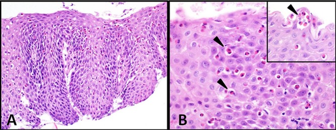
Eosinophilic oesophagitis (EoE) is an inflammatory disorder of the oesophagus which has become increasingly recognised over recent years, although it remains underdiagnosed in many centres. It is characterised histologically by a significant eosinophilic infiltration of the oesophageal mucosa (>15 eosinophils per high powered field), and clinically with features of oesophageal dysfunction such a dysphagia, food impaction, and proton pump inhibitor (PPI) resistant dyspepsia. Fibrosis and oesophageal remodelling may occur and lead to oesophageal strictures. An allergic predisposition is common in the EoE population, which appears to be primarily food antigen driven in children and aeroallergen driven in adults. Evidence suggests that the pathogenesis of EoE is due to a dysregulated immunological response to an environmental allergen, resulting in a T helper type 2 (Th2) inflammatory disease and remodelling of the oesophagus in genetically susceptible individuals. Allergen elimination and anti-inflammatory therapy with corticosteroids are currently the mainstay of treatment; however, an increasing number of studies are now focused on targeting different stages in the disease pathogenesis. A greater understanding of the underlying mechanisms resulting in EoE will allow us to improve the therapeutic options available.
Keywords: HISTOPATHOLOGY; IMMUNOLOGY; PUBLIC HEALTH.
Figures

References
-
- Kumar M, Sweis R, Wong T. Eosinophilic oesophagitis: investigations and management. Postgrad Med J 2014, in press. - PubMed
-
- Straumann A, Spichtin HP, Grize L, et al. Natural history of primary eosinophilic esophagitis: a follow-up of 30 adult patients for up to 11.5 years. Gastroenterology 2003;125:1660–9. - PubMed
-
- Landres RT, Kuster GG, Strum WB. Eosinophilic esophagitis in a patient with vigorous achalasia. Gastroenterology 1978;74:1298–301. - PubMed
-
- Attwood SE, Smyrk TC, Demeester TR, et al. Esophageal eosinophilia with dysphagia. A distinct clinicopathologic syndrome. Dig Dis Sci 1993;38:109–16. - PubMed
-
- Hruz P, Straumann A, Bussmann C, et al. Escalating incidence of eosinophilic esophagitis: a 20-year prospective, population-based study in Olten County, Switzerland. J Allergy Clin Immunol 2011;128:1349–50. - PubMed
Publication types
MeSH terms
Substances
LinkOut - more resources
Full Text Sources
Other Literature Sources
Medical
