. 2014 Mar 19;9(3):e92505.
doi: 10.1371/journal.pone.0092505. eCollection 2014.

Effect of silibinin in reducing inflammatory pathways in in vitro and in vivo models of infection-induced preterm birth

Affiliations

Effect of silibinin in reducing inflammatory pathways in in vitro and in vivo models of infection-induced preterm birth

Ratana Lim et al. PLoS One. .

Abstract

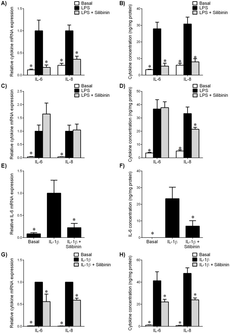
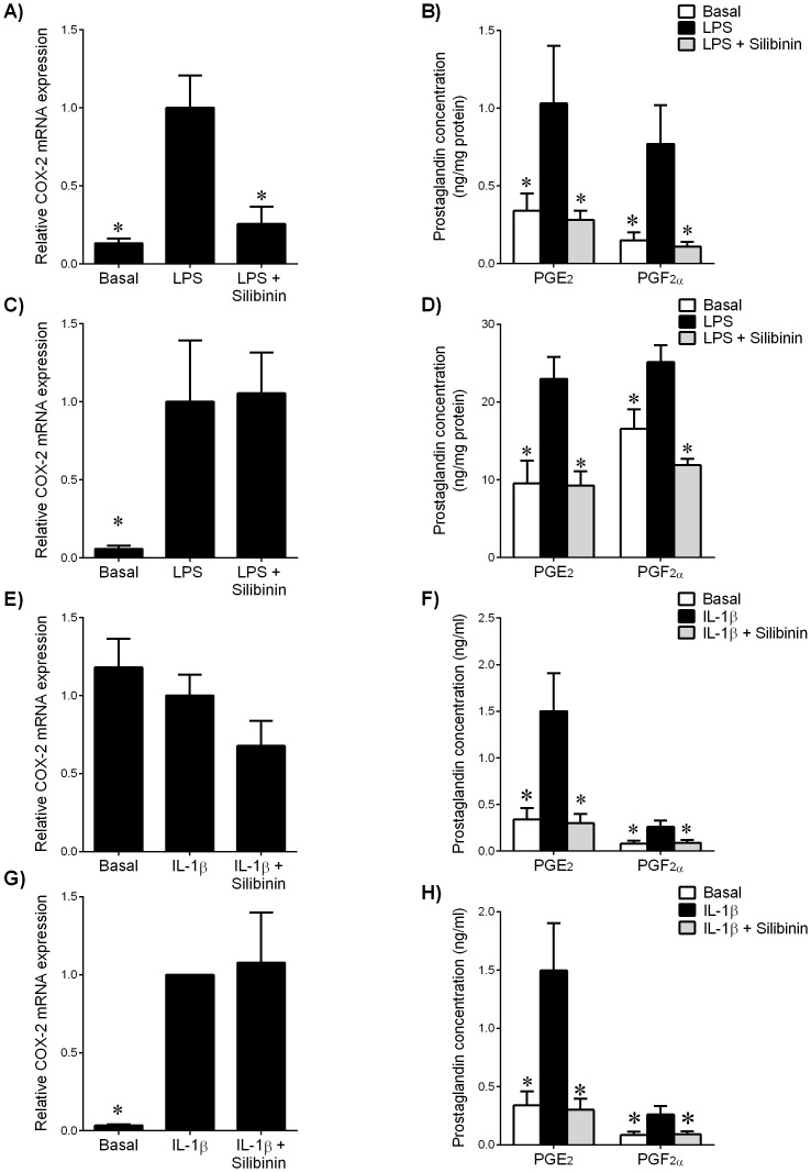
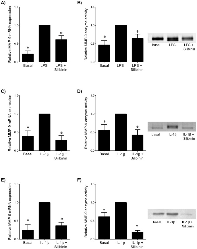
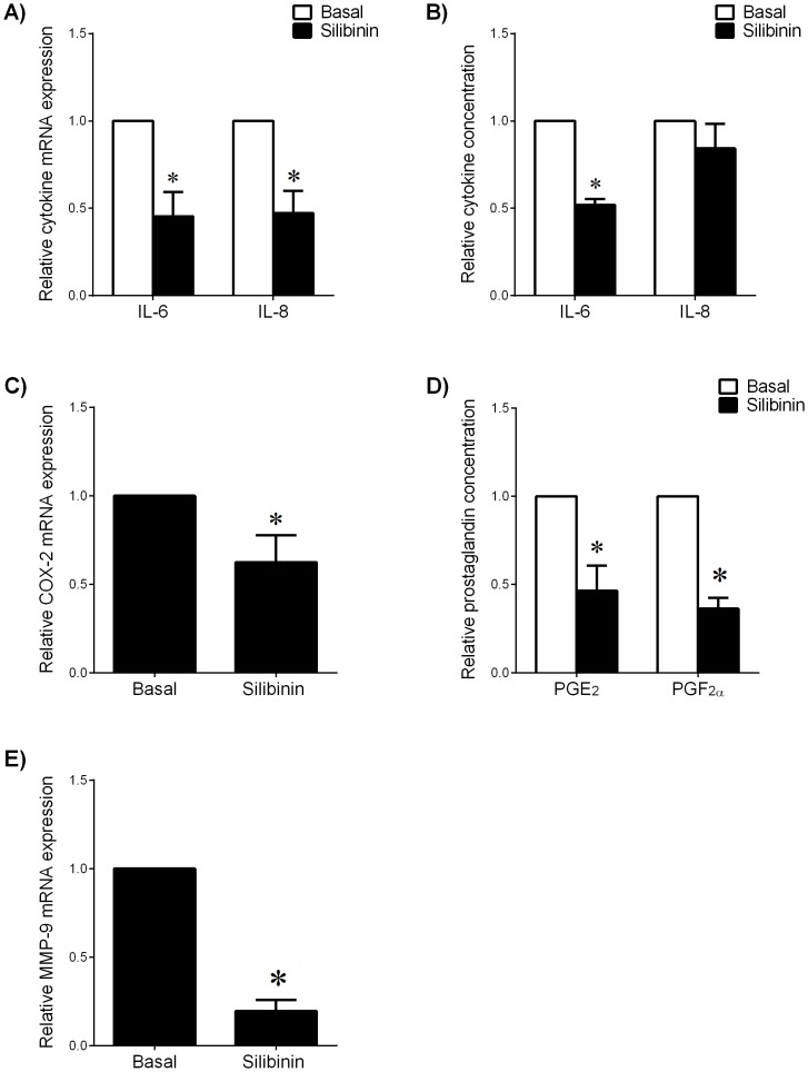
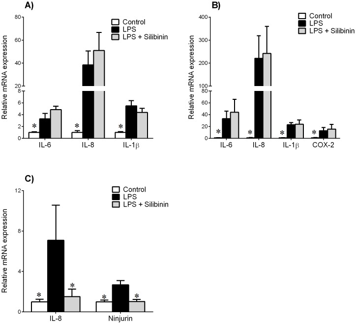
Infection-induced preterm birth is the largest cause of infant death and of neurological disabilities in survivors. Silibinin, from milk thistle, exerts potent anti-inflammatory activities in non-gestational tissues. The aims of this study were to determine the effect of silibinin on pro-inflammatory mediators in (i) human fetal membranes and myometrium treated with bacterial endotoxin lipopolysaccharide (LPS) or the pro-inflammatory cytokine IL-1β, and (ii) in preterm fetal membranes with active infection. The effect of silibinin on infection induced inflammation and brain injury in pregnant mice was also assessed. Fetal membranes and myometrium (tissue explants and primary cells) were treated with 200 μM silibinin in the presence or absence of 10 μg/ml LPS or 1 ng/ml IL-1β. C57BL/6 mice were injected with 70 mg/kg silibinin with or without 50 μg LPS on embryonic day 16. Fetal brains were collected after 6 h. In human fetal membranes, silibinin significantly decreased LPS-stimulated expression of IL-6 and IL-8, COX-2, and prostaglandins PGE2 and PGF2α. In primary amnion and myometrial cells, silibinin also decreased IL-1β-induced MMP-9 expression. Preterm fetal membranes with active infection treated with silibinin showed a decrease in IL-6, IL-8 and MMP-9 expression. Fetal brains from mice treated with silibinin showed a significant decrease in LPS-induced IL-8 and ninjurin, a marker of brain injury. Our study demonstrates that silibinin can reduce infection and inflammation-induced pro-labour mediators in human fetal membranes and myometrium. Excitingly, the in vivo results indicate a protective effect of silibinin on infection-induced brain injury in a mouse model of preterm birth.


Conflict of interest statement

Competing Interests: The authors have declared that no competing interests exist.

Figures

Figure 1
Figure 1. Effect of silibinin on pro-inflammatory cytokines in human gestational tissues.
(A,B) Fetal membranes and (C,D) myometrium were incubated 24 h in the absence or presence of 10 μg/ml LPS with or without 200 μM silibinin (n = 6 patients). (E,F) Human amnion cells and (G,H) myometrial cells were incubated 24 h in the absence or presence of IL-1β with or without silibinin (n = 6 patients). (A,C,E,G) Gene expression for IL-6 and IL-8 was analysed by qRT-PCR. Cytokine mRNA expression was normalized to GAPDH mRNA expression and the fold change was calculated relative to LPS or IL-1β-stimulated expression. Data is displayed as mean ± SEM (one-way ANOVA). *P<0.05 vs. LPS or IL-1β-stimulated expression. (B,D,F,H) The incubation medium was assayed for concentration of IL-6 and IL-8 release by ELISA. Each bar represents mean concentration ± SEM (one-way ANOVA). *P<0.05 vs. LPS or IL-1β-stimulated release.
Figure 2
Figure 2. Effect of silibinin on COX-prostaglandin pathway in human gestational tissues.
(A,B) Fetal membranes and (C,D) myometrium were incubated 24 h in the absence or presence of 10 μg/ml LPS with or without 200 μM silibinin (n = 6 patients). (E,F) Human amnion cells and (G,H) myometrial cells were incubated 24 h in the absence or presence of IL-1β with or without silibinin (n = 6 patients). (A,C,E,G) Gene expression for COX-2 was analysed by qRT-PCR. COX-2 mRNA expression was normalized to GAPDH mRNA expression and the fold change was calculated relative to LPS or IL-1β-stimulated expression. Data is displayed as mean ± SEM (one-way ANOVA). *P<0.05 vs. LPS or IL-1β-stimulated expression. (B,D,F,H) The incubation medium was assayed for concentration of PGE2 and PGF release by ELISA. Each bar represents mean concentration ± SEM (one-way ANOVA). *P<0.05 vs. LPS or IL-1β-stimulated release.
Figure 3
Figure 3. Effect of silibinin on MMP-9 expression in human gestational tissues.
(A,B) Myometrium was incubated 24 h in the absence or presence of 10 μg/ml LPS with and without 200 μM silibinin (n = 6 patients). (C,D) Human amnion cells and (E,F) myometrial cells were incubated 24 h in the absence or presence of IL-1β with and without silibinin (n = 6 patients). (A,C,E) Gene expression for MMP-9 was analysed by qRT-PCR. MMP-9 mRNA expression was normalized to GAPDH mRNA expression and the fold change was calculated relative to LPS or IL-1β-stimulated expression. Data is displayed as mean ± SEM (one-way ANOVA). *P<0.05 vs. LPS or IL-1β-stimulated expression. (B,D,F) The incubation medium was assayed for MMP-9 activity by gelatin zymography. Zymography from one patient per tissue type is shown depicting MMP-9 activity.
Figure 4
Figure 4. Effect of silibinin on pro-labour mediators in preterm fetal membranes.
Preterm fetal membranes with histological chorioamnionitis and following spontaneous preterm labour were incubated for 24 μM silibinin (n = 7 patients). (A,C,E) Gene expression for IL-6, IL-8, COX-2 and MMP-9 were analysed by qRT-PCR. mRNA expression was normalized to GAPDH mRNA expression and the fold change was calculated relative to basal expression. Data is displayed as mean ± SEM (one-way ANOVA). *P<0.05 vs. basal expression. (B,D) The incubation medium was assayed for concentration of IL-6, IL-8, PGE2 and PGF release by ELISA. Data was normalised to untreated (basal) levels, which was set at 1. Each bar represents mean ± SEM (one-way ANOVA). *P<0.05 vs. basal release.
Figure 5
Figure 5. Effect of silibinin in a mouse model of infection-induced preterm birth.
Time mated C57BL/6 mice were intraperitoneally injected with either saline (control, n = 7), 50 μg LPS (n = 6), or LPS with 70 mg/kg silibinin (n = 5). Tissues were collected after 6 h. Gene expression for IL-6, IL-8, IL-1β and COX-2 were analysed by qRT-PCR in (A) placenta and (B) myometrium. Gene expression of IL-8 and ninjurin were analysed in (C) fetal brain. Gene expression was normalized to GAPDH mRNA expression and the fold change was calculated relative to control expression. Data is displayed as mean ± SEM (one-way ANOVA). *P<0.05 vs. control gene expression.

Similar articles

Cited by

References

    1. Beck S, Wojdyla D, Say L, Betran AP, Merialdi M, et al. (2010) The worldwide incidence of preterm birth: a systematic review of maternal mortality and morbidity. Bull World Health Organ 88: 31–38. - PMC - PubMed
    1. Goldenberg RL, Culhane JF, Iams JD, Romero R (2008) Epidemiology and causes of preterm birth. Lancet 371: 75–84. - PMC - PubMed
    1. Yoon BH, Romero R, Moon JB, Shim SS, Kim M, et al. (2001) Clinical significance of intra-amniotic inflammation in patients with preterm labor and intact membranes. Am J Obstet Gynecol 185: 1130–1136. - PubMed
    1. Menon R, Fortunato SJ (2007) Infection and the role of inflammation in preterm premature rupture of the membranes. Best Pract Res Clin Obstet Gynaecol 21: 467–478. - PubMed
    1. Galinsky R, Polglase GR, Hooper SB, Black MJ, Moss TJ (2013) The consequences of chorioamnionitis: preterm birth and effects on development. J Pregnancy 2013: 412831. - PMC - PubMed

Publication types