. 2014 May 7;14(9):1622-31.
doi: 10.1039/c3lc51353j. Epub 2014 Mar 20.

In vitro generation of colonic epithelium from primary cells guided by microstructures

Affiliations

In vitro generation of colonic epithelium from primary cells guided by microstructures

Yuli Wang et al. Lab Chip. .

Abstract

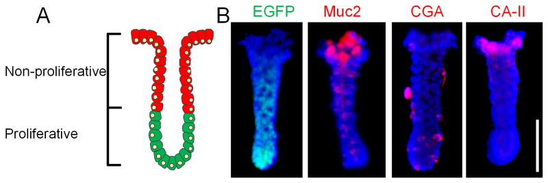
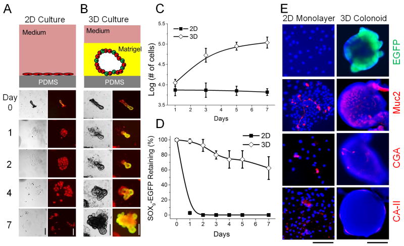
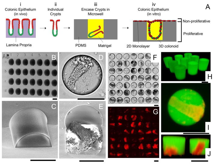
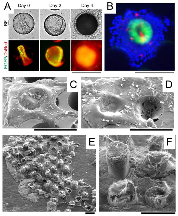
The proliferative compartment of the colonic epithelium in vivo is located in the basal crypt where colonic stem cells and transit-amplifying cells reside and fuel the rapid renewal of non-proliferative epithelial cells as they migrate toward the gut lumen. To mimic this tissue polarity, microstructures composed of polydimethylsiloxane (PDMS) microwells and Matrigel micropockets were used to guide a combined 2-dimensional (2D) and 3-dimensional (3D) hybrid culture of primary crypts isolated from the murine colon. The 2D and 3D culture of crypts on a planar PDMS surface was first investigated in terms of cell proliferation and stem cell activity. 3D culture of crypts with overlaid Matrigel generated enclosed, but highly proliferative spheroids (termed colonoids). 2D culture of crypts produced a spreading monolayer of cells, which were non-proliferative. A combined 2D/3D hybrid culture was generated in a PDMS microwell platform on which crypts were loaded by centrifugation into microwells (diameter = 150 μm, depth = 150 μm) followed by addition of Matrigel that formed micropockets locking the crypts within the microwells. Embedded crypts first underwent 3D expansion inside the wells. After the cells filled the microwells, they migrated onto the surrounding surface forming a 2D monolayer in the array regions without Matrigel. This unique 2D/3D hybrid culture generated a continuous, millimeter-scale colonic epithelial tissue in vitro, which resembled the polarized architecture (i.e. distinct proliferative and non-proliferative zones) and geometry of the colonic epithelium in vivo. This work initiates the construction of a "colon-on-a-chip" using primary cells/tissues with the ultimate goal of producing the physiologic structure and organ-level function of the colon.


Figures

Figure 1
Figure 1
Tissue polarity of colonic crypts. (A) A simplified schematic of colonic crypts possessing distinct proliferative (green cells) and non-proliferative (red cells) zones. (B) EGFP (green) fluorescence and immunofluorescence staining of Muc2, CGA and CA-II (red). Nuclei were stained with Hoechst 33342 (blue). Scale bar = 100 μm.
Figure 2
Figure 2
Comparison between 2D and 3D culture of crypts on or over a PDMS planar surface. (A) 2D culture. (B) 3D culture. (A–B) Top panels are schematics of the culture system. Bottom panels are brightfield (left) and overlaid images of EGFP and DsRed fluorescence (right) of crypt fate over time. (C) Cell number vs. time in 2D and 3D culture systems. Both contained the same number of crypts (1,000) at time point 0. (D) EGFP expression vs. time for crypts cultured in 2D and 3D. (E) EGFP (green) fluorescence image and immunofluorescence staining of Muc2, CGA and CA-II (red) for 2D monolayers (left panel) and 3D colonoids (right panels). Nuclei were stained with Hoechst 33342 (blue). Time in culture was 7 days. Scale bars = 200 μm for images in A, B and F.
Figure 3
Figure 3
Microwell arrays for combined 2D/3D culture of crypts. (A) Schematic of the 2D/3D culture strategy. (i) The colonic epithelium in vivo is composed of crypt subunits each in contact with a lamina propria. (ii) Individual crypts are isolated from a colon. (iii) Crypts are loaded into PDMS microwells and encased within Matrigel micropockets. (iv) In vitro culture of crypts in microwells generates colonic epithelium resembling the polarized architecture of crypts in vivo. (B) SEM image of the PDMS microwell array. The wells were labeled with shallow alphanumeric characters. (C) A close-up SEM image of a disrupted section of a microwell. (D–E) Brightfield (D) and SEM image (E) of a microwell with a loaded crypt. (F) Brightfield image showing 28 out of 49 wells loaded with crypts. (G) DsRed fluorescence image of F. (H) Fluorescence confocal image of Matrigel pockets formed in the microwell array. (I–J) Shown is a standard fluorescence image (I) and a confocal reconstructed Z slice (J) of crypts encapsulated within isolated Matrigel pockets in the microwell array. The panels display overlaid red/green fluorescence images. The Matrigel was mixed with 100 μg/mL fluorescein-dextran in images H through I. Scale bars = 100 μm for images in B–J.
Figure 4
Figure 4
In vitro generation of millimeter-scale, colonic epithelium with a polarized architecture from primary tissue. (A) Culture of colonoids in a PDMS microwell. A crypt was loaded into a microwell on the array. By 48 h, a 3D colonoid formed that filled the microwell. By 96 h, a 2D/3D hybrid culture was formed. The top panels are brightfield images while the bottom panels are overlaid red/green fluorescence images. (B) Overlaid fluorescence image of the 2D/3D hybrid culture at 96 h (green = EGFP, red = Muc2 stained by immunofluorescence, blue = nuclei stained with Hoechst 33342). (C–D) SEM image of colonic epithelial tissues while still on the PDMS microwell array. The 2D/3D hybrid culture was imaged at 96 h (C) and 144 h (D). (E) SEM images of a 1.7 mm2 section of the in vitro-generated colonic epithelial tissue transferred to a tape so that the tissue is viewed from its underside. (F) A close-up of the underside of the tissue shown in (E). Scale bars = 200 μm. The Matrigel plug is not visualized in the SEM images since its protein concentration (~4 mg/mL) is too low to maintain a solid structure during fixation.

Similar articles

Cited by

References

    1. Barker N, van de Wetering M, Clevers H. Genes & Development. 2008;22:1856–1864. - PMC - PubMed
    1. Fuchs E, Chen T. Embo Reports. 2013;14:39–48. - PMC - PubMed
    1. Brittan M, Wright NA. Gut. 2004;53:899–910. - PMC - PubMed
    1. Kosinski C, Li VSW, Chan ASY, Zhang J, Ho C, Tsui WY, Chan TL, Mifflin RC, Powell DW, Yuen ST, Leung SY, Chen X. Proc Natl Acad Sci U S A. 2007;104:15418–15423. - PMC - PubMed
    1. Yen TH, Wright NA. Stem Cell Rev. 2006;2:203–212. - PubMed

Publication types