. 2014 Mar 19;9(3):e92363.
doi: 10.1371/journal.pone.0092363. eCollection 2014.

Pro-inflammatory mediation of myoblast proliferation

Affiliations

Pro-inflammatory mediation of myoblast proliferation

Jeffrey S Otis et al. PLoS One. .

Abstract

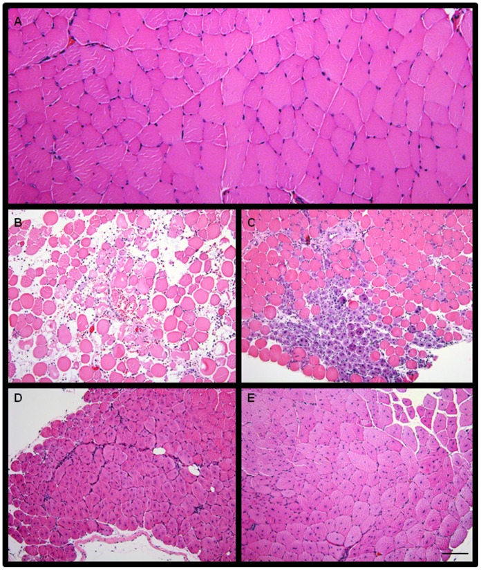
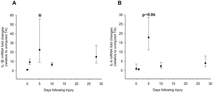
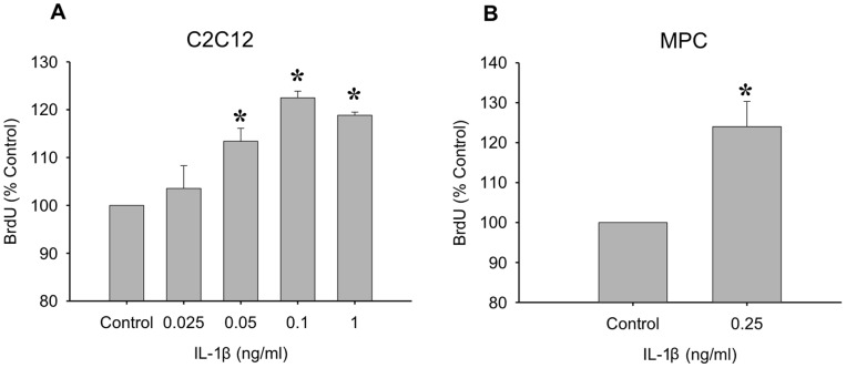
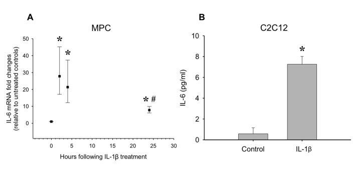
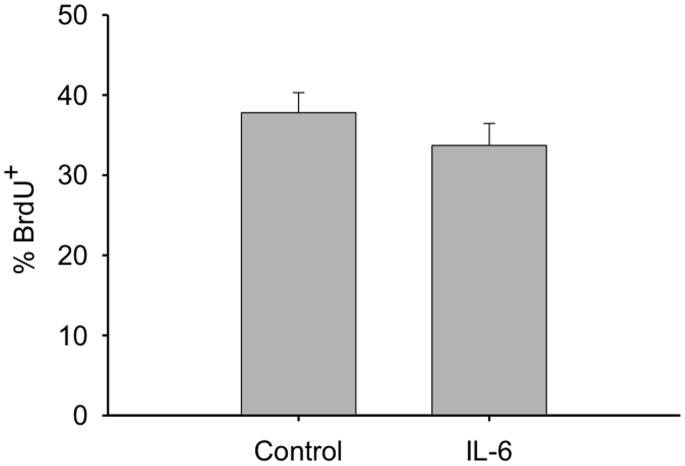
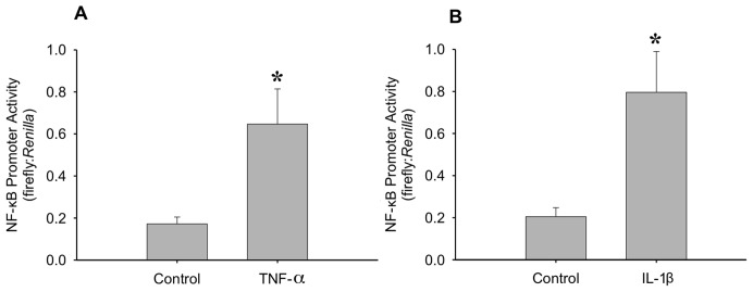
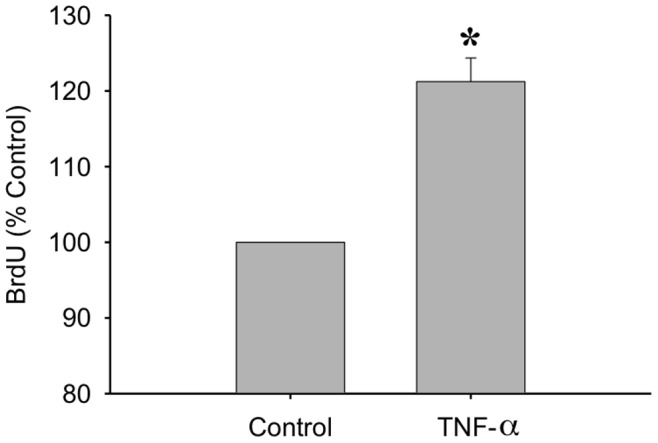
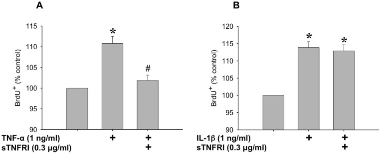
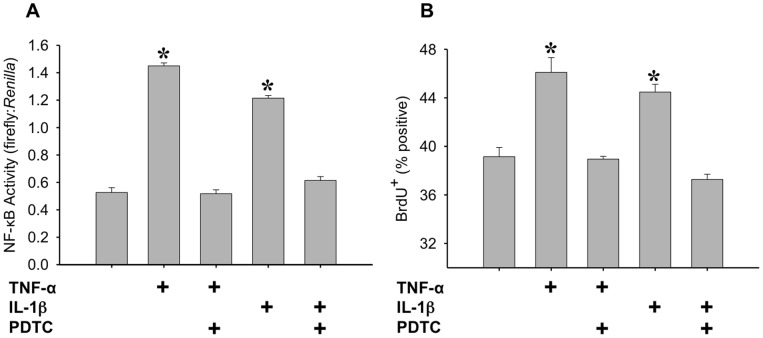
Skeletal muscle satellite cell function is largely dictated by the surrounding environment following injury. Immune cell infiltration dominates the extracellular space in the injured area, resulting in increased cytokine concentrations. While increased pro-inflammatory cytokine expression has been previously established in the first 3 days following injury, less is known about the time course of cytokine expression and the specific mechanisms of cytokine induced myoblast function. Therefore, the expression of IL-1β and IL-6 at several time points following injury, and their effects on myoblast proliferation, were examined. In order to do this, skeletal muscle was injured using barium chloride in mice and tissue was collected 1, 5, 10, and 28 days following injury. Mechanisms of cytokine induced proliferation were determined in cell culture using both primary and C2C12 myoblasts. It was found that there is a ∼20-fold increase in IL-1β (p≤0.05) and IL-6 (p = 0.06) expression 5 days following injury. IL-1β increased proliferation of both primary and C2C12 cells ∼25%. IL-1β stimulation also resulted in increased NF-κB activity, likely contributing to the increased proliferation. These data demonstrate for the first time that IL-1β alone can increase the mitogenic activity of primary skeletal muscle satellite cells and offer insight into the mechanisms dictating satellite cell function following injury.


Conflict of interest statement

Competing Interests: The authors have declared that no competing interests exist.

Figures

Figure 1
Figure 1. Histological representation of the progression of injured muscle through repair and regeneration.
(A) Uninjured tibialis anterior (TA) muscle. Injured TA muscle (B) 1 days, (C) 5 days, (D) 10 days, and (E) 28 days after barium chloride (BaCl2) injection. Bar in E = 100 μm. Images taken at 10X magnification.
Figure 2
Figure 2. IL-1β and IL-6 mRNA expression post-injury to the tibialis anterior (TA).
TA muscle was injured using a barium chloride injection method and muscles were collected 1, 5, 10, and 28 days post-injury and analyzed for (A) IL-1β and (B) IL-6 mRNA expression levels. Data are expressed as fold-increase +/− expected high and low expression relative to the average value . *denotes significance (p≤0.05) compared to uninjured control (n = 3–4 per time point).
Figure 3
Figure 3. The mitogenic effects of IL-1β.
(A) An IL-1β dose response was performed on C2C12 myoblasts. Concentrations of 0.5 ng/ml to 1 ng/ml significantly increased proliferation in myoblasts. (B) An intermediate dose of IL-1β (0.25 ng/ml) was used to test the mitogenic effects of IL-1β on primary muscle precursor cells. Data are expressed relative to control ± SEM. *denotes significance (p≤0.05) compared to control (n = 3–5 per dose).
Figure 4
Figure 4. The effect of IL-1β treatment on IL-6 mRNA and protein levels.
(A) Primary MPCs were treated with IL-1β (0.25 ng/ml) and IL-6 mRNA was determined over 24 hours. *denotes significance (p≤0.05) compared to control. # denotes significance (p≤0.05) compared to the 2 hour time point (n = 4 per time point). (B) IL-1β treatment (1 ng/ml) also increased IL-6 protein released into the media in C2C12 myoblasts. *denotes significance (p≤0.05) compared to control (n = 4–6).
Figure 5
Figure 5. The mitogenic effects of IL-6 on C2C12 myoblasts.
Myoblasts were treated with IL-6 (1 ng/ml) and BrdU incorporation was determined 24 hours after treatment (n = 5).
Figure 6
Figure 6. The effects of TNF-α and IL-1β on nuclear factor-kappa B (NF-κB) activity.
Transfection of myoblasts with NF-κB cis-reporter construct allowed the study of the effects of (A) TNF-α (20 ng/ml) and (B) IL-1β (1 ng/ml) on NF-κB activity. 24 hours after transfection, cells were treated with either TNF-α or IL-1β for an additional 24 hours. Data are reported as the ratio of firefly to Renilla luminescence *denotes significance (p≤0.05) compared to control (n = 7 for TNF-α and n = 6 for IL-1β).
Figure 7
Figure 7. TNF-α promotes proliferation of myoblasts.
BrdU incorporation was measured after a 24 hour TNF-α treatment (20 ng/ml). Data are expressed relative to control ± SEM. *denotes significance (p≤0.05) compared to control (n = 7).
Figure 8
Figure 8. Determining the role of TNF-α in IL-1β mediated proliferation.
(A) The effect of TNF-α on myoblast proliferation is blocked by pre-incubation of TNF-α with soluble TNF receptor I (sTNFRI) (0.3 μg/ml, 2 hours at 37°C). (B) IL-1β induced proliferation of myoblasts is not blocked by pre-incubation with sTNFRI. Data are expressed relative to control ± SEM. *denotes significance (p≤0.05) compared to control. # denotes significance (p<0.05) compared to TNF-α treated.
Figure 9
Figure 9. Determining the role of NF-κB activation in IL-1β and TNF-α induced proliferation.
(A) NF-κB activity was determined using the NF-κB cis-reporter construct and data are reported as the ratio of firefly to Renilla luminescence. C2C12 myoblasts were transfected with the NF-κB cis-reporter construct, pretreated with 50 μM PDTC for 1.5 hours, and treated with either 1 ng/ml IL-1β or 20 ng/ml TNF-α for four hours. *denotes significance (p≤0.05) compared to control (n = 4). (B) Proliferation of myoblasts was measured through BrdU incorporation in C2C12 myoblasts following a 50 μM PDTC pretreatment for 1.5 hours, and a 20 hour treatment with either 1 ng/ml IL-1β or 20 ng/ml TNF-α. *denotes significance (p≤0.05) compared to control (n = 4).

References

    1. Mauro A (1961) Satellite cell of skeletal muscle fibers. JBiophysBiochemCytol 9: 493–495. - PMC - PubMed
    1. Hawke TJ, Garry DJ (2001) Myogenic satellite cells: physiology to molecular biology. J Appl Physiol 91: 534–551. - PubMed
    1. Tedesco FS, Dellavalle A, Diaz-Manera J, Messina G, Cossu G (2010) Repairing skeletal muscle: regenerative potential of skeletal muscle stem cells. J Clin Invest 120: 11–19. - PMC - PubMed
    1. Parise G, McKinnell IW, Rudnicki MA (2008) Muscle satellite cell and atypical myogenic progenitor response following exercise. Muscle Nerve 37: 611–619. - PubMed
    1. Sambasivan R, Yao R, Kissenpfennig A, Van Wittenberghe L, Paldi A, et al. (2011) Pax7-expressing satellite cells are indispensable for adult skeletal muscle regeneration. Development 138: 3647–3656. - PubMed

Publication types