Comparative Study
. 2014 May 15;306(10):F1198-209.
doi: 10.1152/ajprenal.00699.2013. Epub 2014 Mar 19.

Cells of renin lineage take on a podocyte phenotype in aging nephropathy

Affiliations
Comparative Study

Cells of renin lineage take on a podocyte phenotype in aging nephropathy

Jeffrey W Pippin et al. Am J Physiol Renal Physiol. .

Abstract

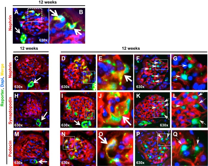
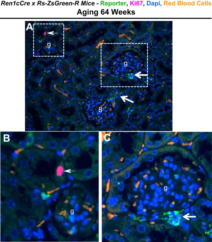
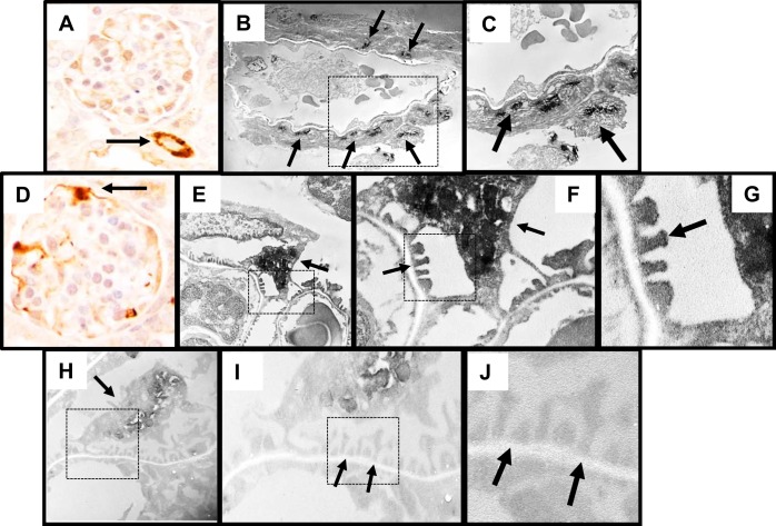
Aging nephropathy is characterized by podocyte depletion accompanied by progressive glomerulosclerosis. Replacement of terminally differentiated podocytes by local stem/progenitor cells is likely a critical mechanism for their regeneration. Recent studies have shown that cells of renin lineage (CoRL), normally restricted to the kidney's extraglomerular compartment, might serve this role after an abrupt depletion in podocyte number. To determine the effects of aging on the CoRL reserve and if CoRL moved from an extra- to the intraglomerular compartment during aging, genetic cell fate mapping was performed in aging Ren1cCre × Rs-ZsGreen reporter mice. Podocyte number decreased and glomerular scarring increased with advanced age. CoRL number decreased in the juxtaglomerular compartment with age. There was a paradoxical increase in CoRL in the intraglomerular compartment at 52 and 64 wk of age, where a subset coexpressed the podocyte proteins nephrin, podocin, and synaptopodin. Transmission electron microscopy studies showed that a subset of labeled CoRL in the glomerulus displayed foot processes, which attached to the glomerular basement membrane. No CoRL in the glomerular compartment stained for renin. These results suggest that, despite a decrease in the reserve, a subpopulation of CoRL moves to the glomerulus after chronic podocyte depletion in aging nephropathy, where they acquire a podocyte-like phenotype. This suggests that they might serve as adult podocyte stem/progenitor cells under these conditions, albeit in insufficient numbers to fully replace podocytes depleted with age.

Keywords: cells of renin lineage; focal segmental glomerulosclerosis; glomerulus; podocyte; regeneration.


Figures

Fig. 1.
Fig. 1.
Changes in podocyte number in aging nephropathy. Ren1cCre × Rs-ZsGreen-R reporter mice were studied with advancing age at 4, 12, 52 and 64 wk. A: glomerular tuft area increased progressively and significantly at 52 and 64 wk of age. B: podocyte density, measured by the number of p57-stained cells/glomerular tuft area was significantly reduced at 52 and 64 wk compared with younger ages. C–F: examples of p57 staining (brown, nuclear) at ages 4 (C), 12 (D), 52 (E), and 64 (F) wk respectively.
Fig. 2.
Fig. 2.
Glomerulosclerosis in aging nephropathy. A: glomerulosclerosis, quantitated on PAS-stained sections in aging Ren1cCre × Rs-ZsGreen-R reporter mice, increased significantly at 52 and 64 wk compared with younger ages. B–E: representative images of glomeruli at 4 (B), 12 (C), 52 (F), and 64 wk (G) show decreased podocyte number, glomerulosclerosis, and synchiae (indicated by arrows), features consistent with aging nephropathy.
Fig. 3.
Fig. 3.
Cells of renin lineage increase in the intraglomerular compartment and glomerular injury is reduced in glomeruli containing cells of renin lineage in aged Ren1cCre × Rs-ZsGreen-R mice. AC: representative pictures of kidneys from mice aged 12, 52, and 64 wk. Glomeruli with no labeled cells of renin lineage (CoRL) are indicated by solid circles, glomeruli containing labeled CoRL are indicated by dashed circles. D: quantification of the percentage of glomeruli containing labeled CoRL, percentage of glomeruli with injury, percentage of glomeruli with labeled CoRL with injury, and percentage of glomeruli with no labeled CoRL with injury.
Fig. 4.
Fig. 4.
Labeled CoRL decrease in the extraglomerular vascular smooth muscle compartment, and increase in the intraglomerular compartment in aging nephropathy. A–E: representative pictures of kidneys from mice aged 4, 12, 52, and 64 wk. A: low-magnification image (×100) from a 4-wk-old kidney shows that CoRL are permanently labeled with ZsGreen reporter in the extraglomerular vascular smooth muscle compartment. They were rarely detected in the intraglomerular compartment and are faintly detected in the tubular compartment. B: low magnification image (×100) from a 12 wk kidney shows a similar distribution of reporter labeled CoRL to 4 wk kidneys shown in A. C: at 52 wk, reporter labeled CoRL decrease in the extraglomerular vascular smooth muscle compartment. Labeled reporter cells were still rarely detected in glomeruli and in tubules. D: at 64 wk, reporter labeled CoRL remained decreased in the extraglomerular vascular smooth muscle compartment. However, there was a paradoxical increase in the number of reporter labeled CoRL in the intraglomerular compartment (labeled g). E: glomerulus from a 64-wk-old kidney viewed at higher magnification (×630) shows a number of reporter-labeled cells (green) in the intraglomerular compartment, which were in a characteristic podocyte distribution pattern. F: number of ZsGreen reporter-labeled CoRL was quantitated in Ren1cCre × Rs-ZsGreen-R reporter mice. Compared with 4 and 12 wk of age, the number of ZsGreen-labeled cells decreased significantly in the extraglomerular vascular smooth muscle compartment at 52 and 64 wk (shaded bars). In contrast, there was an increase in the number of reporter labeled CoRL in the glomerular tuft (black bars) at 64 wk. G–L: representative images of double staining for renin and ZsGreen reporter. G: lower magnification shows the distribution of CoRL reporter (green) was restricted to the extraglomerular vascular smooth muscle compartment (arrows indicate examples). H: distribution of renin staining (red) was also restricted to the extraglomerular vascular smooth muscle compartment (arrows indicate examples). I: merged image shows clear overlap of renin staining and the reporter (yellow, arrows indicate examples). Red blood cell autofluorescence appears orange in color. J–L: higher magnification images of the glomerulus indicated by the white boxes in G–I, respectively. These data show that renin staining is restricted to the extraglomerular compartment and that when labeled CoRL migrate to the glomerulus they do not stain for renin.
Fig. 5.
Fig. 5.
Quantification of Renin staining in aging nephropathy. A: quantitation of renin staining was expressed as a percentage of cortical area in aging Ren1cCre × Rs-ZsGreen-R reporter mice. No changes were detected between 4, 12, and 52 wk. The percentage area of renin staining decreased in mice with aging nephropathy at 64 wk. B–E: typical renin staining in 4-, 12-, 52-, and 64-wk-old mice. F and G: higher magnification images of the areas indicated in C and E, respectively, show renin staining was confined to the extraglomerular compartment and not present in the intraglomerular compartment (g).
Fig. 6.
Fig. 6.
Labeled CoRL costain for podocyte proteins and reporter in glomeruli in aging nephropathy. A: at 12 wk, reporter-labeled cells (green) were detected in the extraglomerular vascular smooth muscle compartment (white arrow), and a subset of reporter labeled cells present in the intraglomerular compartment costained for nephrin (yellow, represented by square). Cell nuclei were marked with DAPI (blue) B: portion of the glomerulus represented by the white square shows costaining of the reporter and nephrin at higher magnification, indicated by the arrows. C: typical glomerulus at 12 wk, where reporter labeled cells (green) were detected in the extraglomerular vascular smooth muscle compartment (white arrow), and nephrin (red) was limited to the glomerulus in a typical podocyte distribution. Cell nuclei were marked with DAPI (blue). D: at 64 wk, an increased subset of reporter labeled cells present in the intraglomerular compartment costained for nephrin (yellow, represented by square). E: portion of the glomerulus represented by the white square shows costaining of the reporter and nephrin at higher magnification, indicated by the arrow. F and G: lack of costaining for the reporter and nephrin. F: at 64 wk of age, a subset of reporter-labeled cells present in the intraglomerular compartment did not costain for nephrin (green, represented by square). G: portion of the glomerulus represented by the white square shows costaining of the reporter and nephrin at higher magnification, indicated by the thin arrows. H: typical glomerulus at 12 wk, reporter-labeled cells (green) were detected in the extraglomerular vascular smooth muscle compartment (white arrow), and synaptopodin (red) was limited to the glomerulus in a typical podocyte distribution. I: at 64 wk, increased reporter-labeled cells present in the intraglomerular compartment costained for synaptopodin (yellow, represented by square). J: portion of the glomerulus represented by the white square shows costaining at higher magnification, indicated by the arrow. K and L: lack of synaptopodin-reporter costaining. E: At 64 wk, a subset of reporter-labeled cells present in the intraglomerular compartment did not costain for synaptopodin (green, represented by square). F: portion of the glomerulus represented by the white square shows costaining of the reporter and synaptopodin at higher magnification, indicated by the thin arrows. M-O: podocin-reporter costaining. M: typical glomerulus at 12 wk, reporter-labeled cells (green) were detected in the extraglomerular vascular smooth muscle compartment (white arrow), and podocin (red) was limited to the glomerulus in a typical podocyte distribution. N: at 64 wk, increased reporter-labeled cells present in the intraglomerular compartment costained for podocin (yellow, represented by square). O: portion of the glomerulus represented by the white square shows costaining at higher magnification, indicated by the arrow. These data show that a subset of reporter-labeled CoRL in glomeruli of aged mice coexpress the podocyte proteins nephrin, synaptopodin, and podocin. P and Q: lack of costaining for the reporter and podocin. P: at 64 wk, a subset of reporter-labeled cells present in the intraglomerular compartment did not costain for podocin (green, represented by square). Q: portion of the glomerulus represented by the white square shows costaining of the reporter and podocin at higher magnification, indicated by the thin arrows.
Fig. 7.
Fig. 7.
Reporter and Ki67 staining in aging nephropathy. A: lower power image of ZsGreen reporter (green, arrows) and Ki67 staining (purple, arrow) in aging Ren1cCre × Rs-ZsGreen-R reporter mice. Red blood cell autofluorescence appears orange in color. B and C: higher magnification images of the 2 areas indicated by the white boxes in A show a Ki67-positive cell within a tubule (B, arrow) and reporter staining in the extraglomerular vascular smooth muscle compartment (C, arrow); glomeruli are indicated with (g).
Fig. 8.
Fig. 8.
Labeled CoRL acquire ultrastructural features of podocytes. To assess ultrastructural features labeled CoRL, immunoperoxidase staining for ZsGreen reporter was performed. A: image of immunoperoxidase staining for the ZsGreen reporter (brown) in the vascular smooth muscle compartment (indicated by arrow, positive control). B: immunoperoxidase staining for the ZsGreen reporter (black) on ultrathin section viewed by transmission electron microscopy (TEM) was detected in the vascular smooth muscle compartment, used as a positive control (arrows indicate examples). C: higher magnification image of reporter staining in the area indicated by the dashed square shown in B. D: immunoperoxidase staining for reporter (brown) in the intraglomerular compartment appears in a podocyte distribution (indicated by arrow). E: TEM image of podocyte with reporter staining (black) in both the cell body and within foot processes. F: higher magnification image of the area indicated by the dashed square shown in E. Arrows indicate staining in foot processes. G: even higher magnification image of the area indicated by the dashed square shown in F. Arrow indicates reporter staining in foot processes. H: lower magnification image of podocyte that lacked reporter staining in the cell body or within foot processes. I: higher magnification image of reporter staining (black) in the area indicated by the dashed square shown in H. Arrows indicate the lack of staining in foot processes. J: higher magnification image of the area indicated by the dashed square shown in I. Arrows indicate the lack of staining in foot processes.

Comment in

References

    1. Ahn SY, Ryu J, Baek SH, Kim S, Na KY, Kim KW, Chae DW, Chin HJ. Incident chronic kidney disease and newly developed complications related to renal dysfunction in an elderly population during 5 years: a community-based elderly population cohort study. PLos One 8: e84467, 2013 - PMC - PubMed
    1. Alpers CE, Hudkins KL, Floege J, Johnson RJ. Human renal cortical interstitial cells with some features of smooth muscle cells participate in tubulointerstitial and crescentic glomerular injury. J Am Soc Nephrol 5: 201–209, 1994 - PubMed
    1. Angelotti ML, Lazzeri E, Lasagni L, Romagnani P. Only anti-CD133 antibodies recognizing the CD133/1 or the CD133/2 epitopes can identify human renal progenitors. Kidney Int 78: 620–621, 2010 - PubMed
    1. Benigni A, Morigi M, Rizzo P, Gagliardini E, Rota C, Abbate M, Ghezzi S, Remuzzi A, Remuzzi G. Inhibiting angiotensin-converting enzyme promotes renal repair by limiting progenitor cell proliferation and restoring the glomerular architecture. Am J Pathol 179: 628–638, 2011 - PMC - PubMed
    1. Brinkkoetter PT, Wu JS, Ohse T, Krofft RD, Schermer B, Benzing T, Pippin JW, Shankland SJ. p35, the non-cyclin activator of Cdk5, protects podocytes against apoptosis in vitro and in vivo. Kidney Int 77: 690–699, 2010 - PubMed

Publication types

MeSH terms

LinkOut - more resources