. 2014 Mar 19;34(12):4200-13.
doi: 10.1523/JNEUROSCI.4172-13.2014.

Single-neuron NMDA receptor phenotype influences neuronal rewiring and reintegration following traumatic injury

Affiliations

Single-neuron NMDA receptor phenotype influences neuronal rewiring and reintegration following traumatic injury

Tapan P Patel et al. J Neurosci. .

Abstract

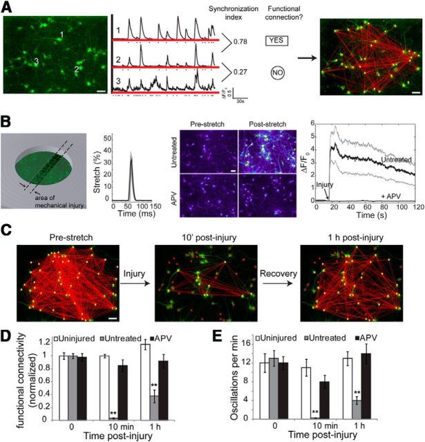
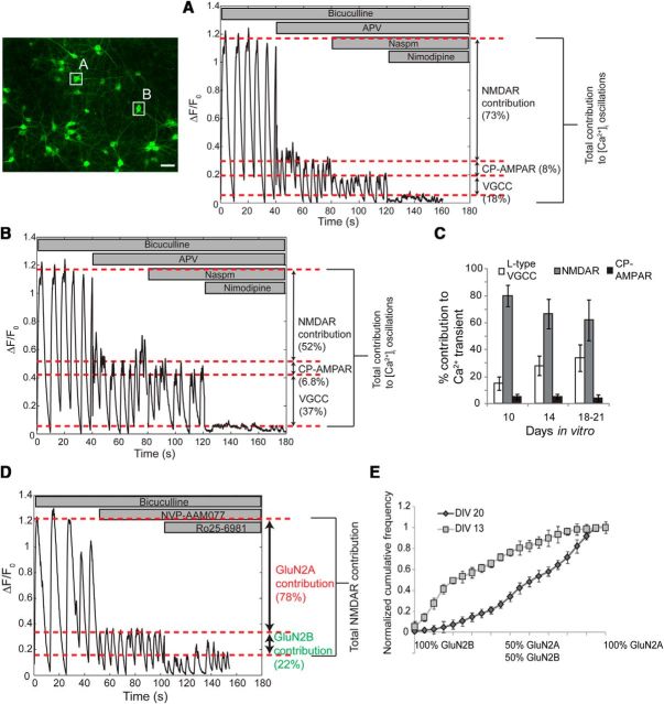
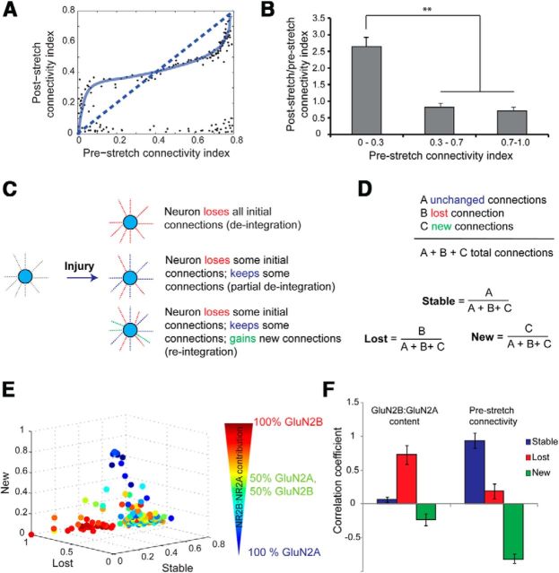
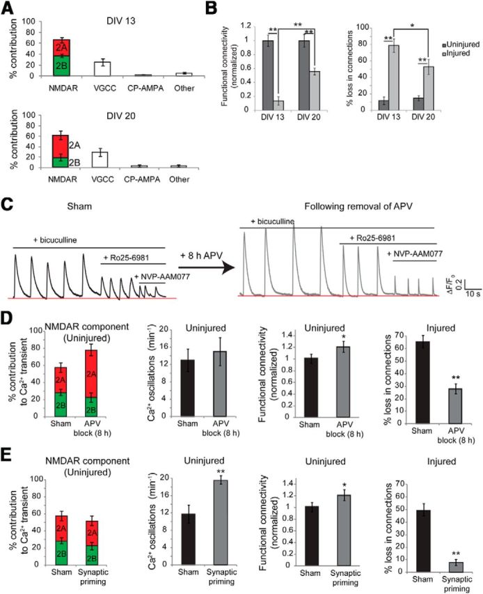
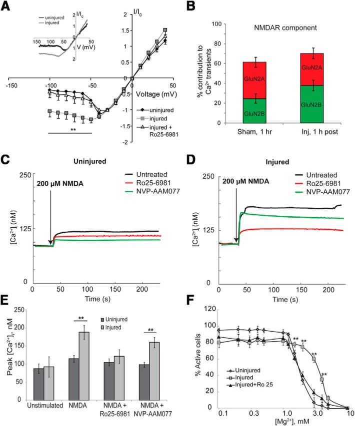
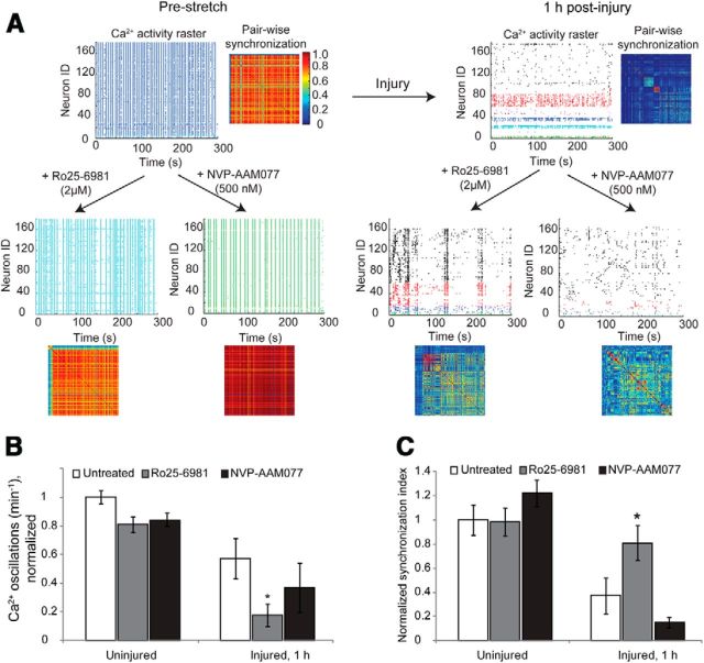
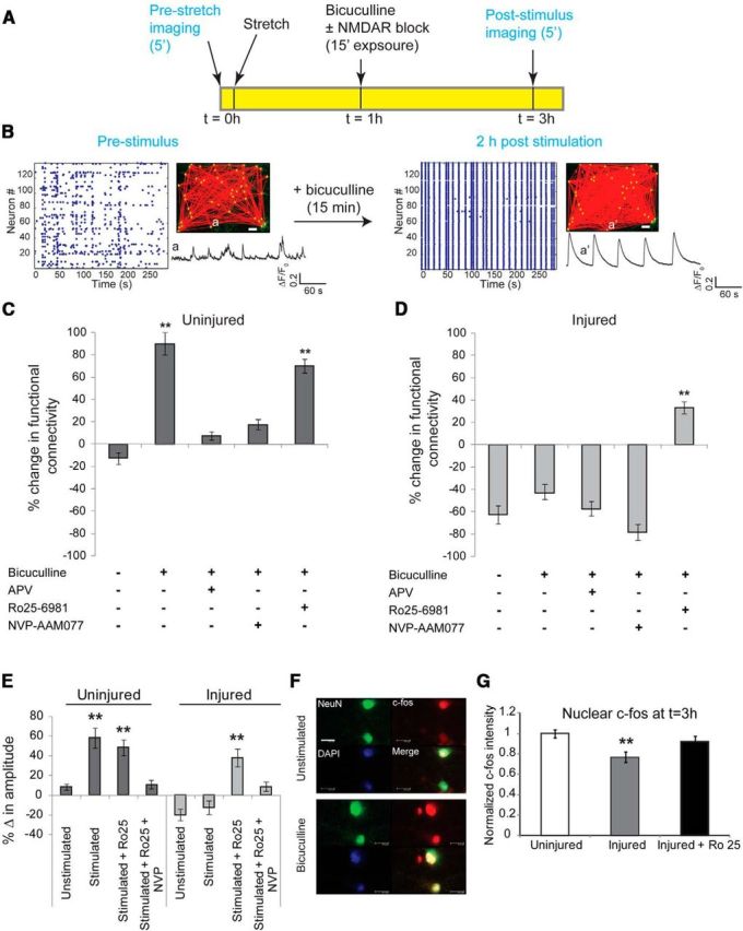
Alterations in the activity of neural circuits are a common consequence of traumatic brain injury (TBI), but the relationship between single-neuron properties and the aggregate network behavior is not well understood. We recently reported that the GluN2B-containing NMDA receptors (NMDARs) are key in mediating mechanical forces during TBI, and that TBI produces a complex change in the functional connectivity of neuronal networks. Here, we evaluated whether cell-to-cell heterogeneity in the connectivity and aggregate contribution of GluN2B receptors to [Ca(2+)]i before injury influenced the functional rewiring, spontaneous activity, and network plasticity following injury using primary rat cortical dissociated neurons. We found that the functional connectivity of a neuron to its neighbors, combined with the relative influx of calcium through distinct NMDAR subtypes, together contributed to the individual neuronal response to trauma. Specifically, individual neurons whose [Ca(2+)]i oscillations were largely due to GluN2B NMDAR activation lost many of their functional targets 1 h following injury. In comparison, neurons with large GluN2A contribution or neurons with high functional connectivity both independently protected against injury-induced loss in connectivity. Mechanistically, we found that traumatic injury resulted in increased uncorrelated network activity, an effect linked to reduction of the voltage-sensitive Mg(2+) block of GluN2B-containing NMDARs. This uncorrelated activation of GluN2B subtypes after injury significantly limited the potential for network remodeling in response to a plasticity stimulus. Together, our data suggest that two single-cell characteristics, the aggregate contribution of NMDAR subtypes and the number of functional connections, influence network structure following traumatic injury.

Keywords: GluN2B; functional connectivity; mechanical injury; network activity; synchrony.


Figures

Figure 1.
Figure 1.
Traumatic mechanical injury in vitro leads to a loss in functional connectivity mediated by NMDAR activation. A, The spontaneous activity of a population of neurons in vitro was recorded using a virally transduced fluorescent calcium indicator (GCaMP5). Middle, Fluorescence time series of three example neurons is depicted; the dot denotes the onset of calcium transient. Phase synchronization index of each pairwise neuron reflects the degree of temporal similarity in spontaneous activity. Statistically significant synchronized interactions are defined as functional connections and yield a functional connectivity map for the network (right). B, For stretch injury, neurons were plated and cultured on a SILASTIC membrane, and a small area of the membrane was dynamically stretched to peak at 35% strain within 15 ms. This level of stretch injury resulted in the immediate influx of Ca2+and could be prevented by pretreating neurons with the NMDA receptor antagonist APV. The solid black line is the average somatic calcium fluorescence trace of 122 neurons, with the 95% confidence interval shown in gray. C, Stretch injury causes an immediate drop in functional connectivity by 10 min that slowly recovers by 1 h. D, Functional connectivity of the network, defined as the average number of functional connections per neuron normalized by the number of neurons, was significantly reduced with injury by 10 min and 1 h. The effect of injury on network functional connectivity was dependent on NMDAR activation since APV pretreatment abolished the injury response. E, The frequency of calcium activity was also significantly reduced by 10 min and 1 h poststretch, and was dependent on NMDAR activation. Scale bar, 50 μm.
Figure 2.
Figure 2.
Profiling the contributions of NMDA receptor subtypes to intracellular calcium oscillations. We extended the calcium imaging methodology to profile the source of calcium for each neuron in the network. Inhibitory currents were blocked with bicuculline. The contribution of synaptically activated NMDA receptors to [Ca2+]I oscillations was assessed by comparing calcium oscillation amplitude in bicuculline to that in bicuculline plus APV. Similarly, the sensitivity to naspm and nimodipine was used to assess the contributions of calcium-permeable AMPA receptors and l-type voltage-gated channels to [Ca2+]i oscillations. An example of calcium receptor profiling of two neurons from the same field of view is illustrated in A and B. C, In general, the contribution of NMDARs and voltage-gated Ca2+ channels (VGCCs) to [Ca2+]I varied with developmental maturation of neurons. The NMDA receptor component of [Ca2+]i oscillations was further subdivided in fractional contribution from GluN2A and GluN2B subtypes with Ro25–6981 and NVP-AAM077, respectively (D). E, The cumulative distribution of NMDA receptor subtype-specific source of calcium influx varied across neurons from the same network but did not differ among networks from the same DIV. Immature neurons at DIV 13 had significantly different relative contributions of GluN2A and GluN2BN subtypes to [Ca2+]i oscillations (average cumulative distribution at DIV 13 and DIV 20, n = 10 wells each; N = 843 neurons at DIV 13 and N = 964 neurons at DIV 20; Kolmogorov–Smirnov test, p < 0.001). Scale bar, 50 μm. CP-AMPAR, Calcium-permeable AMPA receptors.
Figure 3.
Figure 3.
NMDAR subtype composition and connectivity influence functional rewiring of individual neurons following injury. A, The normalized functional connectivity of a neuron (i.e., its level of integration in the network) before stretch influenced post-stretch connectivity. B, Sparsely connected neurons (normalized CI, <0.3) showed a significant increase in functional connectivity following stretch injury compared with moderately connected neurons (CI, >0.3 and <0.7) or highly connected neurons (CI, >0.7). C, D, As a result of injury, a neuron could undergo one of three changes in its functional connectivity. Of the set of all functional targets, a fraction of connections may be lost as a result of injury, a fraction of connections may remain stable following injury, or there may be some newly formed functional connections that were not present in the uninjured network. The “lost,” “stable,” and “new” fractions for a given neuron compactly describe its extent of functional rewiring in the injured network. E, For each neuron, we plotted the fraction of total connections that were lost, remained stable, or appeared new as a result of injury on three independent axes. The contributions of GluN2A- and GluN2B-containing NMDARs to somatic calcium oscillations for each neuron before injury were projected on the 3D plot of its functional rewiring. F, We found a significant positive correlation between prestretch GluN2B contribution and deintegration from the network following injury. Additionally, highly connected neurons retained many of their functional targets following stretch injury, whereas sparsely connected neurons participated in network remodeling by forming new functional targets (F).
Figure 4.
Figure 4.
Manipulating GluN2B NMDAR signaling and NMDAR mechanosensitivity influences the network structure after injury. A, The contribution of NMDAR subtypes to somatic calcium oscillations shifted from GluN2B dominant at DIV 13 to GluN2A dominant at DIV 21. B, The same level of stretch injury yielded a greater deficit in functional connectivity in GluN2B-dominant young (DIV 13) neurons relative to the GluN2A-dominant mature neurons (DIV 20). C, Chronic inhibition of NMDA receptors (25 μm APV for 8 h) led to homeostatic enhancement of the GluN2A contribution to calcium oscillations in the young DIV 13 network upon release from inhibition. The frequency of spontaneous calcium activity and the functional connectivity of the network did not change as a result of APV treatment. D, However, stretch injury attenuated the loss in functional connectivity in DIV 13 neurons treated with APV relative to age-matched untreated cultures, suggesting that single-cell prestretch GluN2B contribution influences deintegration from the network. E, The mechanosensitivity of GluN2B receptors was reduced by inducing a brief period of heightened synaptic activity and acute synaptic priming. The relative contributions of GluN2A and GluN2B subtypes did not change as a result of synaptic priming. Compared with age-matched untreated cultures, cultures treated with bicuculline were protected against stretch-induced loss in network functional connectivity.
Figure 5.
Figure 5.
GluN2B-containing NMDA receptors undergo stretch induced reduction in Mg2+ block and results in a selective enhancement of GluN2B NMDAR calcium influx. NMDA-activated I–V relationship of stretch-injured neurons was more linear compared with the typical J shape of uninjured neurons. A, Blocking the GluN2B subtypes (Ro25–6981) restored the J curve, suggesting that injury specifically reduces the voltage-dependent Mg2+ block of GluN2B-containing NMDA receptors (average response of n = 10 neurons; inset, raw traces of representative single uninjured and injured neurons). B, Consistent with partial relief of GluN2B Mg2+ block, stretch-injured neurons also had a significantly greater calcium influx from GluN2B receptors during spontaneous network activity. C, D, To further quantify this change, all sources of calcium except the NMDAR were blocked, and calcium entry in response to NMDA (200 μm) was measured with the fluorescent ratiometric calcium indicator fura-2. NMDA stimulation yielded a significantly greater peak [Ca2+]i in injured neurons compared with age-matched uninjured samples (102% rise over baseline injured vs 36% uninjured). E, Blocking the GluN2B subtypes in injured neurons significantly attenuated the NMDA-induced increase in [Ca2+]i to uninjured levels. F, In a separate set of experiments, the spontaneous activity of cultures was recorded in increasing [Mg2+]e ranging from 0 to 10 mm and the percentage of active neurons at each [Mg2+]e value computed. In uninjured cultures, raising [Mg2+]e from physiologic 0.8 to 3.0 mm almost completely abolished spontaneous activity, whereas the effect of [Mg2+]e block was significantly reduced in injured cultures. Sensitivity to [Mg2+]e was restored in injured cultures by blocking GluN2B subtypes. **p < 0.01.
Figure 6.
Figure 6.
Stretch-induced reduction in GluN2B NMDAR Mg2+ block causes asynchronous circuit activity. A–C, The pattern of spontaneous calcium activity in mature cultures was characterized by rhythmic, synchronized oscillations and was unaffected by antagonism of either GluN2A or GluN2B subtypes. In representative raster plots, unique temporal patterns of calcium activity are color coded for visualization; a pairwise synchronization matrix depicts the level of temporal correlation between the calcium activities of neuronal pairs across the network. Blocking either GluN2A or GluN2B subtypes separately did not change either the frequency or the synchronization of calcium activity in uninjured cultures. In contrast, 1 h following stretch injury, both the frequency of calcium activity of individual neurons and the temporal pattern of activity across the network were significantly reduced. B, C, Inhibiting GluN2B subtypes in an injured network further reduced the amount of calcium activity (B); however, the remaining spontaneous activity was more synchronized (C), suggesting that the activation of GluN2B subtypes underlies asynchronous firing. Antagonism of GluN2A subtypes reduced both the frequency and the synchronization; however, they were not significantly different from untreated injured cultures (p > 0.05).
Figure 7.
Figure 7.
Activation of GluN2B NMDARs in injured neurons reduces network augmentation induced with synchronization. A, Plasticity through recurrent excitation was induced by blocking inhibitory currents with bicuculline for 15 min. Calcium activity of the network was recorded before injury, the network was subjected to stretch injury and stimulated with bicuculline, and the calcium activity of the same field of view was recorded again 2 h after bicuculline washout. B, In uninjured cultures, a brief period of pharmacological blockade of inhibitory neurons resulted in a persistent increase in synchronized activity and functional connectivity, and an augmentation in single-cell somatic calcium amplitude. Inset, GCaMP5 trace of the same neuron before (a) and 2 h after bicuculline stimulus (a′), illustrating the emergence of rhythmic, large-amplitude calcium oscillations in a′ compared with a. Scale bar, 50 μm. C, We used the percentage change in functional connectivity as one of three metrics to explore the plasticity of the circuit [change in somatic calcium amplitude (E) and nuclear localization of c-fos (G) are the other two metrics]. In uninjured cultures, 15 min of bicuculline treatment led to a significant increase in functional connectivity relative to no stimulus (change in functional connectivity following bicuculline stimulus, 89 ± 9.2%; change in functional connectivity untreated, −12 ± 5.4%; t test, p < 0.001, df = 4). Enhancement in connectivity was abolished by blocking either the activation of NMDARs or specifically targeting GluN2A-containing NMDARs (bicuculline plus APV, 8.2 ± 3.1%; bicuculline plus NVP-AAM077, 17.8 ± 4.7%). D, Injury resulted in a significant decrease in network connectivity (injured, −62 ± 7.3%; uninjured, −12 ± 5.4%; p < 0.001), which could not be restored with bicuculline stimulus (−44 ± 8.5%). However, blocking GluN2B NMDARs during the period induced synchronization led to a significant enhancement in global connectivity (treatment with bicuculline plus Ro25–6981, 37 ± 4.1%; treatment with bicuculline, −44 ± 8.5%; p < 0.001). E, Amplitude of somatic calcium transients increased significantly following washout of bicuculline stimulus in uninjured cultures, which is indicative of enhanced excitability (unstimulated change in amplitude, 8.5 ± 2.2%; change in amplitude following bicuculline stimulus, 58.4 ± 9.1%; p < 0.001). This was dependent on activation of GluN2A-containing NMDARs since exposure to NVP-AAM077 during the period of bicuculline stimulus abolished the change in somatic calcium amplitude (10.2 ± 4.7%). In comparison, the amplitude of somatic calcium transients was significantly lower following injury (−20.2 ± 5.5%) and did not recover following bicuculline treatment (−13.8 ± 6.2%). However, antagonism of GluN2B-containing NMDARs during bicuculline stimulus recovered somatic amplitude to near the levels observed in uninjured cultures (change after bicuculline plus Ro25 stimulus, 37.7 ± 7.7%; change after bicuculline stimulus, −13.8 ± 6.2%; p < 0.001). F, G, We probed the localization of the immediate early transcription factor c-fos and found increased nuclear localization within 2 h of bicuculline treatment in uninjured cultures. Nuclear localization of c-fos was significantly reduced in injured cultures (normalized injured c-fos intensity, 0.77 ± 0.04; normalized uninjured c-fos intensity, 1.0 ± 0.03; p < 0.01) but could be restored by blocking GluN2B-containing NMDARs during the period of bicuculline stimulation (0.91 ± 0.05). Scale bar, 100 μm.

Similar articles

Cited by

References

    1. Akerboom J, Chen TW, Wardill TJ, Tian L, Marvin JS, Mutlu S, Calderón NC, Esposti F, Borghuis BG, Sun XR, Gordus A, Orger MB, Portugues R, Engert F, Macklin JJ, Filosa A, Aggarwal A, Kerr RA, Takagi R, Kracun S, et al. Optimization of a GCaMP calcium indicator for neural activity imaging. J Neurosci. 2012;32:13819–13840. doi: 10.1523/JNEUROSCI.2601-12.2012. - DOI - PMC - PubMed
    1. Alexander MP. Mild traumatic brain injury: pathophysiology, natural history, and clinical management. Neurology. 1995;45:1253–1260. doi: 10.1212/WNL.45.7.1253. - DOI - PubMed
    1. Arnold FJ, Hofmann F, Bengtson CP, Wittmann M, Vanhoutte P, Bading H. Microelectrode array recordings of cultured hippocampal networks reveal a simple model for transcription and protein synthesis-dependent plasticity. J Physiol. 2005;564:3–19. doi: 10.1113/jphysiol.2004.077446. - DOI - PMC - PubMed
    1. Arrondo G, Alegre M, Sepulcre J, Iriarte J, Artieda J, Villoslada P. Abnormalities in brain synchronization are correlated with cognitive impairment in multiple sclerosis. Mult Scler. 2009;15:509–516. doi: 10.1177/1352458508101321. - DOI - PubMed
    1. Auberson YP, Allgeier H, Bischoff S, Lingenhoehl K, Moretti R, Schmutz M. 5-Phosphonomethylquinoxalinediones as competitive NMDA receptor antagonists with a preference for the human 1A/2A, rather than 1A/2B receptor composition. Bioorg Med Chem Lett. 2002;12:1099–1102. doi: 10.1016/S0960-894X(02)00074-4. - DOI - PubMed

Publication types

Substances

LinkOut - more resources