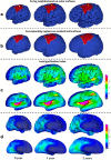
Mapping longitudinal development of local cortical gyrification in infants from birth to 2 years of age
- PMID: 24647943
- PMCID: PMC3960466
- DOI: 10.1523/JNEUROSCI.3976-13.2014
Mapping longitudinal development of local cortical gyrification in infants from birth to 2 years of age
Abstract
Human cortical folding is believed to correlate with cognitive functions. This likely correlation may have something to do with why abnormalities of cortical folding have been found in many neurodevelopmental disorders. However, little is known about how cortical gyrification, the cortical folding process, develops in the first 2 years of life, a period of dynamic and regionally heterogeneous cortex growth. In this article, we show how we developed a novel infant-specific method for mapping longitudinal development of local cortical gyrification in infants. By using this method, via 219 longitudinal 3T magnetic resonance imaging scans from 73 healthy infants, we systemically and quantitatively characterized for the first time the longitudinal cortical global gyrification index (GI) and local GI (LGI) development in the first 2 years of life. We found that the cortical GI had age-related and marked development, with 16.1% increase in the first year and 6.6% increase in the second year. We also found marked and regionally heterogeneous cortical LGI development in the first 2 years of life, with the high-growth regions located in the association cortex, whereas the low-growth regions located in sensorimotor, auditory, and visual cortices. Meanwhile, we also showed that LGI growth in most cortical regions was positively correlated with the brain volume growth, which is particularly significant in the prefrontal cortex in the first year. In addition, we observed gender differences in both cortical GIs and LGIs in the first 2 years, with the males having larger GIs than females at 2 years of age. This study provides valuable information on normal cortical folding development in infancy and early childhood.
Keywords: cortical folding; cortical surface; infant; local gyrification; longitudinal development.
Figures


References
-
- Brun CC, Leporé N, Pennec X, Lee AD, Barysheva M, Madsen SK, Avedissian C, Chou YY, de Zubicaray GI, McMahon KL, Wright MJ, Toga AW, Thompson PM. Mapping the regional influence of genetics on brain structure variability—a tensor-based morphometry study. Neuroimage. 2009;48:37–49. doi: 10.1016/j.neuroimage.2009.05.022. - DOI - PMC - PubMed
Publication types
MeSH terms
Grants and funding
- R01 EB006733/EB/NIBIB NIH HHS/United States
- R01 AG041721/AG/NIA NIH HHS/United States
- EB008374/EB/NIBIB NIH HHS/United States
- MH088520/MH/NIMH NIH HHS/United States
- R01 NS055754/NS/NINDS NIH HHS/United States
- EB008760/EB/NIBIB NIH HHS/United States
- EB009634/EB/NIBIB NIH HHS/United States
- R01 MH070890/MH/NIMH NIH HHS/United States
- U01 MH070890/MH/NIMH NIH HHS/United States
- NS055754/NS/NINDS NIH HHS/United States
- MH064065/MH/NIMH NIH HHS/United States
- MH070890/MH/NIMH NIH HHS/United States
- R01 EB008374/EB/NIBIB NIH HHS/United States
- MH100217/MH/NIMH NIH HHS/United States
- HD053000/HD/NICHD NIH HHS/United States
- R03 EB008760/EB/NIBIB NIH HHS/United States
- R01 AG042599/AG/NIA NIH HHS/United States
- P50 MH064065/MH/NIMH NIH HHS/United States
- R01 EB009634/EB/NIBIB NIH HHS/United States
- RC1 MH088520/MH/NIMH NIH HHS/United States
- R01 HD053000/HD/NICHD NIH HHS/United States
- AG041721/AG/NIA NIH HHS/United States
- EB006733/EB/NIBIB NIH HHS/United States
- R01 MH100217/MH/NIMH NIH HHS/United States
LinkOut - more resources
Full Text Sources
Other Literature Sources
Medical
