

Postnatal development of the hippocampus in the Rhesus macaque (Macaca mulatta): a longitudinal magnetic resonance imaging study
- PMID: 24648155
- PMCID: PMC4065201
- DOI: 10.1002/hipo.22271
Postnatal development of the hippocampus in the Rhesus macaque (Macaca mulatta): a longitudinal magnetic resonance imaging study
Abstract
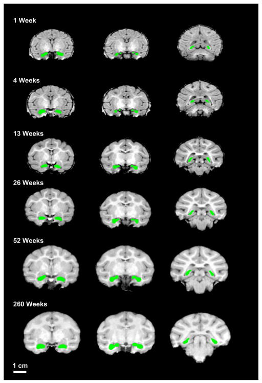
Nonhuman primates are widely used models to investigate the neural substrates of human behavior, including the development of higher cognitive and affective function. Due to their neuroanatomical and behavioral homologies with humans, the rhesus macaque monkey (Macaca mulatta) provides an excellent animal model in which to characterize the maturation of brain structures from birth through adulthood and into senescence. To evaluate hippocampal development in rhesus macaques, structural magnetic resonance imaging scans were obtained longitudinally at 9 time points between 1 week and 260 weeks (5 years) of age on 24 rhesus macaque monkeys (12 males, 12 females). In our sample, the hippocampus reaches 50% of its adult volume by 13 weeks of age and reaches an adult volume by 52 weeks in both males and females. The hippocampus appears to be slightly larger at 3 years than at 5 years of age. Male rhesus macaques have larger hippocampi than females from 8 weeks onward by approximately 5%. Interestingly, there was increased variability in hemispheric asymmetry for hippocampus volumes at younger ages than at later ages. These data provide a comprehensive evaluation of the longitudinal development of male and female rhesus macaque hippocampus across development from 1 week to 5 years of age.
Keywords: Rhesus macaque; hippocampus; longitudinal development; magnetic resonance imaging; primate model.
© 2014 Wiley Periodicals, Inc.
Figures


References
-
- Alvarado MC, Bachevalier J. Selective neurotoxic damage to the hippocampal formation impairs performance of the transverse patterning and location memory tasks in rhesus macaques. Hippocampus. 2005;15:118–131. - PubMed
-
- Amaral DG, Bauman MD, Schumann CM. The amygdala and autism: implications from non-human primate studies. Genes Brain Behav. 2003;2:295–302. - PubMed
-
- Bachevalier J, Alvarado MC, Malkova L. Memory and socioemotional behavior in monkeys after hippocampal damage incurred in infancy or in adulthood. Biol Psychiatry. 1999;46:329–339. - PubMed
Publication types
MeSH terms
Grants and funding
LinkOut - more resources
Full Text Sources
Other Literature Sources
Medical
