

Spatial specificity of working memory representations in the early visual cortex
- PMID: 24648192
- PMCID: PMC4527611
- DOI: 10.1167/14.3.22
Spatial specificity of working memory representations in the early visual cortex
Abstract
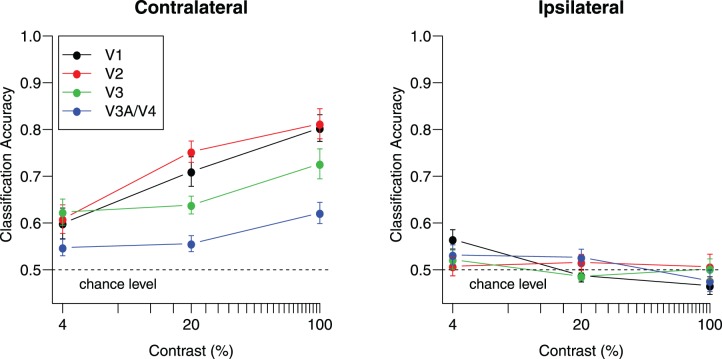
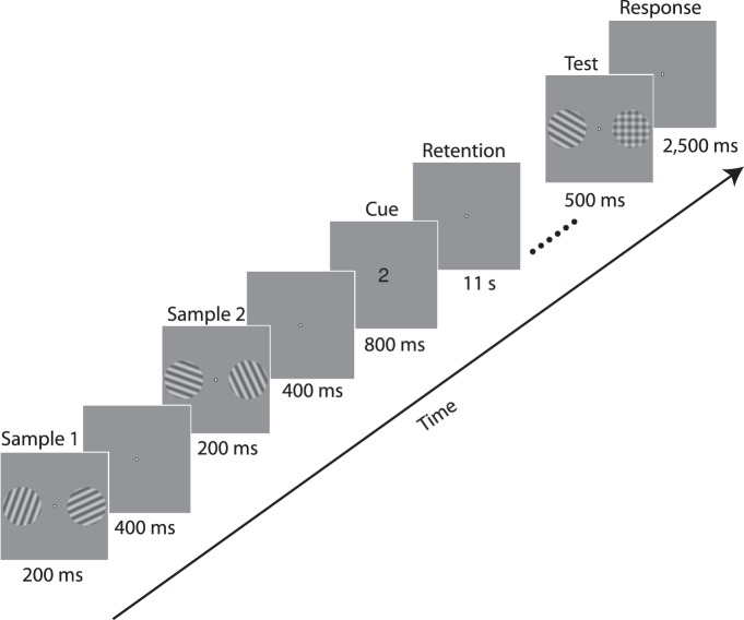
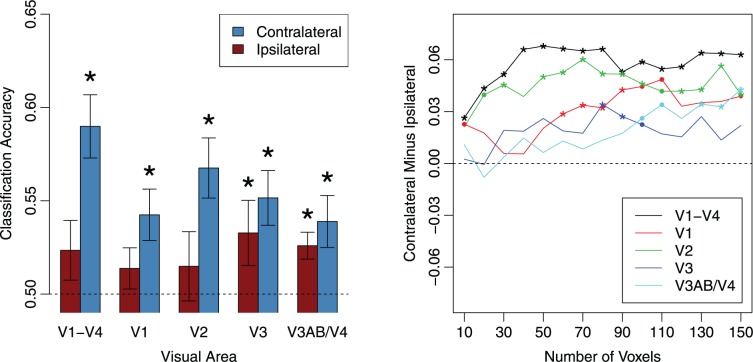
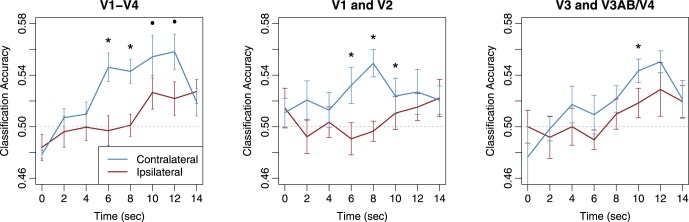
Recent fMRI decoding studies have demonstrated that early retinotopic visual areas exhibit similar patterns of activity during the perception of a stimulus and during the maintenance of that stimulus in working memory. These findings provide support for the sensory recruitment hypothesis that the mechanisms underlying perception serve as a foundation for visual working memory. However, a recent study by Ester, Serences, and Awh (2009) found that the orientation of a peripheral grating maintained in working memory could be classified from both the contralateral and ipsilateral regions of the primary visual cortex (V1), implying that, unlike perception, feature-specific information was maintained in a nonretinotopic manner. Here, we evaluated the hypothesis that early visual areas can maintain information in a spatially specific manner and will do so if the task encourages the binding of feature information to a specific location. To encourage reliance on spatially specific memory, our experiment required observers to retain the orientations of two laterally presented gratings. Multivariate pattern analysis revealed that the orientation of each remembered grating was classified more accurately based on activity patterns in the contralateral than in the ipsilateral regions of V1 and V2. In contrast, higher extrastriate areas exhibited similar levels of performance across the two hemispheres. A time-resolved analysis further indicated that the retinotopic specificity of the working memory representation in V1 and V2 was maintained throughout the retention interval. Our results suggest that early visual areas provide a cortical basis for actively maintaining information about the features and locations of stimuli in visual working memory.
Keywords: decoding; fMRI; feature-based attention; imagery; pattern classification; visual short-term memory.
Figures
Similar articles
-
Spatially global representations in human primary visual cortex during working memory maintenance.J Neurosci. 2009 Dec 2;29(48):15258-65. doi: 10.1523/JNEUROSCI.4388-09.2009. J Neurosci. 2009. PMID: 19955378 Free PMC article.
-
Decoupling of BOLD amplitude and pattern classification of orientation-selective activity in human visual cortex.Neuroimage. 2018 Oct 15;180(Pt A):31-40. doi: 10.1016/j.neuroimage.2017.09.046. Epub 2017 Sep 22. Neuroimage. 2018. PMID: 28951159
-
Visual short-term memory: activity supporting encoding and maintenance in retinotopic visual cortex.Neuroimage. 2012 Oct 15;63(1):166-78. doi: 10.1016/j.neuroimage.2012.06.053. Epub 2012 Jul 6. Neuroimage. 2012. PMID: 22776452
-
Functional analysis of primary visual cortex (V1) in humans.Proc Natl Acad Sci U S A. 1998 Feb 3;95(3):811-7. doi: 10.1073/pnas.95.3.811. Proc Natl Acad Sci U S A. 1998. PMID: 9448245 Free PMC article. Review.
-
Multi-Voxel Decoding and the Topography of Maintained Information During Visual Working Memory.Front Syst Neurosci. 2016 Feb 15;10:2. doi: 10.3389/fnsys.2016.00002. eCollection 2016. Front Syst Neurosci. 2016. PMID: 26912997 Free PMC article. Review.
Cited by
-
Distinct neural mechanisms for spatially lateralized and spatially global visual working memory representations.J Neurophysiol. 2016 Oct 1;116(4):1715-1727. doi: 10.1152/jn.00991.2015. Epub 2016 Jul 20. J Neurophysiol. 2016. PMID: 27440249 Free PMC article.
-
Gaining knowledge mediates changes in perception (without differences in attention): A case for perceptual learning.Behav Brain Sci. 2016 Jan;39:e240. doi: 10.1017/S0140525X15002496. Behav Brain Sci. 2016. PMID: 28355843 Free PMC article.
-
Preoperative Altered Spontaneous Brain Activity and Functional Connectivity Were Independent Risk Factors for Delayed Neurocognitive Recovery in Older Adults Undergoing Noncardiac Surgery.Neural Plast. 2020 Jun 16;2020:9796419. doi: 10.1155/2020/9796419. eCollection 2020. Neural Plast. 2020. PMID: 32617099 Free PMC article.
-
Parietal and Frontal Cortex Encode Stimulus-Specific Mnemonic Representations during Visual Working Memory.Neuron. 2015 Aug 19;87(4):893-905. doi: 10.1016/j.neuron.2015.07.013. Epub 2015 Aug 6. Neuron. 2015. PMID: 26257053 Free PMC article.
-
Predicting Delayed Neurocognitive Recovery After Non-cardiac Surgery Using Resting-State Brain Network Patterns Combined With Machine Learning.Front Aging Neurosci. 2021 Nov 12;13:715517. doi: 10.3389/fnagi.2021.715517. eCollection 2021. Front Aging Neurosci. 2021. PMID: 34867266 Free PMC article.
References
-
- Albers A. M., Kok P., Toni I., Dijkerman H. C., de Lange F. P. (2013). Shared representations for working memory and mental imagery in early visual cortex. Current Biology, 23 (15), 1427–1431. - PubMed
-
- Baddeley A. (1986). Working memory. Oxford, UK: Oxford University Press.
Publication types
MeSH terms
Grants and funding
LinkOut - more resources
Full Text Sources
Other Literature Sources
