. 2014 May 15;74(10):2825-34.
doi: 10.1158/0008-5472.CAN-13-3157. Epub 2014 Mar 19.

EGFR-mediated chromatin condensation protects KRAS-mutant cancer cells against ionizing radiation

Affiliations

EGFR-mediated chromatin condensation protects KRAS-mutant cancer cells against ionizing radiation

Meng Wang et al. Cancer Res. .

Abstract

Therapeutics that target the epidermal growth factor receptor (EGFR) can enhance the cytotoxic effects of ionizing radiation (IR). However, predictive genomic biomarkers of this radiosensitization have remained elusive. By screening 40 non-small cell lung cancer cell (NSCLC) lines, we established a surprising positive correlation between the presence of a KRAS mutation and radiosensitization by the EGFR inhibitors erlotinib and cetuximab. EGFR signaling in KRAS-mutant NSCLC cells promotes chromatin condensation in vitro and in vivo, thereby restricting the number of DNA double-strand breaks (DSB) produced by a given dose of IR. Chromatin condensation in interphase cells is characterized by an unexpected mitosis-like colocalization of serine 10 phosphorylation and lysine 9 trimethylation on histone H3. Aurora B promotes this process in a manner that is codependent upon EGFR and protein kinase C α (PKCα). PKCα, in addition to MEK/ERK signaling, is required for the suppression of DSB-inducible premature senescence by EGFR. Blockade of autophagy results in a mutant KRAS-dependent senescence-to-apoptosis switch in cancer cells treated with IR and erlotinib. In conclusion, we identify EGFR as a molecular target to overcome a novel mechanism of radioresistance in KRAS-mutant tumor cells, which stands in contrast to the unresponsiveness of KRAS-mutant cancers to EGFR-directed agents in monotherapy. Our findings may reposition EGFR-targeted agents for combination with DSB-inducing therapies in KRAS-mutant NSCLC.


Conflict of interest statement

Conflicts of Interest: Jeff Settleman, Ph.D., is employed by Genentech Inc., San Francisco, CA

Figures

Figure 1
Figure 1
Lung cancer cell line screening identifies a positive correlation between KRAS mutation and radiosensitization by EGFR-directed agents. A, Left panel, pie charts illustrate percentage of cell lines radiosensitized in the syto60 short-term assay by either erlotinib (2 µM) or cetuximab (100 nM) (see supplementary data, Fig. S1). Right panel, display of selected genomic profile of the top 11 cell lines radiosensitized by both agents, with mutations highlighted by a dark shade (curated from references Broad/Sanger). B, Scatter dot plots compare short-term radiosensitization factors (SRF2Gy) for erlotinib or cetuximab in KAS mutant and wild-type NSCLC cell lines. Dots represent averages of at least 3 biological repeats and horizontal lines indicate the overall mean. Statistical comparisons were performed with the unpaired T-test (two-tailed). C, Analogous to panel B, SRF2Gy comparisons were performed in isogenic cell pairs with wild-type (wt) or mutant (mut) KRAS, or cells expressing shKRAS (+) or a scrambled control (0). D, Clonogenic survival of NCI-H1703 cells after single dose irradiation with or without erlotinib (2 µM) treatment initiated 1 hour before irradiation. Statistical comparison was carried out using the F-test.
Figure 2
Figure 2
EGFR suppresses the production of ionizing radiation (IR) induced DNA double-strand breaks in KRAS-mutant cells. A, A549 cells following irradiation with 1 Gy with or without cetuximab treatment (100 nM) initiated 1 hour before irradiation. Percentage of cells with ≥ 20 foci/nucleus over time post-irradiation. P-value (T-test) for data points at 0.5 hours (h). B, Left, percentage of A549 cells with ≥ 20 53BP1 foci/nucleus 30 minutes after 1 Gy +/− erlotinib (2 µM). Right, result of single-cell gel electrophoresis. C, Percentage of A549 cells with ≥ 20 IR-induced γ-H2AX foci +/− erlotinib and +/− DNA-PKcs inhibitor NU7026 (10 µM). D, Fraction of nuclei with IR-induced EGFR translocation as determined by immunofluorescence microscopy (15 minutes after 8 Gy). E, Percentage of A549 cells with ≥ 20 γ-H2AX foci with erlotinib treatment initiated 1 hour before irradiation (left) or immediately after irradiation (right) and in each case maintained for 1.5 hours before staining. F, Analogous comparison of γ-H2AX foci in non-isogenic and isogenic cell lines with or without mutant K-Ras expression. In all panels, bars represent mean +/− standard error based on typically 2–3 biological repeats with EGFR-dependent fold-suppression indicated. Where indicated, IR-induced foci were calculated by subtracting baseline foci in non-irradiated cells.
Figure 3
Figure 3
EGFR promotes chromatin condensation in KRAS-mutant cells in-vitro and in-vivo. A, Left, representative transmission electron microscopy images illustrating a reduction of dense chromatin by erlotinib. Right, fraction of nuclei with high, intermediate, or low chromatin density in A549 cells treated with or without erlotinib for 1 hour. B, Western blot for A549 cells treated for 1 hour with or without EGF (100 ng/ml), erlotinib (2 µM), or cetuximab (100 nM). Representative gel from 2 independent experiments is shown. C, Left, representative histological and anti-H3K9me3 immunofluorescence images (40×) from A549 xenografts following treatment of mice with erlotinib alone or no treatment. Right, quantification of H3K9me3 signal based on 10 images. Statistical comparison was performed with the T-test. D, Analogous to panel C, the nuclear H3K9me3 signal was assessed in A549 cells in-vitro. E, Percentage of A549 cells with ≥ 20 IR-induced γ-H2AX foci with or without treatment with erlotinib or the histone methyltransferase inhibitor cheatocin. Western blot insert indicates the abrogation of H3K9me3 signal by chaetocin (100nM).
Fig. 4
Fig. 4
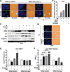
Regulation of interphase chromatin condensation by Aurora B kinase and PKCα. A, Whole-cell lysates from A549 cells with or without treatment with EGF (100 ng/ml) or erlotinib (2 µM) were subjected to Western blotting with antibodies against proteins as shown. B, Left, representative immunofluorescence images showing co-localized phospho-H3S10 and H3K9me3 using a specific dual antibody. Metaphase-type (m) and interphase-type (i) staining patterns are indicated. Right, percentage of cells with any phospho-H3S10/H3K9me3 signal. C, Percentage of nuclei with metaphase- or interphase-type phospo-H3S10/H3K9me3 staining with or without double thymidine block (TdR x2) to reduce the fraction of G2/M phase cells. D, Percentage of A549 cells with a high γ-H2AX staining intensity in G1- and G2/M phases based on FACS analysis 30 minutes after 8 Gy irradiation +/− erlotinib or +/− the Aurora B kinase inhibitor hesperadin. E, Percentage of NCI-H1703 cells with or without mutant K-Ras displaying ≥ 20 IR-induced γ-H2AX foci analogous to Fig. 2. F, Percentage of cells with γ-H2AX foci analogous to Fig. 2 +/− erlotinib or +/− the specific PKCα/β inhibitor Gö6976 (10 µM). G, Upper panel, Western blot of A549 cells transfected with scrambled control (CON) siRNA or siRNA against PKCα. Lower panel, analogous to Fig. 2B percentage of cells with 53BP1 foci +/− Gö6976 or siRNA transfections as shown. H, Upper panel, lysates from A549 cells following 1 hour treatment with or without EGF (100 ng/ml), erlotinib (2 µM), Gö6976 (10 µM), or the specific PKCα inhibitor Ro-32-0432 (100 nM) were subjected to Western blotting with antibodies against proteins as shown. Lower panel, percentage of cells with co-localized p-H3S10/H3K9me3 +/− erlotinib or +/− Gö6976. I, Percentage of H1703 cells with wild-type (wt) or mutant (mut) KRAS status displaying co-localized p-H3S10/H3K9me3 analogous to panel H. In all panels, bars represent mean +/− standard error based on typically 2–3 biological repeats.
Figure 5
Figure 5
A senescence-to-apoptosis switch in KRAS-mutant cancer cells. A, Representative images (40×) showing staining for DAPI and senescence-associated β-galactosidase (SA-β-gal) 3 days following 2 Gy irradiation in DLD1 (KRAS wt/mut) or DWT7 (wt/−) cells. B, Percentage of A549 cells staining for SA-β-gal staining 7 days following 2 Gy irradiation +/− erlotinib +/− the MEK inhibitor AZD6244 (250 nM), or Gö6976. C, Whole cell lysates from A549 cells 72 hours after irradiation (8 Gy) +/− erlotinib (2 µM) or +/− chloroquine (CQ; 25 mM) were subjected to Western blotting with antibodies against proteins as shown. D, Representative images for DAPI and SA-β-gal staining obtained in parallel to the data shown in panel D. E, Percentage of sub-G1 cells as determined by FACS in KRAS mutant DLD-1 and wild-type DWT7 cells for the treatments indicated. F, SRF2Gy values for DLD-1 and DWT7 tumor spheres treated as indicated. Statistical comparisons by one-sample or unpaired T-test. In all panels, bars represent mean +/− standard error based on typically 2–3 biological repeats.

References

    1. Ward JF. DNA damage produced by ionizing radiation in mammalian cells: identities, mechanisms of formation, and reparability. Prog Nucleic Acid Res Mol Biol. 1988;35:95–125. - PubMed
    1. Kegel P, Riballo E, Kuhne M, Jeggo PA, Lobrich M. X-irradiation of cells on glass slides has a dose doubling impact. DNA Repair (Amst) 2007;6:1692–1697. - PubMed
    1. Willers H, Held KD. Introduction to clinical radiation biology. Hematol Oncol Clin North Am. 2006;20:1–24. - PubMed
    1. Efimova EV, Mauceri HJ, Golden DW, Labay E, Bindokas VP, Darga TE, Chakraborty C, Barreto-Andrade JC, Crawley C, Sutton HG, Kron SJ, Weichselbaum RR. Poly(ADP-ribose) polymerase inhibitor induces accelerated senescence in irradiated breast cancer cells and tumors. Cancer Res. 2010;70:6277–6282. - PMC - PubMed
    1. Toulany M, Kasten-Pisula U, Brammer I, Wang S, Chen J, Dittmann K, Baumann M, Dikomey E, Rodemann HP. Blockage of epidermal growth factor receptor-phosphatidylinositol 3-kinase-AKT signaling increases radiosensitivity of K-RAS mutated human tumor cells in vitro by affecting DNA repair. Clin Cancer Res. 2006;12:4119–4126. - PubMed

Publication types

MeSH terms